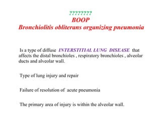
This document discusses various lung conditions including bronchiolitis obliterans, organizing pneumonia, and interstitial lung disease. It defines key terms and describes the symptoms, causes, diagnosis, and treatment of these conditions. Bronchiolitis obliterans involves scarring and obstruction of the bronchioles. Organizing pneumonia refers to unresolved pneumonia where exudates persist and cause fibrosis. Interstitial lung disease is a class of diffuse lung diseases involving inflammatory responses with varying degrees of pulmonary fibrosis. Specific forms like BOOP, COP, and IPF are mentioned along with their clinical presentation and pathological features.
4. Bronchiolitis
• Is an inflammatory reaction that follows damage to the
bronchiolar epithelium of the small conducting airways
Subsequent healing leads to excessive proliferation of granulation
tissue within the airway walls , lumen or both.
Depending on disease stage the repair process may cause
narrowing and distortion of the small airway (constrictive
bronchiolitis ) or complete obliteration ( bronchiolitis obliterans)
5. BRONCHIOLITIS OBLITERANS
• Rare form of non-reversible Obstructive lung disease
• Scarring
• Polyploidy obliteration of the lumen of the bronchioles without involving of
the distal lung parenchyma.
• Infants - adenovirus
6. BRONCHIOLITIS OBLITERANS
• Rare form of non-reversible Obstructive lung disease
• Scarring
• Polyploidy obliteration of the lumen of the bronchioles without
involving of the distal lung parenchyma.
• Infants - adenovirus
12. HISTORY
• HIPPOCRATES 460-370 BC
• 12 November world pneumonia day
• 450 million /year
• 4 million deaths /year
• 7% world’s total deaths
• Greatest < 5 >75 yrs
• 8th
leading death in the states 2009
13. PNEUMONIA IMAGING
• CXR does not always match the severity of pneumonia
• Not reliably separate between bacterial infection and
viral infection
• CXR typically clear within one month
17. Interstitial lung disease
• Is a class of diffuse lung diseases .
• It is a common pathological response of the lung to a wide variety
of known & unknown cause .
• It is often referred to interstitial pneumonia , IP or interstitial lung
disease ILD
• Until 1960 IPF remained largely a clinical diagnosis considered as
a single entity
• Has been felt to be a terminal disease with poor prognosis
because of lack of response to immunosuppressive therapy only
30% respond
• only 30% histopathology done
18. ILD
Several acute and chronic lung disorders with variable degrees of
pulmonary fibrosis ,variable degree of disease progression &
variable response to treatment.
Retrospective review of clinical & histopathology data allows
IPF to be classified into distinct subsets that differ in clinical
course , prognosis & response to treatment .
19. ILD
• NSIP , LIP , DIP ,RBI lung disease , UIP , OP
• In some ILDs , the small airways- the respiratory and terminal
bronchioles- are primarily affected .
respiratory bronchiolitis-associated interstitial lung disease
(RBILD)
idiopathic , smoking related in contrast to BO .
20. Known factors or insults lead to
Interstitial lung disease
• Drugs : antineoplastic (bleomycin , methotrexate )
: antimicrobial (nitrofrantoin , penicillin )
:amidorone
: hydroclorothiazide
• Hypersensitivity pneumonia
Farmer’s lung , pigeon breeder’s lung
. Dusts:
Asbestos , silica
Viral agents … mycoplasma pneumonia
. CD
21. ILD
.
It is important to differentiate terms used to define clinical entities
and terms describing the pathologic lesions of these disorders .
Clinical syndromes IPF,CFA ,idiopathic BOOP,COP,BO,BOS
,Hamman-Rich syndrome .
Pathological:UIP , LIP , giant cell interstitial pneumonitis ,
diffuse alveolar damage, non classifiable interstitial pneumonia ,
BOOP .
22. DD
ILD
• Clinical setting:-
• First: requires consideration of a variety of other pathological processes that
mimic the interstitial disorders in appropriate clinical setting
• Second: Physician should be aware of at least 150 other clinical entities &
situations associated with ILD
• Third: Owing the broad DD & the availability of various evolving invasive &
non-invasive diagnostic techniques, the best approach to use is to establish a
specific diagnosis which is frequently difficult to determine
• Fourth: In a significant proportion of patients a conclusive cause can't be
ascertained even when the most invasive diagnostic pathways are taken.
• Finally , even when a specific diagnosis is made, an effective therapeutic
regimen is not available for many patients with ILD
23. ????????
BOOP
Bronchiolitis obliterans organizing pneumonia
Is a type of diffuse INTERSTITIAL LUNG DISEASE that
affects the distal bronchioles , respiratory bronchioles , alveolar
ducts and alveolar wall.
Type of lung injury and repair
Failure of resolution of acute pneumonia
The primary area of injury is within the alveolar wall.
24. BOOP Hx
• As a distinct clinical entity 1st described by Davison in 1983
• Gary Epler 1985 gave the name BOOP
• Inflammation of the bronchioles & surrounding tissue in the
lung.
• Non infectious- infectious
• Complication of an existing chronic inflammatory disease such as
RA
• Side effect of certain medication such as amidarone
• MF 1:1
• Age 50-60 20-80
• No relation to smoking
25. Organizing pneumonia
• Refers to unresolved pneumonia in which alveolar exudates
persists & eventually undergo fibrosis .
. presence of buds of granulation tissue progressing from fibrin
exudates to loose collagen containing fibroblast.
Accessory finding in other inflammatory diseases such as
vasculitis
. It may also be a feature of organizing stage of ARDS
• A complication of an existing chronic inflammatory disease
or a side effect of certain medication .
26. • organizing Pneumonia of determined causes
• organizing Pneumonia of unknown cause occurring in a
specific context causes
• Cryptogenic organizing pneumonia
27. Undetermined causes of organizing
Pneumonia
• RA , Sjorgen syndrome---------- SLE ,SS
• Sweet”s disease
• HCV thyroiditis
• Crhons disease…..UC
• Behcets”s …. Wegener”s granulomatosis ….PN
• Polymyalgia Rheumatica
• Myelodysplasia
• Leukemia ….. Cancer
• Mesangiocapillary GN
• Chronic rejection …. Graft versus host disease
29. • It was long been known that radiation may induce radiation
pneumonitis
• However a syndrome similar to COP has been identified recently
• It differs strikingly from usual radiation pneumonitis in that the
pulmonary infiltrate occur or migrate outside the radiation fields
add respond good to steroids.
• Interestingly, unilateral breast irradiation, whether or not it results
in pneumonitis , has been shown to induce bilateral lymphocytic
alveolitis with activated CD4+Tcells
30. Infectious causes of organising
pneumonia
• Bacteria
Chlamydia pneumoniae
Coxiella burnetii
Legionella pneumophila
Mycoplasma pneumoniae
Nocardia asteroides
Pseudomonas aeruginosa
Serratia marcescens
Staphylococcus aureus
Streptococcus group B (newborn treated by extracorporeal oxygenation)
Streptococcus pneumoniae
31. Infectious causes of organising pneumonia
cont…..
• Viruses
Herpes virus
Human immunodeficiency virus
Influenza virus
Parainfluenza virus
• Parasites
Plasmodium vivax
• Fungi
Cryptococcus neoformans
Penicillium janthinellum
Pneumocystis carinii (in AIDS )
32. Clinical approach
• Medical Hx , environmental factors, occupational exposures,
medication & drug usage, FH
• Age , smoking, sex
• Onset & Duration of symptoms
• Rate of disease progression
• Extra pulmonary symptoms
34. BOOP diagnosis
• Diagnosis is suspected after no response to multiple antibiotics & blood &
sputum cultures are –ve for organisms
. Clinical picture & radiological images resemble infectious pneumonia
• CXR similar to an extensive pneumonia wide spread white patches
small nodular opacities 50% & large nodules in 15%
• Preservation of normal lung volume
• Migrate
• Persists or progresses
35. BOOP – CT
• CT may be used to confirm the diagnosis ( findings are typical enough to allow
the doctor to make the diagnosis without ordering additional tests)
• …airspace consolidation with airbronchogram , lower zone predominance ,
sub pleural or peribronchial distribution 50%.
• Ground glass appearance in most pt , honeycombing in some
• Diffuse bilateral infiltratation
• as a solitary focal lesion associated usually with subacute or chronic
inflammatory illness, it occurs in the upper lobes and may cavitate.
• .
36. BOOP - histopathology
• Confirmation
• Broncoscopic biopsy !
• VATS
• Histology: presence of polyploidy plugs of loose organizing connective tissue
( Masson bodies ) within the alveolar ducts , alveoli , bronchioles .
• Intraluminal polyps in respiratory bronchioles , alveolar ducts , alveolar spaces .
• Accompanied by organizing pneumonia in the more distant parenchyma
( proliferative bronchitis)
37. Diagnosis of exclusion
• COP is a diagnosis of exclusion .
• Once confirmation done , physician has to rule out :-
Chronic esinophilic pneumonia
Hypersensitivity pneumonia
Bacterial …. Viral
CD
Drug reaction