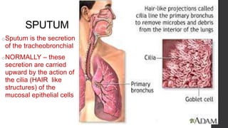
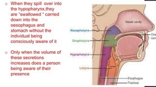
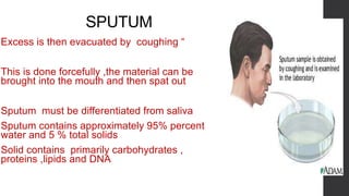
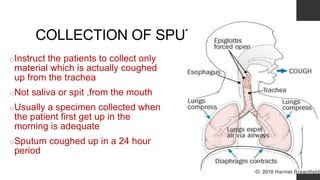
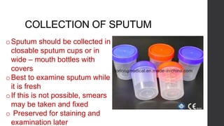
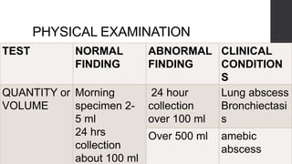
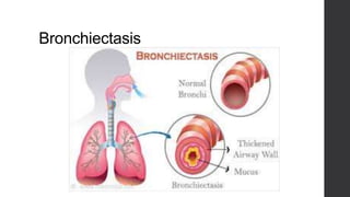
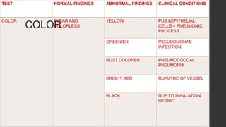
EXAMINATION OF
SPUTUM
SPUTUM
oSputum is the secretion
of the tracheobronchial
oNORMALLY – these
secretion are carried
upward by the action of
the cilia (HAIR like
structures) of the
mucosal epithelial cells
o When they spill over into
the hypopharynx,they
are “swallowed “ carried
down into the
oesophagus and
stomach without the
individual being
consciously aware of it
o Only when the volume of
these secretions
increases does a person
being aware of their
presence
SPUTUM
oExcess is then evacuated by coughing “
oThis is done forcefully ,the material can be
brought into the mouth and then spat out
oSputum must be differentiated from saliva
oSputum contains approximately 95% percent
water and 5 % total solids
oSolid contains primarily carbohydrates ,
proteins ,lipids and DNA
COLLECTION OF SPUTUM
oInstruct the patients to collect only
material which is actually coughed
up from the trachea
oNot saliva or spit ,from the mouth
oUsually a specimen collected when
the patient first get up in the
morning is adequate
oSputum coughed up in a 24 hour
period
COLLECTION OF SPUTUM
oSputum should be collected in
closable sputum cups or in
wide – mouth bottles with
covers
oBest to examine sputum while
it is fresh
oIf this is not possible, smears
may be taken and fixed
o Preserved for staining and
examination later
PHYSICAL EXAMINATION
TEST NORMAL
FINDING
ABNORMAL
FINDING
CLINICAL
CONDITION
S
QUANTITY or
VOLUME
Morning
specimen 2-
5 ml
24 hrs
collection
about 100 ml
24 hour
collection
over 100 ml
Lung abscess
Bronchiectasi
s
Over 500 ml amebic
abscess
Bronchiectasis
Bronchiectasis
LUNG ABSCESS
QUANTITY or VOLUME
Even if they have a cough , it is a dry cough without any
sputum –(NON – PRODUCTIVE COUGH)
Large amount of sputum – PRODUCTIVE COUGH
Some patients do not bring up much sputum during the
day
But in the morning after a night lying down
They may bring up a large volume of sputum
COLOR
TEST NORMAL FINDINGS ABNORMAL FINDINGS CLINICAL CONDITIONS
COLOR CLEAR AND
COLORLESS
YELLOW PUS &EPITHELIAL
CELLS – PNEUMONIC
PROCESS
GREENISH PSEUDOMONAS
INFECTION
RUST COLORED PNEUMOCOCCAL
PNEUMONIA
BRIGHT RED RUPUTRE OF VESSEL
BLACK DUE TO INHALATION
OF DIRT
PSEUDOMONAS INFECTION
RUST COLORED SPUTUM
CONSISTENCY AND APPEARANCE
When saliva is watery ,sputum always contains mucus which
gives an uneven or lumpy consistency
Sputum may be classified into various types according to its
consistency and appearance
CONSISTENCY AND APPEARANCE
Test Normal findings Abnormal findings Clinical conditions
Consistency and
appearance
Colorless, watery and
opalescent
Mucoid Acute bronchitis
Lobar pneumonia
Purulent Ruputred emphyema
bronchiectasis
Bloody Mitral stenosis
serous Frothy colorless or
yellow found in
pulmonary edema
Acute bronchitis
Lobar pneumonia
MITRAL STENOSIS
pulmonary edema
Miscellaneous findings
Occasionally various objects large
enough to be visible macroscopically
may be found in the sputum
BRONCHIAL CASTS
BRONCHOLITHS
FOREIGN BODIES
SULPHUR GRANULES
PARASITES
BRONCHIAL CASTS
oBranching structures of fibrin called
“fibrinous casts “
oFormed inside the bronchial tree in
some kinds of bronchitis and
pneumonia
oFound in the sputum when they
expelled by coughing
BRONCHOLITHS (LUNG STONES)
These are calcified bits of dead
or infected lung tissue
when found most likely would be
associated with chronic
tuberculosis or chronic fungus
infection in the lung
Sulphur granules
• Small yellowish lumps
• granules in the sputum
• Which are actually made up of colonies of
a fungus
• The yellow colour of Sulphur granules
• Seen in infection of the lungs by
actinomycosis species in actinomycosis
of the lung
parasites
• Misplaced intestinal parasites such
as round worm (Ascaris lumbricoides
) or the trematode
• ‘lung fluke’(paragoniums westermani
)
•In such countries as Japan,are
occasionally found in the sputum
FOREIGN BODIES
•These include any object a pateint,
especially a young child, has swallowed
and then aspirated by mistakes
including
• Pins
• Small glass marbles or beads
• Ground nuts etc
Chemical examination
•Usually no chemical tests are
done for sputum
•Occasionally test for occult blood
may be requested
•Same way as urine or stool
specimen
Concentration method for tubercle
bacilli in sputum
• Usually done on a 24 hour sputum
specimen collected in a sterile bottle
• Mix equal volume of sputum and 6%
sulphuric acid
• Let stand for 20 minutes
• Centrifuge at 3000 rpm for 30 minutes
Concentration method for tubercle
bacilli in sputum
•Decant the supernatant and carefully
wash the sediment with distilled water,
doing this three times
•Make a smear of the deposit
•Dry and fix the smear
•Stain it with ziel Neelsen stain
•The deposit may be cultured if desired
MICROSCOPIC EXAMINATION
• UNSTAINED SPUTUM
• A smear of unstained sputum ,on which a cover slip has been placed
• Usually examinaed first for any of the following features
• ELASTIC FIBRES
• CURSCHMANNS SPIRALS
• CHARCOT LEYDEN CRYSTALS
• PIGMENTED CELLS
• FUNGI
• PARASITES
Unstained sputum
• A smear of unstained sputum, on which a cover slip has been
placed
Usually examined first for any of the following features
1 .ELASTIC FIBRES
Appear ad wavy fibres in bundles
May be found by examining a number of wet coverslip
preparations
Also found in the concentration techniques
ELASTIC FIBRES
o10 ml of the sputum is mixed with
an equal volume of 10 g/dl sodium
hydroxide
oWhen the sputum is completely
emulsified it is centrifuges and the
sediment or deposit is examined
for the fibres
ELASTIC FIBRES
•Presence of elastic fibres in
sputum indicates the destructive
disease process in the lungs
CRUSCHMANN,S SPIRALS
•Appear as wiry spiral structures
with a central thread
•Structures are found in the
sputum of patients with asthma
CHARCOT – LEYDEN CRYSTALS
Fine needle- shaped Or hexagonal
Colourless crystals
Usually about 20 – 30 micron meter
in length called charcot – leyden
crystals
Often seen in the sputum of patients
with asthma
CHARCOT – LEYDEN CRYSTALS
• May not be present in freshly collected
sputum
• Seems to form as the sputum sites for
some time
• They arise from the disintegration of
eosinophils
• Stain strongly with eosin
CHARCOT – LEYDEN CRYSTALS
PIGMENTED CELLS
• LARGE MONONUCLEAR CELLS
CONTAINING PIGMENTS
• USUALLY MACROPHAGES
• TISSUE MONOCYTES
• SHOW DARK BLOOD PIGMENT OR
BLACK CARBON PIGMENT
FUNGI
• Colonies of
branching mycelia of
fungi
• Sulphur granules are
seen grossly
• Should be examined
microscopically to
see the branching
mycelia of fungus
PARASITES
• PARASITIC WORM LARVAE OR OVA
• OVA OF THE LUNG FLUKE - PARAGONIMUS
WESTERMANI
• HOOKLETS OF THE LARVAE FORM -
ECHINOCOCCUS GRANULOSA
• FILARIFORM LARVAE - STRONGYLOIDES
STERCORALIS
STAINED SPUTUM SMEAR
• SMEARS ARE MADE ON CLEAN ,DRY SLIDES
• SMEARS ARE DRIED AND FIXED OVER A FLAME
• STAINED WITH GRAMS STAIN FOR COCCOI OR BACILLI
• ZIEHL NEELSON STAIN FOR ACID FAST ORGANISMS
• FLUORESCENT STAINS - TUBERCLE BACILLI
• SPECIAL STAINS USED FOR FUNGI OR BACTERIA
• PAPANICOLAOU STAIN – DETAILED CYTOLOGIC STUDY FOR CANCER
CELLS
• WRIGHTS STAIN OR LEISHMAN STAIN – IDENTIFYING VARIOUS CELLS
INCLUDING
• POLYMORPHONUCLEAR LEUKOCYTES
• EOSINOPHILS
• LYMPHOCYTES
• LARGE MONONUCLEAR CELLS
• EPITHELIAL CELLS
• ERYTHROCYTES
POLYMORPHONUCLEAR LEUKOCYTES
• USUALLY NEUTROPHILS
• OCCASIONALLY EOSINOPHILS ARE SEEN
• FEW NEUTROPHILS MAY BE SSEN IN ANY CONDITIONS IN WHICH
SPUTUM IS INCREASED
• FRANK PUS – LUNG ABSCESS
• MANY NEUTROPHILS – PYOGENIC BACTERIA
EOSINOPHILS
• SEEN IN ASTHMATIC CONDITIONS
• EOSINOPHILIC LUNG
• PARASITIC INFESTATIONS IN THE LUNG
LYMPHOCYTES
LARGE NUMBER OF LYMPHOCYTES ARE FOUND IN EARLY TUBERCULOSIS
FEW LYMPHOCYTES MAY BE SEEN ALONG WITH THE NEUTROPHIL
POLYMORPHS
LARGE MONONUCLEAR CELLS
• MACROPHAGES COME FROM THE ALVEOLI
• LOWER BRONCHIAL TRACT
• THEIR PRESENCE CONFIRMS THAT THE SPECIMEN IS FROM THE LUNG
• NOT FROM THE MOUTH
EPITHELIAL CELLS
• DERIVED FROM THE MUCOSAL LINING OF THE TRACHEOBRONCHIAL
TREE
• EVEN FROM THE PULMONARY ALVEOLI
• COLUMNAR CELLS WITH CILIA AT ONE END
• FLAT CELLS WITH A CENTRAL CIRCULAR NUCLEUS
• SQUAMOUS CELLS ARE VERY SMALL DARK NUCLEI AND ABUNDANT
CYTOPLASM
• MATERIAL BEING EXAMINED IS FROM THE THROAT OR MOUTH
ERYTHROCYTES
• FEW ERYTHROCYTES MAY BE PRESENT IN THE SPUTUM
• MANY ERYTHROCYTES – HAEMORRHAGE INTO THE LUNGS OR
BRONCHI
 sputum

sputum

  • 2.
  • 3.
    SPUTUM oSputum is thesecretion of the tracheobronchial oNORMALLY – these secretion are carried upward by the action of the cilia (HAIR like structures) of the mucosal epithelial cells
  • 4.
    o When theyspill over into the hypopharynx,they are “swallowed “ carried down into the oesophagus and stomach without the individual being consciously aware of it o Only when the volume of these secretions increases does a person being aware of their presence
  • 5.
    SPUTUM oExcess is thenevacuated by coughing “ oThis is done forcefully ,the material can be brought into the mouth and then spat out oSputum must be differentiated from saliva oSputum contains approximately 95% percent water and 5 % total solids oSolid contains primarily carbohydrates , proteins ,lipids and DNA
  • 6.
    COLLECTION OF SPUTUM oInstructthe patients to collect only material which is actually coughed up from the trachea oNot saliva or spit ,from the mouth oUsually a specimen collected when the patient first get up in the morning is adequate oSputum coughed up in a 24 hour period
  • 7.
    COLLECTION OF SPUTUM oSputumshould be collected in closable sputum cups or in wide – mouth bottles with covers oBest to examine sputum while it is fresh oIf this is not possible, smears may be taken and fixed o Preserved for staining and examination later
  • 8.
    PHYSICAL EXAMINATION TEST NORMAL FINDING ABNORMAL FINDING CLINICAL CONDITION S QUANTITYor VOLUME Morning specimen 2- 5 ml 24 hrs collection about 100 ml 24 hour collection over 100 ml Lung abscess Bronchiectasi s Over 500 ml amebic abscess
  • 9.
  • 10.
  • 11.
  • 12.
    QUANTITY or VOLUME Evenif they have a cough , it is a dry cough without any sputum –(NON – PRODUCTIVE COUGH) Large amount of sputum – PRODUCTIVE COUGH Some patients do not bring up much sputum during the day But in the morning after a night lying down They may bring up a large volume of sputum
  • 13.
    COLOR TEST NORMAL FINDINGSABNORMAL FINDINGS CLINICAL CONDITIONS COLOR CLEAR AND COLORLESS YELLOW PUS &EPITHELIAL CELLS – PNEUMONIC PROCESS GREENISH PSEUDOMONAS INFECTION RUST COLORED PNEUMOCOCCAL PNEUMONIA BRIGHT RED RUPUTRE OF VESSEL BLACK DUE TO INHALATION OF DIRT
  • 14.
  • 15.
  • 18.
  • 19.
    CONSISTENCY AND APPEARANCE Whensaliva is watery ,sputum always contains mucus which gives an uneven or lumpy consistency Sputum may be classified into various types according to its consistency and appearance
  • 20.
    CONSISTENCY AND APPEARANCE TestNormal findings Abnormal findings Clinical conditions Consistency and appearance Colorless, watery and opalescent Mucoid Acute bronchitis Lobar pneumonia Purulent Ruputred emphyema bronchiectasis Bloody Mitral stenosis serous Frothy colorless or yellow found in pulmonary edema
  • 22.
  • 23.
  • 24.
  • 25.
  • 26.
    Miscellaneous findings Occasionally variousobjects large enough to be visible macroscopically may be found in the sputum BRONCHIAL CASTS BRONCHOLITHS FOREIGN BODIES SULPHUR GRANULES PARASITES
  • 27.
    BRONCHIAL CASTS oBranching structuresof fibrin called “fibrinous casts “ oFormed inside the bronchial tree in some kinds of bronchitis and pneumonia oFound in the sputum when they expelled by coughing
  • 28.
    BRONCHOLITHS (LUNG STONES) Theseare calcified bits of dead or infected lung tissue when found most likely would be associated with chronic tuberculosis or chronic fungus infection in the lung
  • 29.
    Sulphur granules • Smallyellowish lumps • granules in the sputum • Which are actually made up of colonies of a fungus • The yellow colour of Sulphur granules • Seen in infection of the lungs by actinomycosis species in actinomycosis of the lung
  • 30.
    parasites • Misplaced intestinalparasites such as round worm (Ascaris lumbricoides ) or the trematode • ‘lung fluke’(paragoniums westermani ) •In such countries as Japan,are occasionally found in the sputum
  • 31.
    FOREIGN BODIES •These includeany object a pateint, especially a young child, has swallowed and then aspirated by mistakes including • Pins • Small glass marbles or beads • Ground nuts etc
  • 32.
    Chemical examination •Usually nochemical tests are done for sputum •Occasionally test for occult blood may be requested •Same way as urine or stool specimen
  • 33.
    Concentration method fortubercle bacilli in sputum • Usually done on a 24 hour sputum specimen collected in a sterile bottle • Mix equal volume of sputum and 6% sulphuric acid • Let stand for 20 minutes • Centrifuge at 3000 rpm for 30 minutes
  • 34.
    Concentration method fortubercle bacilli in sputum •Decant the supernatant and carefully wash the sediment with distilled water, doing this three times •Make a smear of the deposit •Dry and fix the smear •Stain it with ziel Neelsen stain •The deposit may be cultured if desired
  • 35.
    MICROSCOPIC EXAMINATION • UNSTAINEDSPUTUM • A smear of unstained sputum ,on which a cover slip has been placed • Usually examinaed first for any of the following features • ELASTIC FIBRES • CURSCHMANNS SPIRALS • CHARCOT LEYDEN CRYSTALS • PIGMENTED CELLS • FUNGI • PARASITES
  • 36.
    Unstained sputum • Asmear of unstained sputum, on which a cover slip has been placed Usually examined first for any of the following features 1 .ELASTIC FIBRES Appear ad wavy fibres in bundles May be found by examining a number of wet coverslip preparations Also found in the concentration techniques
  • 37.
    ELASTIC FIBRES o10 mlof the sputum is mixed with an equal volume of 10 g/dl sodium hydroxide oWhen the sputum is completely emulsified it is centrifuges and the sediment or deposit is examined for the fibres
  • 38.
    ELASTIC FIBRES •Presence ofelastic fibres in sputum indicates the destructive disease process in the lungs
  • 39.
  • 40.
    CRUSCHMANN,S SPIRALS •Appear aswiry spiral structures with a central thread •Structures are found in the sputum of patients with asthma
  • 41.
    CHARCOT – LEYDENCRYSTALS Fine needle- shaped Or hexagonal Colourless crystals Usually about 20 – 30 micron meter in length called charcot – leyden crystals Often seen in the sputum of patients with asthma
  • 42.
    CHARCOT – LEYDENCRYSTALS • May not be present in freshly collected sputum • Seems to form as the sputum sites for some time • They arise from the disintegration of eosinophils • Stain strongly with eosin
  • 43.
  • 44.
    PIGMENTED CELLS • LARGEMONONUCLEAR CELLS CONTAINING PIGMENTS • USUALLY MACROPHAGES • TISSUE MONOCYTES • SHOW DARK BLOOD PIGMENT OR BLACK CARBON PIGMENT
  • 45.
    FUNGI • Colonies of branchingmycelia of fungi • Sulphur granules are seen grossly • Should be examined microscopically to see the branching mycelia of fungus
  • 46.
    PARASITES • PARASITIC WORMLARVAE OR OVA • OVA OF THE LUNG FLUKE - PARAGONIMUS WESTERMANI • HOOKLETS OF THE LARVAE FORM - ECHINOCOCCUS GRANULOSA • FILARIFORM LARVAE - STRONGYLOIDES STERCORALIS
  • 47.
    STAINED SPUTUM SMEAR •SMEARS ARE MADE ON CLEAN ,DRY SLIDES • SMEARS ARE DRIED AND FIXED OVER A FLAME • STAINED WITH GRAMS STAIN FOR COCCOI OR BACILLI • ZIEHL NEELSON STAIN FOR ACID FAST ORGANISMS • FLUORESCENT STAINS - TUBERCLE BACILLI • SPECIAL STAINS USED FOR FUNGI OR BACTERIA • PAPANICOLAOU STAIN – DETAILED CYTOLOGIC STUDY FOR CANCER CELLS • WRIGHTS STAIN OR LEISHMAN STAIN – IDENTIFYING VARIOUS CELLS INCLUDING
  • 48.
    • POLYMORPHONUCLEAR LEUKOCYTES •EOSINOPHILS • LYMPHOCYTES • LARGE MONONUCLEAR CELLS • EPITHELIAL CELLS • ERYTHROCYTES
  • 49.
    POLYMORPHONUCLEAR LEUKOCYTES • USUALLYNEUTROPHILS • OCCASIONALLY EOSINOPHILS ARE SEEN • FEW NEUTROPHILS MAY BE SSEN IN ANY CONDITIONS IN WHICH SPUTUM IS INCREASED • FRANK PUS – LUNG ABSCESS • MANY NEUTROPHILS – PYOGENIC BACTERIA
  • 50.
    EOSINOPHILS • SEEN INASTHMATIC CONDITIONS • EOSINOPHILIC LUNG • PARASITIC INFESTATIONS IN THE LUNG
  • 51.
    LYMPHOCYTES LARGE NUMBER OFLYMPHOCYTES ARE FOUND IN EARLY TUBERCULOSIS FEW LYMPHOCYTES MAY BE SEEN ALONG WITH THE NEUTROPHIL POLYMORPHS
  • 52.
    LARGE MONONUCLEAR CELLS •MACROPHAGES COME FROM THE ALVEOLI • LOWER BRONCHIAL TRACT • THEIR PRESENCE CONFIRMS THAT THE SPECIMEN IS FROM THE LUNG • NOT FROM THE MOUTH
  • 53.
    EPITHELIAL CELLS • DERIVEDFROM THE MUCOSAL LINING OF THE TRACHEOBRONCHIAL TREE • EVEN FROM THE PULMONARY ALVEOLI • COLUMNAR CELLS WITH CILIA AT ONE END • FLAT CELLS WITH A CENTRAL CIRCULAR NUCLEUS • SQUAMOUS CELLS ARE VERY SMALL DARK NUCLEI AND ABUNDANT CYTOPLASM • MATERIAL BEING EXAMINED IS FROM THE THROAT OR MOUTH
  • 54.
    ERYTHROCYTES • FEW ERYTHROCYTESMAY BE PRESENT IN THE SPUTUM • MANY ERYTHROCYTES – HAEMORRHAGE INTO THE LUNGS OR BRONCHI