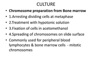
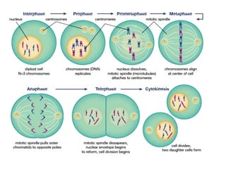
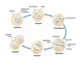
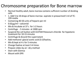
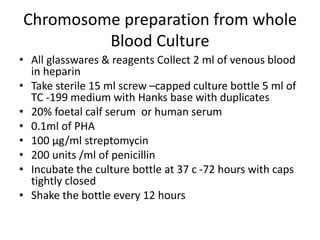
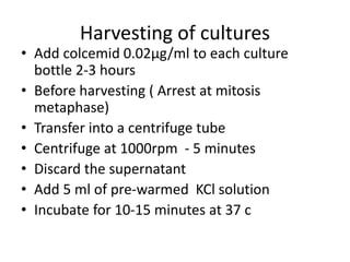
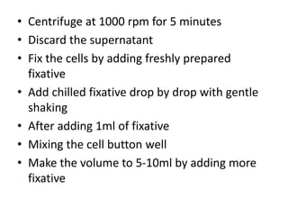
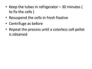
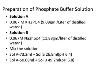
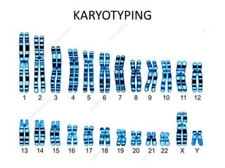
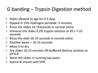
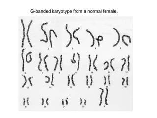
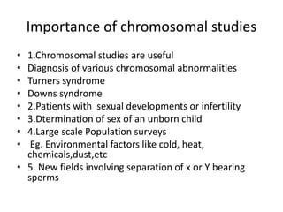
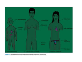
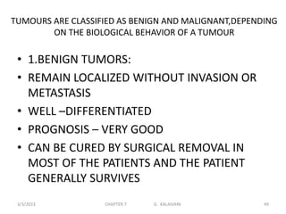
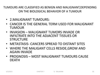
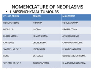
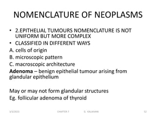
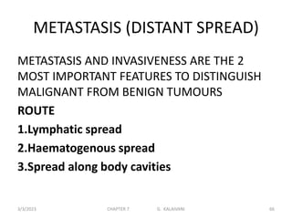
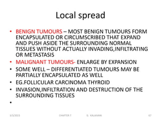
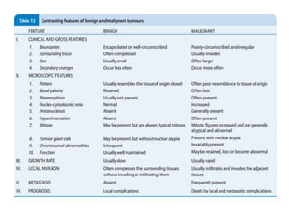
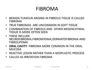
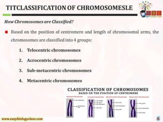
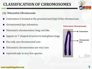
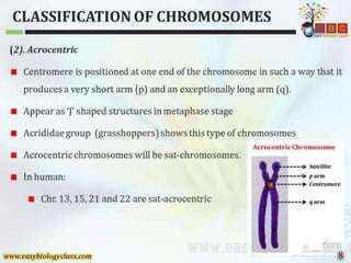
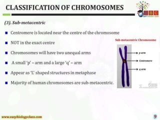
Cytogenetics
INTRODUCTION
• Study of chromosomes of man
• 23 pairs of chromosomes
• 44 –autosomes
• 2 – sex chromosomes
• Female –XX
• Male –XY
• Approximately 1 % of new born found to have
chromosomes defect
• Many cases prevention of recurrence of the
disease with counseling
• Prenatal diagnosis
• Mutation: a permanent transmissible change
in the genetic material, usually in a single gene
• Polymorphism: two or more genetically
determined, proportionally represented
phenotypes in the same population
• Genomic: abnormal chromosome number
(monosomy, polysomy, aneuploidy)
• Chromosomal: abnormal chromosome
structure
• Gene: DNA sequence changes in specific
genes
Method of karyotyping
• Specific applications like prenatal diagnosis,
detection of microdeletions, etc.
• More rapid
• Sensitive
• using a DNA probe of specific length that
binds to the specific DNA sequence (a part of
the gene) in the chromosomes. .
• Tagging of a fluorescent label to the probe
facilitates the visualization of the signal that is
produced due to the binding of the probe to
its complementary region.
• FISH technique include comparative
• genome hybridization (CGH) and Spectral
Karyotyping (SKY)
• used for characterizing chromosomal
imbalance in solid tumors
• complex chromosomal rearrangements
• The method involves denaturation as a first step
to separate the complementary strands within
the chromosome followed by a hybridization step
to facilitate the site specific ending of the probe
to its complementary specific region on the
chromosome.
• FISH probes are of 4 different types
• gene specific probes
• repetitive sequence probes
• whole genomic DNA probes
• chromosome painting probes.
• FISH signals are visualized by fluorescent
microscope that uses a light source (mercury
vapor and xenon lamps) to excite the
fluorochromes with which the probes are
labelled.
• A variety of filter sets are provided with the FISH
microscope that are specific for the different
fluorochromes.
• Routine cytogenetic methods to prepare an
ideogram(graphic symbol ) the images are captured in
high magnification and printed.
• specific chromosomes are cut neatly from the print and
stuck in a specific format.
• the images are captured by a digital imaging system
such as a CCD camera provided with the microscope
• then analyzed using specific systems and are also
stored for future purposes.
• Automation is the key note that adds on to the
advantage of FISH over conventional method.
Reagents required
• 20X SSC
• 2X SSC
• Methanol
• 100% Ethanol 1
• 2N HCL
• 1N NaOH Formamide
• ultra pure grade
• Autoclaved filtered
• milli Q water
Procedure
1. A slide with good metaphase spread was taken.
2. Humidified box prepared and kept in 37°C incubator an
hour prior to beginning of the procedure.
3. Place the jars containing the denaturation solution in a
73±1°C water bath approximately 30 minutes prior to start of
experiment.
4. Mark hybridization areas with a diamond tipped scribe
under the specimen slide.
5. Ensure that the denaturation solution is 73±1°C.
6. To age the slides, immerse the slides in 2X SSC for 3
minutes.
7. Dehydrate slides for 1 minute in 70% ethanol, followed by 1
minute in 85% ethanol and 1 minute in 100% ethanol.
8. Immerse the slides in the denaturation solution for 5
minutes.
• Preparation and denaturation of the probe
mixture
• Add to the following for each target area, to a
micro centrifuge tube at room temperature
• 7μl LS /WCP Hybridization Buffer
• 1μl probe
• 2 μL purified water
• 1. Centrifuge tube for 1–3 seconds.
• 2. Vortex and then centrifuge again.
• 3. Hold tube in a 73±1°C waterbath for 5 minutes
to denature the probe.
• 4. Remove tube from water bath.
Hybridizing the probe to the specimen
target
• 1. Remove the slide from 100% ethanol.
• 2. Dry slides by touching the bottom edge of the slides
to a blotting sheet and wiping the underside with a
tissue paper.
• 3. Apply 10 μL of probe mixture to one target area and
immediately apply coverslip without any air bobble.
• Repeat for additional target area.
• Seal the edges of the coverslip with clay.
• Place slides in the prewarmed humidified box, cover
with aluminium foil and replace in the 37°C incubator
and incubate for 12–16 hours.
Washing procedure:
 Prepare wash solutions
 Keep it in 46°C the water bath 45 minutes prior to end of incubation.
 At the end of the incubation, remove the humidified chamber from the 37°C
incubator, carefully take the slide from the chamber. 137 Genetics
 Remove the clay and the cover slip is lifted up carefully using the surgical blade.
 Immerse the slide in wash solution “1” of 50% formamide/2X SSC Agitate slides for
1–3 seconds and incubate for 10 minutes.
 The slide is blotted onto a tissue paper and transferred to the wash solution
 Agitate slides for 1–3 seconds and incubate for 10 minutes.
 Immerse slides in jar “3” of wash solution.
 Agitate slides for 1–3 seconds.
 Remove slides after 10 minutes.
 Immerse slides in 2X SSC.
 Agitate slides for 1–3 seconds.
 Remove slides after 10 minutes.
 Immerse slides in 2X SSC/0.1NP-40.
 Agitate slides for 1–3 seconds.
 Remove slides after 5 minutes.
Visualizing the hybridization
1. Air-dry slides completely in darkness.
2. Apply 10 μL of DAPI II to the center of the
hybridized area
apply coverslip without any air bubble.
3. Seal the corner of the coverslip with nail
polish. 4. Store at room temperature for 45–60
minutes in darkness before viewing.
5. First view the cells under the 10X using filter 2
(DAPI), then to view the signal use filter 5 under
oil immersion.
Results of Hybridization
normal metaphase spread, signals are observed
on both chromatids on the two homologous
chromosomes.
abnormal chromosomes show either single
signal (deletion or more than two signal on gain
of a chromosome.)
Chromosomal translocations are also visible by
this method.
CULTURE
• Chromosome preparation from Bone marrow
• 1.Arresting dividing cells at metaphase
• 2.Treatment with hypotonic solution
• 3.Fixation of cells in acetomethanol
• 4.Spreading of chromosomes on slide surface
• Commonly used for peripheral blood
lymphocytes & bone marrow cells - mitotic
chromosomes
Chromosome preparation for Bone marrow
• Normal Healthy adult, bone marrow contains sufficient number of dividing
cells
• 1. Add 5 to 10 drops of bone marrow aspirate in prewarmed 5 ml of TC-
199 media
• Containing 40-50 units of heparin per ml
• 0.02µg/ml – colcemid
• Mix & Incubate at 37 c for 1-2 hours
• Centrifuge - 5 minutes at 1000 rpm
• Suspend the cell button with 0.075M Potassium chloride for hypotonic
treatment for 10-15 minutes
• Centrifuge & discard the supernatant
• Add methanol :glacial acetic acid (3:1)fixative,
• Mix & kept at 4 c for 5 minutes
• Change fixative at least 3-4 times
• Prepare slides by air –dry method
• Stain with Giemsa
• Mount with DPX
Chromosome preparation from whole
Blood Culture
• All glasswares & reagents Collect 2 ml of venous blood
in heparin
• Take sterile 15 ml screw –capped culture bottle 5 ml of
TC -199 medium with Hanks base with duplicates
• 20% foetal calf serum or human serum
• 0.1ml of PHA
• 100 µg/ml streptomycin
• 200 units /ml of penicillin
• Incubate the culture bottle at 37 c -72 hours with caps
tightly closed
• Shake the bottle every 12 hours
Harvesting of cultures
• Add colcemid 0.02µg/ml to each culture
bottle 2-3 hours
• Before harvesting ( Arrest at mitosis
metaphase)
• Transfer into a centrifuge tube
• Centrifuge at 1000rpm - 5 minutes
• Discard the supernatant
• Add 5 ml of pre-warmed KCl solution
• Incubate for 10-15 minutes at 37 c
• Centrifuge at 1000 rpm for 5 minutes
• Discard the supernatant
• Fix the cells by adding freshly prepared
fixative
• Add chilled fixative drop by drop with gentle
shaking
• After adding 1ml of fixative
• Mixing the cell button well
• Make the volume to 5-10ml by adding more
fixative
• Keep the tubes in refrigerator – 30 minutes (
to fix the cells )
• Resuspend the cells in fresh fixative
• Centrifuge as before
• Repeat the process until a colorless cell pellet
is obtained
Preparation of Chromosome Slide
• Discard the supernatant completely without
distributing the cell button
• Add 0.5 to 1.0 ml of freshly prepared fixative
• Final concentration of the cell suspension has
to be adjusted depending on the cellular
concentration
• Keep the pre-cleaned slides in absolute
alcohol
• Place 2-3 drops of the cell suspension with a
Pasteur pipette on the slide from a distance to
facilitate better spreading
• Allow the slides for air drying on heat drying
• Stain with buffered Giemsa solution (1:10) at
pH 6.8
• Dry & pass the slides through xylene and
mount in DPX
Cleaning & Preparation of Slides
• New slides are kept in concentrated nitric acid
for overnight
• Keep the slides in horizontal coplin jar under
• Running tap water 2-3 hours
• Rinse in distilled water
• Store in 90% ethyl alcohol
• Wipe off & dry it with a clean cloth or tissue
paper
Procedure of Giemsa Staining
• 1.Giemsa solution is diluted 1:10 in phosphate
buffer (pH 6.8)
• 2.Add few drops of freshly prepared stain on
the slide - for 5-10 minutes
• 3.Rinse in distilled water
• 4.Air dry @ RT
• 5.Xylene & Mount in DPX
Preparation of Phosphate Buffer Solution
• Solution A
• 0.067 M KH2PO4 (9.08gm /Liter of distilled
water )
• Solution B
• 0.067M Na2hpo4 (11.88gm/liter of distilled
water )
• Mix the solution
• Sol A-73.2ml + Sol B 26.8ml(pH 6.4)
• Sol A-50.08ml + Sol B 49.2ml(pH 6.8)
KARYOTYPING
• Arm lengths are unequal
• Shorter arm – Upwards
• Longer arm – Downwards
• 22pairs can be arranged in 7 groups
• 1. A group 1 to 3 pairs – longest metacentric
• 2.B group 4 to 5 pairs – submetacentric
• 3.C group 6 to 12 pairs –submetacentric
• 4. D group 13 to 15 pairs –acrocentric
• 5.E group 16 to 18 pairs – submetacentric
• 6. F group 19 to 20 pairs -metacentric
• 7.G group 21 to 22 pairs – shortest acrocentric
Characterization of human chromosomes by various
banding techniques
Parameters used to characterise a
chromosome
• 1.Shape
• 2.Length
• 3 Centromeric index
• 4.Proportion of the arms
G banding – Trypsin Digestion method
• Slides allowed to age for 4-5 days
• Dipped in 15% Hydrogen peroxide -5 minutes
• Rinse the slides 10-15Seconds in normal saline
• Immerse the slides 0.2% trypsin solution at 20 c 7-10
seconds
• Rinse the slide 10-15 seconds in normal saline
• Distilled water – 10-15 seconds
• Allow it to dry
• Dry slides 10-15 minutes 5% buffered Giemsa solution at
pH 6.8
• Wash the slides in running tap water
• Xylene & mount with DPX
Q-Banding ( Quinacrine Banding )
• Reveals bright fluorescence bands throughout
the entire length of chromosomes
• Long arm of Y chromosomes shows bright
fluorescence
• Identification of each chromosomes
• Region is possible by its characteristics
fluorescence bands
• 1.Slides are allowed to age 2-3 days
• 2.Down grade ethyl alcohol to distilled water (
3 minutes in each)
3.Immerse the slide in Phosphate buffer pH 5.5
4.Rinse the slide in buffer solution – 5 minutes
5.Mount the slide in buffer with a clean cover
glass
6.Seal & screen the slides under fluorescence
microscopes
C –Banding
• Used for demonstrating centromeric
heterochromatin
• Air dried or heat dried chromosomes slides
are allowed to age for 7-10 days
• Slide in 0.2 N HCl -1 hour at RT
• Rinse the Slide in distilled water
• Rinse the slides in 5%aqueous solution of
barium hydroxide at 50 c for 5-7 minutes in
water bath
• Wash the slides
• Incubate the slide in 2 x SSC (1.754 gm of
sodium chloride & 0.882gm of trisodium
citrate in 100 ml of distilled water) – 1 hour at
60 c
• Rinse in distilled water ,70% ,90% & aloow it
to dry
• Stain with 5% buffered Giemsa solution – Ph
6.8 (10-15 minutes)
• Rinse with distilled water, xylene & mount in
DPX
Importance of chromosomal studies
• 1.Chromosomal studies are useful
• Diagnosis of various chromosomal abnormalities
• Turners syndrome
• Downs syndrome
• 2.Patients with sexual developments or infertility
• 3.Dtermination of sex of an unborn child
• 4.Large scale Population surveys
• Eg. Environmental factors like cold, heat,
chemicals,dust,etc
• 5. New fields involving separation of x or Y bearing
sperms
Barr body analysis Buccal Smear for
staining sex chromatin Mass
NORMAL VALUES NUMBER OF
BARR BODIES
Normal female ( xx) 1
Normal male (XO) 0
Turner syndrome ( female) 0
Klinefelter ‘s syndrome (male)(XXY) 1
Klinefelter ‘s syndrome (male)(48 XXXY) 2
Klinefelter ‘s syndrome (male) (49XXXYY) 2
Klinefelter ‘s syndrome (male) (49XXXYXY) 3
• Screening for sex chromosome abnormalities
PREPARATION
• Rinse the mouth with mouth wash
• Obtain a metal spatula saline, two slides &
preservatives
PROCEDURE
• Gently scrape the buccal mucosa with the metal
spatula dipped in saline
• Clean the spatula
• Repeat the procedure gently but firmly
• Smear the material on the two slides
• Place them in the preservation
POSTPROCEDURE CARE
• Label the container of the slides with the
client’s name, the date & the contents
OTHER DATA
• Barr bodies do not give any information
about Y chromosomes
• Human chromosome analysis ,rather than
buccal smears
• Should be used for evaluations of newborns
with ambiguous genitalia
NEOPLASIA(new growth or tumour )
• All new growth are not neoplasms
• Proliferation and maturation of cells in normal
adults is controlled as a result of which some
cells proliferate throughout life – labile cells
• Limited proliferation – stable cells
• Donot replicate – permanent cells
• Neoplastic cells lose control and regulation of
replication and form an abnormal mass of
tissue
3/3/2023 CHAPTER 7 G. KALAIVANI 44
Some Common tumours are
• Melanoma – carcinoma of melanocytes
• Hepatoma – carcinoma of hepatocytes
• Lymphoma – malignant tumour of lymphoid
tissue
• Seminoma – malignant tumour of the testis
• Leukemia – cancer of blood forming cells
3/3/2023 CHAPTER 7 G. KALAIVANI 45
Special categories of tumours
1.mixed tumours – adenosquamous carcinoma –
mixture of adenocarcinoma and Squamous cell
carcinoma
2.teratomas – encapsulated tumour with tissue
or organ eg. Hair,teeth,bone
3.blastomas –group of malignant tumours which
arise from embryonal. such tumours occur more
frequently in infants and children under 5 years
of age .eg retinoblastoma
3/3/2023 CHAPTER 7 G. KALAIVANI 46
Special categories of tumours
4.Hamartoma – tumour like malformation made
up of an abnormal mixture of cells and tissues
• Considered as a developmental error
3/3/2023 CHAPTER 7 G. KALAIVANI 47
3/3/2023 CHAPTER 7 G. KALAIVANI 48
TUMOURS ARE CLASSIFIED AS BENIGN AND MALIGNANT,DEPENDING
ON THE BIOLOGICAL BEHAVIOR OF A TUMOUR
• 1.BENIGN TUMORS:
• REMAIN LOCALIZED WITHOUT INVASION OR
METASTASIS
• WELL –DIFFERENTIATED
• PROGNOSIS – VERY GOOD
• CAN BE CURED BY SURGICAL REMOVAL IN
MOST OF THE PATIENTS AND THE PATIENT
GENERALLY SURVIVES
3/3/2023 CHAPTER 7 G. KALAIVANI 49
TUMOURS ARE CLASSIFIED AS BENIGN AND MALIGNANT,DEPENDING
ON THE BIOLOGICAL BEHAVIOR OF A TUMOUR
• 2.MALIGNANT TUMOURS:
• CANCER IS THE GENERAL TERM USED FOR MALIGNANT
TUMOUR
• INVASION – MALIGNANT TUMORS INVADE OR
INFILTRATE INTO THE ADJACENT TISSUES OR
STRUCTURE
• METASTASIS- CANCERS SPREAD TO DISTANT SITES
• WHERE THE MALIGANT CELLS RESIDE,GROW AND
AGAIN INVADE
• PROGNOSIS – MOST MALIGNANT TUMOURS CAUSE
DEATH
3/3/2023 CHAPTER 7 G. KALAIVANI 50
NOMENCLATURE OF NEOPLASMS
• 1.MESENCHYMAL TUMOURS
3/3/2023 CHAPTER 7 G. KALAIVANI 51
CELL OF ORIGIN BENIGN MALIGNANT
FIBROUS TISSUE FIBROMA FIBROSARCOMA
FAT CELLS LIPOMA LIPOSARCOMA
BLOOD VESSEL HEMANGIOMA ANGIOSARCOMA
CARTILAGE CHONDROMA CHONDROSARCOMA
SMOOTH MUSCLE LEIOMYOMA LEIOMYOSARCOMA
BONE OSTEOMA OSTEOGENIC SARCOMA
SKELETAL MUSCLE RHABDOMYOMA RHABDOMYOSARCOMA
NOMENCLATURE OF NEOPLASMS
• 2.EPITHELIAL TUMOURS NOMENCLATURE IS NOT
UNIFORM BUT MORE COMPLEX
• CLASSIFIED IN DIFFERENT WAYS
A. cells of origin
B. microscopic pattern
C. macroscopic architecture
Adenoma – benign epithelial tumour arising from
glandular epithelium
May or may not form glandular structures
Eg. follicular adenoma of thyroid
3/3/2023 CHAPTER 7 G. KALAIVANI 52
NOMENCLATURE OF NEOPLASMS
PAPILLOMA- BENIGN EPITHELIAL NEOPLASM
THAT PRODUCES MICROSCOPICALLY OR
MACROSCOPICALLY VISIBLE FINGER LIKE
Eg. SQUAMOUS PAPILLOMA
CYSTADENOMA-TUMOUR FORMING LARGE
CYSTIC MASSES
EG.SEROUS CYSTADENOMA OF OVARY
3/3/2023 CHAPTER 7 G. KALAIVANI 53
MALIGNANT TUMOUR
• 1.SARCOMA
• 2.CARCINOMA
3/3/2023 CHAPTER 7 G. KALAIVANI 54
CARCINOMA
• UNDIFFERENTIATED MALIGNANT TUMOUR –
• MALIGNANT TUMOUR COMPOSED OF
UNDIFFERENTIATED CELLS
• CELLS OF ORIGIN CANNOT BE MADE OUT ON
LIGHT MICROSCOPIC EXAMINATION
• INAPPROPRIATE TERMINOLOGY FOR
MALIGNANT TUMOUR – THE TERM
SUFFIX’OMA’ IS INAPPROPRIATELY USED AND
SOUNDS LIKE BENIGN TUMOR
3/3/2023 CHAPTER 7 G. KALAIVANI 55
CARCINOMA
• MALIGNANT NEOPLASMS ARISING FROM
EPITHELIAL CELL
• MAY BE DERIVED FROM ANY OF THE 3 GERM
LAYERS
• NOMENCLATURE OF CARCINOMAS
3/3/2023 CHAPTER 7 G. KALAIVANI 56
GERM LAYER TISSUE/CELL MALIGNANT TUMOUR
ECTODERM EPIDERMIS SQUAMOUS CELL
CARCINOMA
MESODERM RENAL TUBULES ADENOCARCINOMA
ENDODERM LINING OF THE GIT ADENOCARCINOMA
CARCINOMA
INAPPROPRIATE TERMINOLOGY FOR
MALIGNANT TUMOUR
SITE
HEPATOMA LIVER
MELANOMA SKIN
SEMINOMA/DYSGERMINOMA TESTIS/OVARY
LYMPHOMA LYMPH NODES AND EXTRANODAL
LYMPHOID TISSUE
3/3/2023 CHAPTER 7 G. KALAIVANI 57
SARCOMAS
• Malignant tumour arising in mesenchymal
tissue
• Tumours have little connective tissue stroma
and fleshy
• Eg,fibrosarcoma, liposarcoma, osteosarcoma
• Malignant tumours arising from blood forming
cells are called leukemias
3/3/2023 CHAPTER 7 G. KALAIVANI 58
GRADING AND STAGING OF CANCER
• ‘Grading’ and ‘staging’ are the two systems
• to predict tumour behaviour and guide
therapy after a malignant tumour is detected.
• Grading is defined as the gross appearance
and microscopic degree of differentiation of
the tumour
• staging - extent of spread of the tumour
within the patient.
.
3/3/2023 CHAPTER 7 G. KALAIVANI 59
BRODERS GRADING ARE
 THE DEGREE OF ANAPLASIA (cell with poor
differentiation)
 THE RATE OF GROWTH
 BASED ON THESE FEATURES,CANCERS ARE
CATEGORISED FROM
3/3/2023 CHAPTER 7 G. KALAIVANI 60
GRADING
• GRADE I - WELL –DIFFERENTIATED (LESS THAN
25% ANAPLASTIC CELLS)
• GRADE II - MODERATELY –
DIFFERENTIATED(25-50% ANAPLASTIC CELLS)
• GRADE III - MODERATELY –
DIFFERENTIATED(50 -75% ANAPLASTIC CELLS )
• GRADE IV- POORLY DIFFERENTIATED (MORE
THAN 75% ANAPLASTIC CELLS)
3/3/2023 CHAPTER 7 G. KALAIVANI 61
STAGING
• EXTENT OF SPREAD OF CANCERS CAN BE
ASSESSED BY 3 WAYS
• CLINICAL EXAMINATION
• INVESTIGATIONS
• PATHOLOGIC EXAMINATION OF THE TISSUE
REMOVED
.
3/3/2023 CHAPTER 7 G. KALAIVANI 62
TNM STAGING – UNION INTERNATIONALE CENTRE
CAANCER,GENEVA
• T - PRIMARY TUMOUR
• N - REGIONAL NODAL INVOLVEMENT
• M – DISTANT METASTASES
• EACH OF 3 COMPONENTS ARE ADDED TO INDICATE EXTENT
OF INDIVIDUAL
• T0 to T 4- LARGEST AND MOST EXTENSIVE PRIMARY
TUMOUR
• N0 to N3 – NO NODAL INVOLVEMENT TO WIDESPREAD
LYMPHNODE INVOLVEMENT
• M0 to M2 – NO METASTASIS TO DISSEMINATED
HAEMATOGENOUS METASTASIS
3/3/2023 CHAPTER 7 G. KALAIVANI 63
AJC STAGING -AMERICAN JOINT COMMITTEE
STAGING
• DIVIDES ALL CANCERS INTO STAGE 0 to IV
• TAKES INTO ACCOUNT ALL THE 3
COMPONENTS OF THE PRECEEDING SYSTEM
• (PRIMARY TUMOUR,NODULAR INVOLVEMENT
AND DISTANT METASTASES) IN EACH STAGE
3/3/2023 CHAPTER 7 G. KALAIVANI 64
• ROUTINE –RADIOGRAPHY (X-RAY,ULTRASOUND)
• -EXPLORATORY SURGERY
• MODERN TECHNIQUE - BASED ON TISSUE DENSITY
• - CT (COMPUTED TOMOGRAPHY)
• -MRI (MAGNETIC RESONANCE IMAGING
-PET(POSITRON EMISSION TOMOGRAPHY)-DISTINCTION
OF BENIGN AND MALIGNANT TUMOUR ON THE BASIS OF
BIOCHEMICAL AND MOLECULAR PROCESSES IN
TUOMOUR
RADIOACTIVE TRACER STUDIES –USE OF IODINE ISOTOPE
125 BOUND TO SPECIFIC TUMOUR ANTIBODIES
3/3/2023 CHAPTER 7 G. KALAIVANI 65
METASTASIS (DISTANT SPREAD)
METASTASIS AND INVASIVENESS ARE THE 2
MOST IMPORTANT FEATURES TO DISTINGUISH
MALIGNANT FROM BENIGN TUMOURS
ROUTE
1.Lymphatic spread
2.Haematogenous spread
3.Spread along body cavities
3/3/2023 CHAPTER 7 G. KALAIVANI 66
Local spread
• BENIGN TUMOURS – MOST BENIGN TUMOURS FORM
ENCAPSULATED OR CIRCUMSCRIBED THAT EXPAND
AND PUSH ASIDE THE SURROUNDING NORMAL
TISSUES WITHOUT ACTUALLY INVADING,INFILTRATING
OR METASTASIS
• MALIGNANT TUMOURS- ENLARGE BY EXPANSION
• SOME WELL – DIFFERENTIATED TUMOURS MAY BE
PARTIALLY ENCAPSULATED AS WELL
• EG.FOLLICULAR CARCINOMA THYROID
• INVASION,INFILTRATION AND DESTRUCTION OF THE
SURROUNDING TISSUES
•
3/3/2023 CHAPTER 7 G. KALAIVANI 67
3/3/2023 CHAPTER 7 G. KALAIVANI 68
FIBROMA
• BENIGN TUMOUR ARISING IN FIBROUS TISSUE IS CALLED
FIBROMA
• TRUE FIBROMAS ARE UNCOMMON IN SOFT TISSUE
• COMBINATION OF FIBROUS AND OTHER MESENCHYMAL
TISSUE IS MORE OFTEN SEEN
• THESE INCLUDE
NEUROFIBROMA,FIBROMYOMA,DERMATOFIBROMA AND
FIBROLIPOMA
• ORAL CAVITY- FIBROMA MORE COMMON IN THE ORAL
MUCOSA
• REACTIVE LESION RATHER THAN A NEOPLASTIC PROCESS
• CALLED AS IRRITATION FIBROMA
3/3/2023 CHAPTER 7 G. KALAIVANI 69
FIBROMA
• GROSS- OCCURS AS A SUBMUCOSAL
NODULAR MASS PRIMARILY ON THE BUCCAL
MUCOSA ALONG THE BITE LINE OR THE
GINGIVA
• MICROCOPY – SHOWS FIBROUS CONNECTIVE
TISSUE STROMA(consists of basement
membrane, extracellular matrix ,immune cells
& vasculature)
3/3/2023 CHAPTER 7 G. KALAIVANI 70
FIBROSARCOMA
• SLOW – GROWING TUMOUR
• AFFECTING ADULTS BETWEEN 4 AND 7 DECADES OF
LIFE
• COMMON LOCATIONS ARE THIGH,KNEE,TRUNK,HEAD
,NECK AND RETROPERITONEUM (anatomical space in
the abdominal cavity behind the peritoneum)
• GROSSLY – GREY – WHITE,FIRM,LOBULATED
• CUT SURFACE OF THE TUMOUR IS SOFT,FISHFLESH –
LIKE,WITH FOCI OF NECROSIS AND HAEMORRHAGES
• HISTOLOGICALLY – TUMOUR IS COMPOSED OF
UNIFORM ,SPINDLE – SHAPED FIBROBLASTS
• WELL DIFFERENTIATED – HERRING – BONE PATTERN
• POORLY DIFFERENTIATED – HIGHLY PLEOMORPHIC
3/3/2023 CHAPTER 7 G. KALAIVANI 71
TERATOMAS
• TUMOURS COMPOSED OF DIFFERENT TYPES OF
TISSUES DERIVED FROM THE 3 GERM CELL LAYERS
• ECTODERM, MESODERM AND ENDODERM IN
DIFFERENT COMBINATIONS
DIVIDED INTO 3 TYPES
• MATURE (BENIGN)
• IMMATURE (MALIGNANT)
• MONODERMAL
3/3/2023 CHAPTER 7 G. KALAIVANI 72
TERATOMAS(germ cell tumour arises
from egg or sperm)
• MATURE (BENIGN)- OVARIAN TERATOMAS ARE BENIGN
AND CYSTIC
• PREDOMINAT ECTODERMAL ELEMENTS – DERMOID CYST
• MATURE TERATOMA MAY BE SOLID AND BENIGN
• BENIGN CYSTIC – FREQUENT IN YOUNG WOMEN DURING
THEIR ACTIVE REPRODUCTIVE LIFE
• TUMOUR IS BILATERAL IN 10% OF CASES
3/3/2023 CHAPTER 7 G. KALAIVANI 73
TERATOMAS
• PATHOLOGIC CHANGES
• GROSSLY – UNILOCULAR CYST
• 10 -15 cm in diameter, USUALLY LINED BY SKIN
• ON SECTIONING,THE CYST IS FILLED WITH PASTE –LIKE
SEBACEOUS SECRETIONS
• DESQUAMATED KERATIN ADMIXED WITH MASSES OF HAIR
• CYST WALL IS THIN
• OPAQUE GREY –WHITE
• GENERALLY, IN ONE AREA OF THE CYST WALL,A SOLID
PROMINENCE IS SEEN (ROKITANSKY’S PROTUBERANCE)
• TISSUE ELEMENTS SUCH AS TOOTH,BONE,CARTILAGE
3/3/2023 CHAPTER 7 G. KALAIVANI 74
TERATOMAS
• MICROSCOPICALLY – LINING OF THE CYST
WALL BY STRATIFIED SQUAMOUS EPITHELIUM
• ADNEXAL(ACESSORY VISUAL) STRUCTURES
SUCH AS SEBACEOUS GLANDS,SWEAT GLANDS
AND HAIR FOLLICLES
3/3/2023 CHAPTER 7 G. KALAIVANI 75
ONCOGENES
expression causes cells to exhibit the properties of
cancer cells
Most known oncogenes are derived from normal
cellular genes.
precursor genes are best described as
protooncogenes
they are sometimes loosely referred to as
oncogenes
Activation of a single oncogene is not usually
sufficient for cell transformation
NORMAL ROLE OF ONCOGENES
• The cellular protooncogenes have shown great
evolutionary stability and presumably have an
important role in normal development and function.
• ras genes have been identified in yeast.
• There is activation of c-myc and ras genes in liver
regeneration after partial hepatectomy.
• Protooncogene expression is not necessarily associated
with growth.
• Differentiation of HL60 cells is accompanied by a
decrease in c-myc expression but there is an increase in
the expression of c-fos.
CELLULAR LOCATIONS AND
FUNCTIONS OF ONCOGENE PRODUCTS
• The protein products of oncogenes differ in their
cellular locations.
• They may be associated with the cell membrane,
the cytoplasm or the nucleus.
• Oncogene products have been described with
one of the following functions Growth factor
Growth factor receptor Signal transduction factor
GTP-binding plasma membrane protein Tyrosine-
specific protein kinase Serine/threonine-specific
protein kinase Transcription factor
CELLULAR LOCATIONS AND
FUNCTIONS OF ONCOGENE PRODUCTS
• The most intensively studied oncogenes include: bcl 2
Mitochondrial membrane-associated protein.
• Inhibits apoptosis erb B Truncated EGF receptor
protein - tyrosine kinase fos Forms a dimer with c-jun
to act as the AP-1 transcription factor jun met Soluble
truncated receptor-like protein-tyrosine kinase mos
Cytoplasmic protein-serine kinase myc Sequence-
specific DNA-binding protein raf Cytoplasmic protein-
serine kinase ras Membrane associated GTP
binding/GTPase sis PDGF B-chain growth factor src
Membrane associated non-receptor protein-tyrosine
kinase
MAJOR GENE MUTATIONS REQUIRED
FOR PRIMARY TUMORS AND
METASTASIS
Robert Weinberg has suggested that different gene
mutations are important for the establishment of primary
tumors and the later progression to a metastatic tumor.
Genes mutations important for primary tumors: Ras, Rb,
p53, hTERT, PP2A Gene mutations important for the
development of metastases: Twist, Goosecoid, Slug,
FOXC2. These genes code for transcription factors that
have a role in normal embryological development. The
interaction of tumor cells with stromal cells may be
critical for the establishment of metastases. An important
protein is SDF-1 (stroma derived factor). Twist is
expressed in more aggressive tumors. It is an inducer of
epithelial-mesenchymal transition.
CYTOGENETICS.pptx
CYTOGENETICS.pptx
CYTOGENETICS.pptx
CYTOGENETICS.pptx
CYTOGENETICS.pptx
CYTOGENETICS.pptx

CYTOGENETICS.pptx

  • 1.
  • 2.
    INTRODUCTION • Study ofchromosomes of man • 23 pairs of chromosomes • 44 –autosomes • 2 – sex chromosomes • Female –XX • Male –XY • Approximately 1 % of new born found to have chromosomes defect • Many cases prevention of recurrence of the disease with counseling • Prenatal diagnosis
  • 3.
    • Mutation: apermanent transmissible change in the genetic material, usually in a single gene • Polymorphism: two or more genetically determined, proportionally represented phenotypes in the same population • Genomic: abnormal chromosome number (monosomy, polysomy, aneuploidy) • Chromosomal: abnormal chromosome structure • Gene: DNA sequence changes in specific genes
  • 4.
    Method of karyotyping •Specific applications like prenatal diagnosis, detection of microdeletions, etc. • More rapid • Sensitive • using a DNA probe of specific length that binds to the specific DNA sequence (a part of the gene) in the chromosomes. .
  • 5.
    • Tagging ofa fluorescent label to the probe facilitates the visualization of the signal that is produced due to the binding of the probe to its complementary region. • FISH technique include comparative • genome hybridization (CGH) and Spectral Karyotyping (SKY) • used for characterizing chromosomal imbalance in solid tumors • complex chromosomal rearrangements
  • 6.
    • The methodinvolves denaturation as a first step to separate the complementary strands within the chromosome followed by a hybridization step to facilitate the site specific ending of the probe to its complementary specific region on the chromosome. • FISH probes are of 4 different types • gene specific probes • repetitive sequence probes • whole genomic DNA probes • chromosome painting probes. • FISH signals are visualized by fluorescent microscope that uses a light source (mercury vapor and xenon lamps) to excite the fluorochromes with which the probes are labelled.
  • 7.
    • A varietyof filter sets are provided with the FISH microscope that are specific for the different fluorochromes. • Routine cytogenetic methods to prepare an ideogram(graphic symbol ) the images are captured in high magnification and printed. • specific chromosomes are cut neatly from the print and stuck in a specific format. • the images are captured by a digital imaging system such as a CCD camera provided with the microscope • then analyzed using specific systems and are also stored for future purposes. • Automation is the key note that adds on to the advantage of FISH over conventional method.
  • 9.
    Reagents required • 20XSSC • 2X SSC • Methanol • 100% Ethanol 1 • 2N HCL • 1N NaOH Formamide • ultra pure grade • Autoclaved filtered • milli Q water
  • 10.
    Procedure 1. A slidewith good metaphase spread was taken. 2. Humidified box prepared and kept in 37°C incubator an hour prior to beginning of the procedure. 3. Place the jars containing the denaturation solution in a 73±1°C water bath approximately 30 minutes prior to start of experiment. 4. Mark hybridization areas with a diamond tipped scribe under the specimen slide. 5. Ensure that the denaturation solution is 73±1°C. 6. To age the slides, immerse the slides in 2X SSC for 3 minutes. 7. Dehydrate slides for 1 minute in 70% ethanol, followed by 1 minute in 85% ethanol and 1 minute in 100% ethanol. 8. Immerse the slides in the denaturation solution for 5 minutes.
  • 11.
    • Preparation anddenaturation of the probe mixture • Add to the following for each target area, to a micro centrifuge tube at room temperature • 7μl LS /WCP Hybridization Buffer • 1μl probe • 2 μL purified water • 1. Centrifuge tube for 1–3 seconds. • 2. Vortex and then centrifuge again. • 3. Hold tube in a 73±1°C waterbath for 5 minutes to denature the probe. • 4. Remove tube from water bath.
  • 12.
    Hybridizing the probeto the specimen target • 1. Remove the slide from 100% ethanol. • 2. Dry slides by touching the bottom edge of the slides to a blotting sheet and wiping the underside with a tissue paper. • 3. Apply 10 μL of probe mixture to one target area and immediately apply coverslip without any air bobble. • Repeat for additional target area. • Seal the edges of the coverslip with clay. • Place slides in the prewarmed humidified box, cover with aluminium foil and replace in the 37°C incubator and incubate for 12–16 hours.
  • 13.
    Washing procedure:  Preparewash solutions  Keep it in 46°C the water bath 45 minutes prior to end of incubation.  At the end of the incubation, remove the humidified chamber from the 37°C incubator, carefully take the slide from the chamber. 137 Genetics  Remove the clay and the cover slip is lifted up carefully using the surgical blade.  Immerse the slide in wash solution “1” of 50% formamide/2X SSC Agitate slides for 1–3 seconds and incubate for 10 minutes.  The slide is blotted onto a tissue paper and transferred to the wash solution  Agitate slides for 1–3 seconds and incubate for 10 minutes.  Immerse slides in jar “3” of wash solution.  Agitate slides for 1–3 seconds.  Remove slides after 10 minutes.  Immerse slides in 2X SSC.  Agitate slides for 1–3 seconds.  Remove slides after 10 minutes.  Immerse slides in 2X SSC/0.1NP-40.  Agitate slides for 1–3 seconds.  Remove slides after 5 minutes.
  • 14.
    Visualizing the hybridization 1.Air-dry slides completely in darkness. 2. Apply 10 μL of DAPI II to the center of the hybridized area apply coverslip without any air bubble. 3. Seal the corner of the coverslip with nail polish. 4. Store at room temperature for 45–60 minutes in darkness before viewing. 5. First view the cells under the 10X using filter 2 (DAPI), then to view the signal use filter 5 under oil immersion.
  • 15.
    Results of Hybridization normalmetaphase spread, signals are observed on both chromatids on the two homologous chromosomes. abnormal chromosomes show either single signal (deletion or more than two signal on gain of a chromosome.) Chromosomal translocations are also visible by this method.
  • 17.
    CULTURE • Chromosome preparationfrom Bone marrow • 1.Arresting dividing cells at metaphase • 2.Treatment with hypotonic solution • 3.Fixation of cells in acetomethanol • 4.Spreading of chromosomes on slide surface • Commonly used for peripheral blood lymphocytes & bone marrow cells - mitotic chromosomes
  • 20.
    Chromosome preparation forBone marrow • Normal Healthy adult, bone marrow contains sufficient number of dividing cells • 1. Add 5 to 10 drops of bone marrow aspirate in prewarmed 5 ml of TC- 199 media • Containing 40-50 units of heparin per ml • 0.02µg/ml – colcemid • Mix & Incubate at 37 c for 1-2 hours • Centrifuge - 5 minutes at 1000 rpm • Suspend the cell button with 0.075M Potassium chloride for hypotonic treatment for 10-15 minutes • Centrifuge & discard the supernatant • Add methanol :glacial acetic acid (3:1)fixative, • Mix & kept at 4 c for 5 minutes • Change fixative at least 3-4 times • Prepare slides by air –dry method • Stain with Giemsa • Mount with DPX
  • 21.
    Chromosome preparation fromwhole Blood Culture • All glasswares & reagents Collect 2 ml of venous blood in heparin • Take sterile 15 ml screw –capped culture bottle 5 ml of TC -199 medium with Hanks base with duplicates • 20% foetal calf serum or human serum • 0.1ml of PHA • 100 µg/ml streptomycin • 200 units /ml of penicillin • Incubate the culture bottle at 37 c -72 hours with caps tightly closed • Shake the bottle every 12 hours
  • 22.
    Harvesting of cultures •Add colcemid 0.02µg/ml to each culture bottle 2-3 hours • Before harvesting ( Arrest at mitosis metaphase) • Transfer into a centrifuge tube • Centrifuge at 1000rpm - 5 minutes • Discard the supernatant • Add 5 ml of pre-warmed KCl solution • Incubate for 10-15 minutes at 37 c
  • 23.
    • Centrifuge at1000 rpm for 5 minutes • Discard the supernatant • Fix the cells by adding freshly prepared fixative • Add chilled fixative drop by drop with gentle shaking • After adding 1ml of fixative • Mixing the cell button well • Make the volume to 5-10ml by adding more fixative
  • 24.
    • Keep thetubes in refrigerator – 30 minutes ( to fix the cells ) • Resuspend the cells in fresh fixative • Centrifuge as before • Repeat the process until a colorless cell pellet is obtained
  • 25.
    Preparation of ChromosomeSlide • Discard the supernatant completely without distributing the cell button • Add 0.5 to 1.0 ml of freshly prepared fixative • Final concentration of the cell suspension has to be adjusted depending on the cellular concentration • Keep the pre-cleaned slides in absolute alcohol
  • 26.
    • Place 2-3drops of the cell suspension with a Pasteur pipette on the slide from a distance to facilitate better spreading • Allow the slides for air drying on heat drying • Stain with buffered Giemsa solution (1:10) at pH 6.8 • Dry & pass the slides through xylene and mount in DPX
  • 27.
    Cleaning & Preparationof Slides • New slides are kept in concentrated nitric acid for overnight • Keep the slides in horizontal coplin jar under • Running tap water 2-3 hours • Rinse in distilled water • Store in 90% ethyl alcohol • Wipe off & dry it with a clean cloth or tissue paper
  • 28.
    Procedure of GiemsaStaining • 1.Giemsa solution is diluted 1:10 in phosphate buffer (pH 6.8) • 2.Add few drops of freshly prepared stain on the slide - for 5-10 minutes • 3.Rinse in distilled water • 4.Air dry @ RT • 5.Xylene & Mount in DPX
  • 29.
    Preparation of PhosphateBuffer Solution • Solution A • 0.067 M KH2PO4 (9.08gm /Liter of distilled water ) • Solution B • 0.067M Na2hpo4 (11.88gm/liter of distilled water ) • Mix the solution • Sol A-73.2ml + Sol B 26.8ml(pH 6.4) • Sol A-50.08ml + Sol B 49.2ml(pH 6.8)
  • 30.
  • 31.
    • Arm lengthsare unequal • Shorter arm – Upwards • Longer arm – Downwards • 22pairs can be arranged in 7 groups • 1. A group 1 to 3 pairs – longest metacentric • 2.B group 4 to 5 pairs – submetacentric • 3.C group 6 to 12 pairs –submetacentric • 4. D group 13 to 15 pairs –acrocentric • 5.E group 16 to 18 pairs – submetacentric • 6. F group 19 to 20 pairs -metacentric • 7.G group 21 to 22 pairs – shortest acrocentric Characterization of human chromosomes by various banding techniques
  • 32.
    Parameters used tocharacterise a chromosome • 1.Shape • 2.Length • 3 Centromeric index • 4.Proportion of the arms
  • 33.
    G banding –Trypsin Digestion method • Slides allowed to age for 4-5 days • Dipped in 15% Hydrogen peroxide -5 minutes • Rinse the slides 10-15Seconds in normal saline • Immerse the slides 0.2% trypsin solution at 20 c 7-10 seconds • Rinse the slide 10-15 seconds in normal saline • Distilled water – 10-15 seconds • Allow it to dry • Dry slides 10-15 minutes 5% buffered Giemsa solution at pH 6.8 • Wash the slides in running tap water • Xylene & mount with DPX
  • 35.
    Q-Banding ( QuinacrineBanding ) • Reveals bright fluorescence bands throughout the entire length of chromosomes • Long arm of Y chromosomes shows bright fluorescence • Identification of each chromosomes • Region is possible by its characteristics fluorescence bands • 1.Slides are allowed to age 2-3 days
  • 36.
    • 2.Down gradeethyl alcohol to distilled water ( 3 minutes in each) 3.Immerse the slide in Phosphate buffer pH 5.5 4.Rinse the slide in buffer solution – 5 minutes 5.Mount the slide in buffer with a clean cover glass 6.Seal & screen the slides under fluorescence microscopes
  • 37.
    C –Banding • Usedfor demonstrating centromeric heterochromatin • Air dried or heat dried chromosomes slides are allowed to age for 7-10 days • Slide in 0.2 N HCl -1 hour at RT • Rinse the Slide in distilled water • Rinse the slides in 5%aqueous solution of barium hydroxide at 50 c for 5-7 minutes in water bath
  • 38.
    • Wash theslides • Incubate the slide in 2 x SSC (1.754 gm of sodium chloride & 0.882gm of trisodium citrate in 100 ml of distilled water) – 1 hour at 60 c • Rinse in distilled water ,70% ,90% & aloow it to dry • Stain with 5% buffered Giemsa solution – Ph 6.8 (10-15 minutes) • Rinse with distilled water, xylene & mount in DPX
  • 39.
    Importance of chromosomalstudies • 1.Chromosomal studies are useful • Diagnosis of various chromosomal abnormalities • Turners syndrome • Downs syndrome • 2.Patients with sexual developments or infertility • 3.Dtermination of sex of an unborn child • 4.Large scale Population surveys • Eg. Environmental factors like cold, heat, chemicals,dust,etc • 5. New fields involving separation of x or Y bearing sperms
  • 41.
    Barr body analysisBuccal Smear for staining sex chromatin Mass NORMAL VALUES NUMBER OF BARR BODIES Normal female ( xx) 1 Normal male (XO) 0 Turner syndrome ( female) 0 Klinefelter ‘s syndrome (male)(XXY) 1 Klinefelter ‘s syndrome (male)(48 XXXY) 2 Klinefelter ‘s syndrome (male) (49XXXYY) 2 Klinefelter ‘s syndrome (male) (49XXXYXY) 3
  • 42.
    • Screening forsex chromosome abnormalities PREPARATION • Rinse the mouth with mouth wash • Obtain a metal spatula saline, two slides & preservatives PROCEDURE • Gently scrape the buccal mucosa with the metal spatula dipped in saline • Clean the spatula • Repeat the procedure gently but firmly • Smear the material on the two slides • Place them in the preservation
  • 43.
    POSTPROCEDURE CARE • Labelthe container of the slides with the client’s name, the date & the contents OTHER DATA • Barr bodies do not give any information about Y chromosomes • Human chromosome analysis ,rather than buccal smears • Should be used for evaluations of newborns with ambiguous genitalia
  • 44.
    NEOPLASIA(new growth ortumour ) • All new growth are not neoplasms • Proliferation and maturation of cells in normal adults is controlled as a result of which some cells proliferate throughout life – labile cells • Limited proliferation – stable cells • Donot replicate – permanent cells • Neoplastic cells lose control and regulation of replication and form an abnormal mass of tissue 3/3/2023 CHAPTER 7 G. KALAIVANI 44
  • 45.
    Some Common tumoursare • Melanoma – carcinoma of melanocytes • Hepatoma – carcinoma of hepatocytes • Lymphoma – malignant tumour of lymphoid tissue • Seminoma – malignant tumour of the testis • Leukemia – cancer of blood forming cells 3/3/2023 CHAPTER 7 G. KALAIVANI 45
  • 46.
    Special categories oftumours 1.mixed tumours – adenosquamous carcinoma – mixture of adenocarcinoma and Squamous cell carcinoma 2.teratomas – encapsulated tumour with tissue or organ eg. Hair,teeth,bone 3.blastomas –group of malignant tumours which arise from embryonal. such tumours occur more frequently in infants and children under 5 years of age .eg retinoblastoma 3/3/2023 CHAPTER 7 G. KALAIVANI 46
  • 47.
    Special categories oftumours 4.Hamartoma – tumour like malformation made up of an abnormal mixture of cells and tissues • Considered as a developmental error 3/3/2023 CHAPTER 7 G. KALAIVANI 47
  • 48.
    3/3/2023 CHAPTER 7G. KALAIVANI 48
  • 49.
    TUMOURS ARE CLASSIFIEDAS BENIGN AND MALIGNANT,DEPENDING ON THE BIOLOGICAL BEHAVIOR OF A TUMOUR • 1.BENIGN TUMORS: • REMAIN LOCALIZED WITHOUT INVASION OR METASTASIS • WELL –DIFFERENTIATED • PROGNOSIS – VERY GOOD • CAN BE CURED BY SURGICAL REMOVAL IN MOST OF THE PATIENTS AND THE PATIENT GENERALLY SURVIVES 3/3/2023 CHAPTER 7 G. KALAIVANI 49
  • 50.
    TUMOURS ARE CLASSIFIEDAS BENIGN AND MALIGNANT,DEPENDING ON THE BIOLOGICAL BEHAVIOR OF A TUMOUR • 2.MALIGNANT TUMOURS: • CANCER IS THE GENERAL TERM USED FOR MALIGNANT TUMOUR • INVASION – MALIGNANT TUMORS INVADE OR INFILTRATE INTO THE ADJACENT TISSUES OR STRUCTURE • METASTASIS- CANCERS SPREAD TO DISTANT SITES • WHERE THE MALIGANT CELLS RESIDE,GROW AND AGAIN INVADE • PROGNOSIS – MOST MALIGNANT TUMOURS CAUSE DEATH 3/3/2023 CHAPTER 7 G. KALAIVANI 50
  • 51.
    NOMENCLATURE OF NEOPLASMS •1.MESENCHYMAL TUMOURS 3/3/2023 CHAPTER 7 G. KALAIVANI 51 CELL OF ORIGIN BENIGN MALIGNANT FIBROUS TISSUE FIBROMA FIBROSARCOMA FAT CELLS LIPOMA LIPOSARCOMA BLOOD VESSEL HEMANGIOMA ANGIOSARCOMA CARTILAGE CHONDROMA CHONDROSARCOMA SMOOTH MUSCLE LEIOMYOMA LEIOMYOSARCOMA BONE OSTEOMA OSTEOGENIC SARCOMA SKELETAL MUSCLE RHABDOMYOMA RHABDOMYOSARCOMA
  • 52.
    NOMENCLATURE OF NEOPLASMS •2.EPITHELIAL TUMOURS NOMENCLATURE IS NOT UNIFORM BUT MORE COMPLEX • CLASSIFIED IN DIFFERENT WAYS A. cells of origin B. microscopic pattern C. macroscopic architecture Adenoma – benign epithelial tumour arising from glandular epithelium May or may not form glandular structures Eg. follicular adenoma of thyroid 3/3/2023 CHAPTER 7 G. KALAIVANI 52
  • 53.
    NOMENCLATURE OF NEOPLASMS PAPILLOMA-BENIGN EPITHELIAL NEOPLASM THAT PRODUCES MICROSCOPICALLY OR MACROSCOPICALLY VISIBLE FINGER LIKE Eg. SQUAMOUS PAPILLOMA CYSTADENOMA-TUMOUR FORMING LARGE CYSTIC MASSES EG.SEROUS CYSTADENOMA OF OVARY 3/3/2023 CHAPTER 7 G. KALAIVANI 53
  • 54.
    MALIGNANT TUMOUR • 1.SARCOMA •2.CARCINOMA 3/3/2023 CHAPTER 7 G. KALAIVANI 54
  • 55.
    CARCINOMA • UNDIFFERENTIATED MALIGNANTTUMOUR – • MALIGNANT TUMOUR COMPOSED OF UNDIFFERENTIATED CELLS • CELLS OF ORIGIN CANNOT BE MADE OUT ON LIGHT MICROSCOPIC EXAMINATION • INAPPROPRIATE TERMINOLOGY FOR MALIGNANT TUMOUR – THE TERM SUFFIX’OMA’ IS INAPPROPRIATELY USED AND SOUNDS LIKE BENIGN TUMOR 3/3/2023 CHAPTER 7 G. KALAIVANI 55
  • 56.
    CARCINOMA • MALIGNANT NEOPLASMSARISING FROM EPITHELIAL CELL • MAY BE DERIVED FROM ANY OF THE 3 GERM LAYERS • NOMENCLATURE OF CARCINOMAS 3/3/2023 CHAPTER 7 G. KALAIVANI 56 GERM LAYER TISSUE/CELL MALIGNANT TUMOUR ECTODERM EPIDERMIS SQUAMOUS CELL CARCINOMA MESODERM RENAL TUBULES ADENOCARCINOMA ENDODERM LINING OF THE GIT ADENOCARCINOMA
  • 57.
    CARCINOMA INAPPROPRIATE TERMINOLOGY FOR MALIGNANTTUMOUR SITE HEPATOMA LIVER MELANOMA SKIN SEMINOMA/DYSGERMINOMA TESTIS/OVARY LYMPHOMA LYMPH NODES AND EXTRANODAL LYMPHOID TISSUE 3/3/2023 CHAPTER 7 G. KALAIVANI 57
  • 58.
    SARCOMAS • Malignant tumourarising in mesenchymal tissue • Tumours have little connective tissue stroma and fleshy • Eg,fibrosarcoma, liposarcoma, osteosarcoma • Malignant tumours arising from blood forming cells are called leukemias 3/3/2023 CHAPTER 7 G. KALAIVANI 58
  • 59.
    GRADING AND STAGINGOF CANCER • ‘Grading’ and ‘staging’ are the two systems • to predict tumour behaviour and guide therapy after a malignant tumour is detected. • Grading is defined as the gross appearance and microscopic degree of differentiation of the tumour • staging - extent of spread of the tumour within the patient. . 3/3/2023 CHAPTER 7 G. KALAIVANI 59
  • 60.
    BRODERS GRADING ARE THE DEGREE OF ANAPLASIA (cell with poor differentiation)  THE RATE OF GROWTH  BASED ON THESE FEATURES,CANCERS ARE CATEGORISED FROM 3/3/2023 CHAPTER 7 G. KALAIVANI 60
  • 61.
    GRADING • GRADE I- WELL –DIFFERENTIATED (LESS THAN 25% ANAPLASTIC CELLS) • GRADE II - MODERATELY – DIFFERENTIATED(25-50% ANAPLASTIC CELLS) • GRADE III - MODERATELY – DIFFERENTIATED(50 -75% ANAPLASTIC CELLS ) • GRADE IV- POORLY DIFFERENTIATED (MORE THAN 75% ANAPLASTIC CELLS) 3/3/2023 CHAPTER 7 G. KALAIVANI 61
  • 62.
    STAGING • EXTENT OFSPREAD OF CANCERS CAN BE ASSESSED BY 3 WAYS • CLINICAL EXAMINATION • INVESTIGATIONS • PATHOLOGIC EXAMINATION OF THE TISSUE REMOVED . 3/3/2023 CHAPTER 7 G. KALAIVANI 62
  • 63.
    TNM STAGING –UNION INTERNATIONALE CENTRE CAANCER,GENEVA • T - PRIMARY TUMOUR • N - REGIONAL NODAL INVOLVEMENT • M – DISTANT METASTASES • EACH OF 3 COMPONENTS ARE ADDED TO INDICATE EXTENT OF INDIVIDUAL • T0 to T 4- LARGEST AND MOST EXTENSIVE PRIMARY TUMOUR • N0 to N3 – NO NODAL INVOLVEMENT TO WIDESPREAD LYMPHNODE INVOLVEMENT • M0 to M2 – NO METASTASIS TO DISSEMINATED HAEMATOGENOUS METASTASIS 3/3/2023 CHAPTER 7 G. KALAIVANI 63
  • 64.
    AJC STAGING -AMERICANJOINT COMMITTEE STAGING • DIVIDES ALL CANCERS INTO STAGE 0 to IV • TAKES INTO ACCOUNT ALL THE 3 COMPONENTS OF THE PRECEEDING SYSTEM • (PRIMARY TUMOUR,NODULAR INVOLVEMENT AND DISTANT METASTASES) IN EACH STAGE 3/3/2023 CHAPTER 7 G. KALAIVANI 64
  • 65.
    • ROUTINE –RADIOGRAPHY(X-RAY,ULTRASOUND) • -EXPLORATORY SURGERY • MODERN TECHNIQUE - BASED ON TISSUE DENSITY • - CT (COMPUTED TOMOGRAPHY) • -MRI (MAGNETIC RESONANCE IMAGING -PET(POSITRON EMISSION TOMOGRAPHY)-DISTINCTION OF BENIGN AND MALIGNANT TUMOUR ON THE BASIS OF BIOCHEMICAL AND MOLECULAR PROCESSES IN TUOMOUR RADIOACTIVE TRACER STUDIES –USE OF IODINE ISOTOPE 125 BOUND TO SPECIFIC TUMOUR ANTIBODIES 3/3/2023 CHAPTER 7 G. KALAIVANI 65
  • 66.
    METASTASIS (DISTANT SPREAD) METASTASISAND INVASIVENESS ARE THE 2 MOST IMPORTANT FEATURES TO DISTINGUISH MALIGNANT FROM BENIGN TUMOURS ROUTE 1.Lymphatic spread 2.Haematogenous spread 3.Spread along body cavities 3/3/2023 CHAPTER 7 G. KALAIVANI 66
  • 67.
    Local spread • BENIGNTUMOURS – MOST BENIGN TUMOURS FORM ENCAPSULATED OR CIRCUMSCRIBED THAT EXPAND AND PUSH ASIDE THE SURROUNDING NORMAL TISSUES WITHOUT ACTUALLY INVADING,INFILTRATING OR METASTASIS • MALIGNANT TUMOURS- ENLARGE BY EXPANSION • SOME WELL – DIFFERENTIATED TUMOURS MAY BE PARTIALLY ENCAPSULATED AS WELL • EG.FOLLICULAR CARCINOMA THYROID • INVASION,INFILTRATION AND DESTRUCTION OF THE SURROUNDING TISSUES • 3/3/2023 CHAPTER 7 G. KALAIVANI 67
  • 68.
    3/3/2023 CHAPTER 7G. KALAIVANI 68
  • 69.
    FIBROMA • BENIGN TUMOURARISING IN FIBROUS TISSUE IS CALLED FIBROMA • TRUE FIBROMAS ARE UNCOMMON IN SOFT TISSUE • COMBINATION OF FIBROUS AND OTHER MESENCHYMAL TISSUE IS MORE OFTEN SEEN • THESE INCLUDE NEUROFIBROMA,FIBROMYOMA,DERMATOFIBROMA AND FIBROLIPOMA • ORAL CAVITY- FIBROMA MORE COMMON IN THE ORAL MUCOSA • REACTIVE LESION RATHER THAN A NEOPLASTIC PROCESS • CALLED AS IRRITATION FIBROMA 3/3/2023 CHAPTER 7 G. KALAIVANI 69
  • 70.
    FIBROMA • GROSS- OCCURSAS A SUBMUCOSAL NODULAR MASS PRIMARILY ON THE BUCCAL MUCOSA ALONG THE BITE LINE OR THE GINGIVA • MICROCOPY – SHOWS FIBROUS CONNECTIVE TISSUE STROMA(consists of basement membrane, extracellular matrix ,immune cells & vasculature) 3/3/2023 CHAPTER 7 G. KALAIVANI 70
  • 71.
    FIBROSARCOMA • SLOW –GROWING TUMOUR • AFFECTING ADULTS BETWEEN 4 AND 7 DECADES OF LIFE • COMMON LOCATIONS ARE THIGH,KNEE,TRUNK,HEAD ,NECK AND RETROPERITONEUM (anatomical space in the abdominal cavity behind the peritoneum) • GROSSLY – GREY – WHITE,FIRM,LOBULATED • CUT SURFACE OF THE TUMOUR IS SOFT,FISHFLESH – LIKE,WITH FOCI OF NECROSIS AND HAEMORRHAGES • HISTOLOGICALLY – TUMOUR IS COMPOSED OF UNIFORM ,SPINDLE – SHAPED FIBROBLASTS • WELL DIFFERENTIATED – HERRING – BONE PATTERN • POORLY DIFFERENTIATED – HIGHLY PLEOMORPHIC 3/3/2023 CHAPTER 7 G. KALAIVANI 71
  • 72.
    TERATOMAS • TUMOURS COMPOSEDOF DIFFERENT TYPES OF TISSUES DERIVED FROM THE 3 GERM CELL LAYERS • ECTODERM, MESODERM AND ENDODERM IN DIFFERENT COMBINATIONS DIVIDED INTO 3 TYPES • MATURE (BENIGN) • IMMATURE (MALIGNANT) • MONODERMAL 3/3/2023 CHAPTER 7 G. KALAIVANI 72
  • 73.
    TERATOMAS(germ cell tumourarises from egg or sperm) • MATURE (BENIGN)- OVARIAN TERATOMAS ARE BENIGN AND CYSTIC • PREDOMINAT ECTODERMAL ELEMENTS – DERMOID CYST • MATURE TERATOMA MAY BE SOLID AND BENIGN • BENIGN CYSTIC – FREQUENT IN YOUNG WOMEN DURING THEIR ACTIVE REPRODUCTIVE LIFE • TUMOUR IS BILATERAL IN 10% OF CASES 3/3/2023 CHAPTER 7 G. KALAIVANI 73
  • 74.
    TERATOMAS • PATHOLOGIC CHANGES •GROSSLY – UNILOCULAR CYST • 10 -15 cm in diameter, USUALLY LINED BY SKIN • ON SECTIONING,THE CYST IS FILLED WITH PASTE –LIKE SEBACEOUS SECRETIONS • DESQUAMATED KERATIN ADMIXED WITH MASSES OF HAIR • CYST WALL IS THIN • OPAQUE GREY –WHITE • GENERALLY, IN ONE AREA OF THE CYST WALL,A SOLID PROMINENCE IS SEEN (ROKITANSKY’S PROTUBERANCE) • TISSUE ELEMENTS SUCH AS TOOTH,BONE,CARTILAGE 3/3/2023 CHAPTER 7 G. KALAIVANI 74
  • 75.
    TERATOMAS • MICROSCOPICALLY –LINING OF THE CYST WALL BY STRATIFIED SQUAMOUS EPITHELIUM • ADNEXAL(ACESSORY VISUAL) STRUCTURES SUCH AS SEBACEOUS GLANDS,SWEAT GLANDS AND HAIR FOLLICLES 3/3/2023 CHAPTER 7 G. KALAIVANI 75
  • 76.
    ONCOGENES expression causes cellsto exhibit the properties of cancer cells Most known oncogenes are derived from normal cellular genes. precursor genes are best described as protooncogenes they are sometimes loosely referred to as oncogenes Activation of a single oncogene is not usually sufficient for cell transformation
  • 77.
    NORMAL ROLE OFONCOGENES • The cellular protooncogenes have shown great evolutionary stability and presumably have an important role in normal development and function. • ras genes have been identified in yeast. • There is activation of c-myc and ras genes in liver regeneration after partial hepatectomy. • Protooncogene expression is not necessarily associated with growth. • Differentiation of HL60 cells is accompanied by a decrease in c-myc expression but there is an increase in the expression of c-fos.
  • 78.
    CELLULAR LOCATIONS AND FUNCTIONSOF ONCOGENE PRODUCTS • The protein products of oncogenes differ in their cellular locations. • They may be associated with the cell membrane, the cytoplasm or the nucleus. • Oncogene products have been described with one of the following functions Growth factor Growth factor receptor Signal transduction factor GTP-binding plasma membrane protein Tyrosine- specific protein kinase Serine/threonine-specific protein kinase Transcription factor
  • 79.
    CELLULAR LOCATIONS AND FUNCTIONSOF ONCOGENE PRODUCTS • The most intensively studied oncogenes include: bcl 2 Mitochondrial membrane-associated protein. • Inhibits apoptosis erb B Truncated EGF receptor protein - tyrosine kinase fos Forms a dimer with c-jun to act as the AP-1 transcription factor jun met Soluble truncated receptor-like protein-tyrosine kinase mos Cytoplasmic protein-serine kinase myc Sequence- specific DNA-binding protein raf Cytoplasmic protein- serine kinase ras Membrane associated GTP binding/GTPase sis PDGF B-chain growth factor src Membrane associated non-receptor protein-tyrosine kinase
  • 80.
    MAJOR GENE MUTATIONSREQUIRED FOR PRIMARY TUMORS AND METASTASIS Robert Weinberg has suggested that different gene mutations are important for the establishment of primary tumors and the later progression to a metastatic tumor. Genes mutations important for primary tumors: Ras, Rb, p53, hTERT, PP2A Gene mutations important for the development of metastases: Twist, Goosecoid, Slug, FOXC2. These genes code for transcription factors that have a role in normal embryological development. The interaction of tumor cells with stromal cells may be critical for the establishment of metastases. An important protein is SDF-1 (stroma derived factor). Twist is expressed in more aggressive tumors. It is an inducer of epithelial-mesenchymal transition.