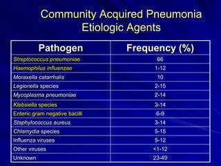
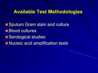
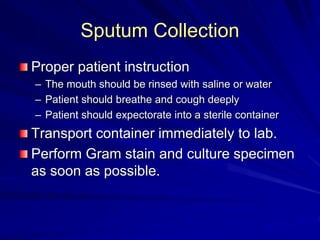
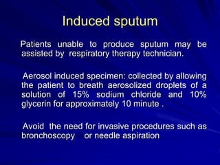
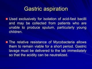
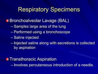
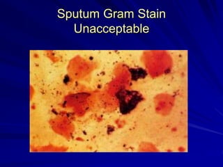
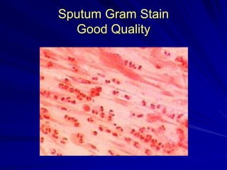
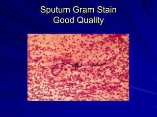
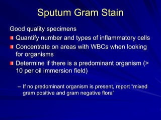
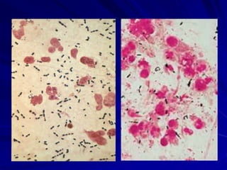
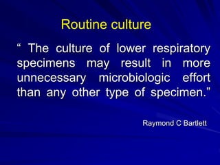
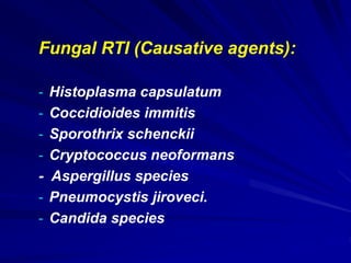
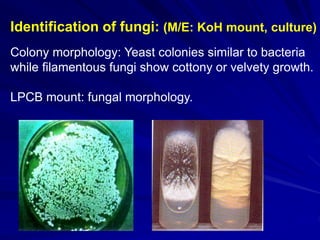
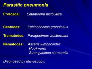
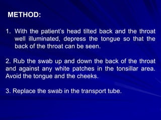
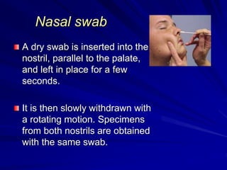
Laboratory Diagnosis of
Respiratory Infections
 Respiratory infections are one of the most
common microbial infections.
 Frequent exposure of respiratory mucosa to
microbes inhaled with air.
Lower Respiratory Tract Infections
Major sections
– Clinical aspects of diseases of LRT
– Specimen collection
– Specimen processing
– Interpretation of bacterial cultures
– Most common pathogens
Categories of Lower Respiratory
Tract Infections
Acute bronchitis
Community acquired pneumonia
Hospital acquired pneumonia
Pneumonia in the immunocompromised
host.
Tuberculosis
Community Acquired Pneumonia
Etiologic Agents
Pathogen Frequency (%)
Streptococcus pneumoniae 66
Haemophilus influenzae 1-12
Moraxella catarrhalis 10
Legionella species 2-15
Mycoplasma pneumoniae 2-14
Klebsiella species 3-14
Enteric gram negative bacilli 6-9
Staphylococcus aureus 3-14
Chlamydia species 5-15
Influenza viruses 5-12
Other viruses <1-12
Unknown 23-49
Available Test Methodologies
Sputum Gram stain and culture
Blood cultures
Serological studies
Nucleic acid amplification tests
Sputum Gram Stain and Culture
Demonstration of predominant morphotype on
Gram stain guides therapy.
Accuracy is good when strict criteria are used
Cheap, rapid
Sputum Collection
Proper patient instruction
– The mouth should be rinsed with saline or water
– Patient should breathe and cough deeply
– Patient should expectorate into a sterile container
Transport container immediately to lab.
Perform Gram stain and culture specimen
as soon as possible.
Induced sputum
Patients unable to produce sputum may be
assisted by respiratory therapy technician.
Aerosol induced specimen: collected by allowing
the patient to breath aerosolized droplets of a
solution of 15% sodium chloride and 10%
glycerin for approximately 10 minute .
Avoid the need for invasive procedures such as
bronchoscopy or needle aspiration
Gastric aspiration
Used exclusively for isolation of acid-fast bacilli
and may be collected from patients who are
unable to produce sputum, particularly young
children.
The relative resistance of Mycobacteria allows
them to remain viable for a short period. Gastric
lavage must be delivered to the lab immediately
so that the acidity can be neutralized.
Respiratory Specimens
Bronchoalveolar Lavage (BAL)
– Samples large area of the lung
– Performed using a bronchoscope
– Saline injected
– Injected saline along with secretions is collected
by aspiration
Transthoracic Aspiration
– Involves percutaneous introduction of a needle.
Sputum Gram Stain
Unacceptable
Sputum Gram Stain
Good Quality
Sputum Gram Stain
Good Quality
Sputum Gram Stain
Good quality specimens
Quantify number and types of inflammatory cells
Concentrate on areas with WBCs when looking
for organisms
Determine if there is a predominant organism (>
10 per oil immersion field)
– If no predominant organism is present, report “mixed
gram positive and gram negative flora”
“ The culture of lower respiratory
specimens may result in more
unnecessary microbiologic effort
than any other type of specimen.”
Raymond C Bartlett
Routine culture
Routine culture:
Sputum sample should be satisfactory otherwise do
not process (Reject the sample).
Do not examine clear, watery saliva.
General guidelines for satisfactory sample:
>25 PMNL and <10 epithelial cells per low power
magnification.
Note: may not be applicable for pt. with
neutropenia.
Routine culture:
 Inoculation of sputum sample on suitable
culture media should be done as soon as
possible, otherwise bacteria like
Haemophilus, Pneumococci may die.
 Should not be refrigerated.
Routine culture
Routinely used media :
5% sheep blood agar
chocolate agar: Neisseria spp. and
Haemophilus spp.
MacConkey agar
SDA (if fungal infection is suspected)
Identification of the isolates:
Identification of bacteria can be done as per
standard methods of identification.
Should be able to differentiate normal flora and
pathogen.
Interpretation of report should be done carefully.
Blood culture
Bacteremia may occur in pt. with pneumonia.
Isolating organism from blood will provide strong
evidence rather than isolation from sputum due to
throat flora contamination.
Fungal RTI (Causative agents):
- Histoplasma capsulatum
- Coccidioides immitis
- Sporothrix schenckii
- Cryptococcus neoformans
- Aspergillus species
- Pneumocystis jiroveci.
- Candida species
Identification of fungi: (M/E: KoH mount, culture)
Colony morphology: Yeast colonies similar to bacteria
while filamentous fungi show cottony or velvety growth.
LPCB mount: fungal morphology.
Parasitic pneumonia
Protozoa: Entamoeba histolytica
Cestodes: Echinococcus granulosus
Trematodes: Paragonimus westermani
Nematodes: Ascaris lumbricoides
Hookworm
Strongyloides stercoralis
Diagnosed by Microscopy.
Viral pneumonia:
- Respiratory Syncytial virus
- Influenza, Parainfluenza virus
- Adenoviruses
- Cytomegalovirus
- Herpes simplex virus
- Severe acute respiratory syndrome virus
- Diagnosis may not be required as viral infections are
self limiting. Microscopy & cell culture.
Laboratory Diagnosis of
Upper Respiratory Tract
Infections
Type of specimens
A variety of specimens are suitable for the
diagnosis of virus infections of the upper
respiratory tract:
Nasal swab
Nasopharyngeal swab
Throat swab
THROAT SWAB FOR CULTURE
MATERIALS NEEDED:
For bacteria: Culture Swab
All others: Virus/Chlamydia/
Mycoplasma Collection Pack
METHOD:
1. With the patient’s head tilted back and the throat
well illuminated, depress the tongue so that the
back of the throat can be seen.
2. Rub the swab up and down the back of the throat
and against any white patches in the tonsillar area.
Avoid the tongue and the cheeks.
3. Replace the swab in the transport tube.
Nasal swab
A dry swab is inserted into the
nostril, parallel to the palate,
and left in place for a few
seconds.
It is then slowly withdrawn with
a rotating motion. Specimens
from both nostrils are obtained
with the same swab.

Lab diag RTI.ppt

  • 1.
  • 2.
     Respiratory infectionsare one of the most common microbial infections.  Frequent exposure of respiratory mucosa to microbes inhaled with air.
  • 3.
    Lower Respiratory TractInfections Major sections – Clinical aspects of diseases of LRT – Specimen collection – Specimen processing – Interpretation of bacterial cultures – Most common pathogens
  • 4.
    Categories of LowerRespiratory Tract Infections Acute bronchitis Community acquired pneumonia Hospital acquired pneumonia Pneumonia in the immunocompromised host. Tuberculosis
  • 5.
    Community Acquired Pneumonia EtiologicAgents Pathogen Frequency (%) Streptococcus pneumoniae 66 Haemophilus influenzae 1-12 Moraxella catarrhalis 10 Legionella species 2-15 Mycoplasma pneumoniae 2-14 Klebsiella species 3-14 Enteric gram negative bacilli 6-9 Staphylococcus aureus 3-14 Chlamydia species 5-15 Influenza viruses 5-12 Other viruses <1-12 Unknown 23-49
  • 6.
    Available Test Methodologies SputumGram stain and culture Blood cultures Serological studies Nucleic acid amplification tests
  • 7.
    Sputum Gram Stainand Culture Demonstration of predominant morphotype on Gram stain guides therapy. Accuracy is good when strict criteria are used Cheap, rapid
  • 8.
    Sputum Collection Proper patientinstruction – The mouth should be rinsed with saline or water – Patient should breathe and cough deeply – Patient should expectorate into a sterile container Transport container immediately to lab. Perform Gram stain and culture specimen as soon as possible.
  • 9.
    Induced sputum Patients unableto produce sputum may be assisted by respiratory therapy technician. Aerosol induced specimen: collected by allowing the patient to breath aerosolized droplets of a solution of 15% sodium chloride and 10% glycerin for approximately 10 minute . Avoid the need for invasive procedures such as bronchoscopy or needle aspiration
  • 10.
    Gastric aspiration Used exclusivelyfor isolation of acid-fast bacilli and may be collected from patients who are unable to produce sputum, particularly young children. The relative resistance of Mycobacteria allows them to remain viable for a short period. Gastric lavage must be delivered to the lab immediately so that the acidity can be neutralized.
  • 11.
    Respiratory Specimens Bronchoalveolar Lavage(BAL) – Samples large area of the lung – Performed using a bronchoscope – Saline injected – Injected saline along with secretions is collected by aspiration Transthoracic Aspiration – Involves percutaneous introduction of a needle.
  • 12.
  • 13.
  • 14.
  • 15.
    Sputum Gram Stain Goodquality specimens Quantify number and types of inflammatory cells Concentrate on areas with WBCs when looking for organisms Determine if there is a predominant organism (> 10 per oil immersion field) – If no predominant organism is present, report “mixed gram positive and gram negative flora”
  • 17.
    “ The cultureof lower respiratory specimens may result in more unnecessary microbiologic effort than any other type of specimen.” Raymond C Bartlett Routine culture
  • 18.
    Routine culture: Sputum sampleshould be satisfactory otherwise do not process (Reject the sample). Do not examine clear, watery saliva. General guidelines for satisfactory sample: >25 PMNL and <10 epithelial cells per low power magnification. Note: may not be applicable for pt. with neutropenia.
  • 19.
    Routine culture:  Inoculationof sputum sample on suitable culture media should be done as soon as possible, otherwise bacteria like Haemophilus, Pneumococci may die.  Should not be refrigerated.
  • 20.
    Routine culture Routinely usedmedia : 5% sheep blood agar chocolate agar: Neisseria spp. and Haemophilus spp. MacConkey agar SDA (if fungal infection is suspected)
  • 21.
    Identification of theisolates: Identification of bacteria can be done as per standard methods of identification. Should be able to differentiate normal flora and pathogen. Interpretation of report should be done carefully.
  • 22.
    Blood culture Bacteremia mayoccur in pt. with pneumonia. Isolating organism from blood will provide strong evidence rather than isolation from sputum due to throat flora contamination.
  • 23.
    Fungal RTI (Causativeagents): - Histoplasma capsulatum - Coccidioides immitis - Sporothrix schenckii - Cryptococcus neoformans - Aspergillus species - Pneumocystis jiroveci. - Candida species
  • 24.
    Identification of fungi:(M/E: KoH mount, culture) Colony morphology: Yeast colonies similar to bacteria while filamentous fungi show cottony or velvety growth. LPCB mount: fungal morphology.
  • 25.
    Parasitic pneumonia Protozoa: Entamoebahistolytica Cestodes: Echinococcus granulosus Trematodes: Paragonimus westermani Nematodes: Ascaris lumbricoides Hookworm Strongyloides stercoralis Diagnosed by Microscopy.
  • 26.
    Viral pneumonia: - RespiratorySyncytial virus - Influenza, Parainfluenza virus - Adenoviruses - Cytomegalovirus - Herpes simplex virus - Severe acute respiratory syndrome virus - Diagnosis may not be required as viral infections are self limiting. Microscopy & cell culture.
  • 27.
    Laboratory Diagnosis of UpperRespiratory Tract Infections
  • 28.
    Type of specimens Avariety of specimens are suitable for the diagnosis of virus infections of the upper respiratory tract: Nasal swab Nasopharyngeal swab Throat swab
  • 29.
    THROAT SWAB FORCULTURE MATERIALS NEEDED: For bacteria: Culture Swab All others: Virus/Chlamydia/ Mycoplasma Collection Pack
  • 30.
    METHOD: 1. With thepatient’s head tilted back and the throat well illuminated, depress the tongue so that the back of the throat can be seen. 2. Rub the swab up and down the back of the throat and against any white patches in the tonsillar area. Avoid the tongue and the cheeks. 3. Replace the swab in the transport tube.
  • 31.
    Nasal swab A dryswab is inserted into the nostril, parallel to the palate, and left in place for a few seconds. It is then slowly withdrawn with a rotating motion. Specimens from both nostrils are obtained with the same swab.