INTRODUCTION
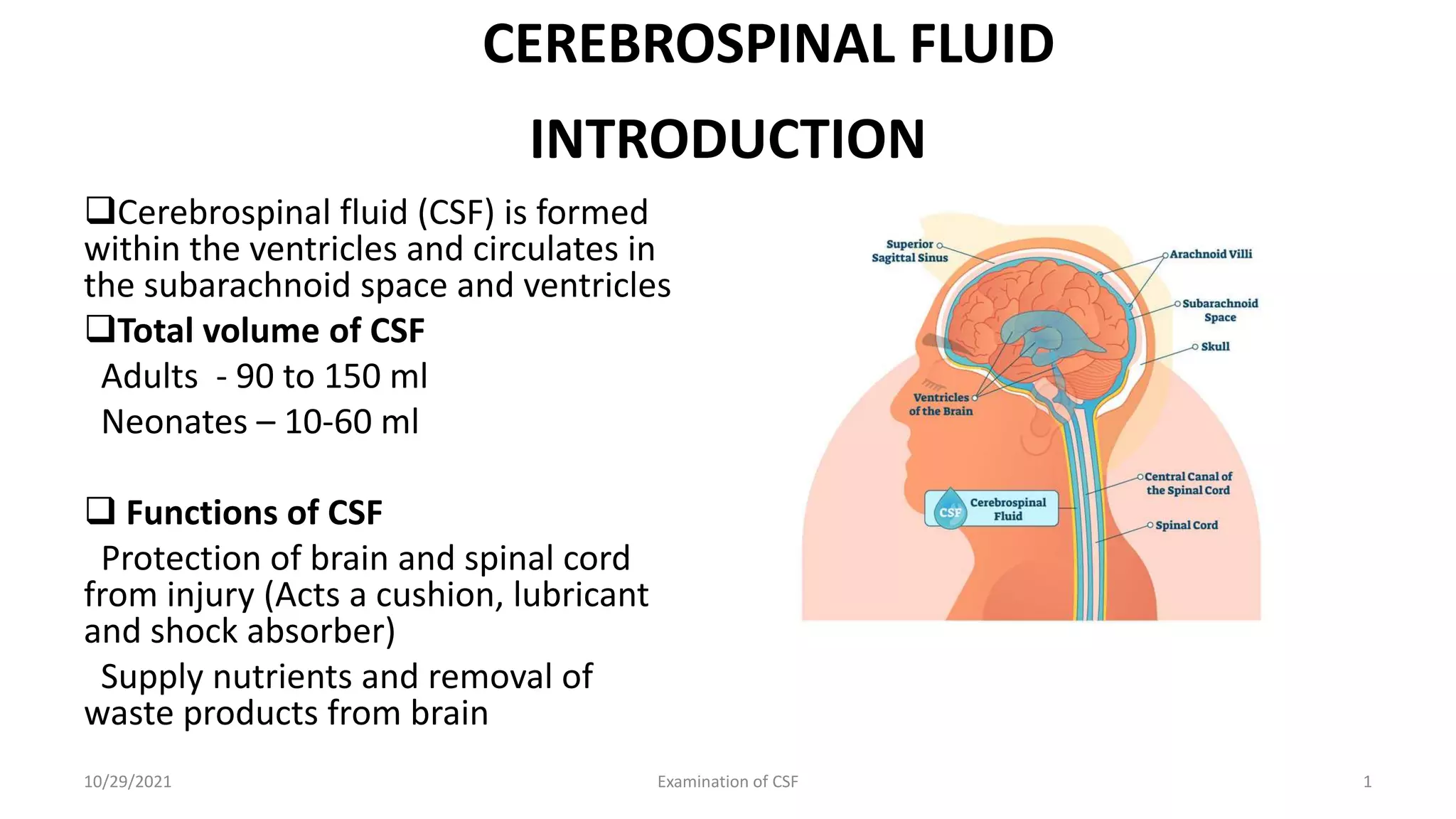
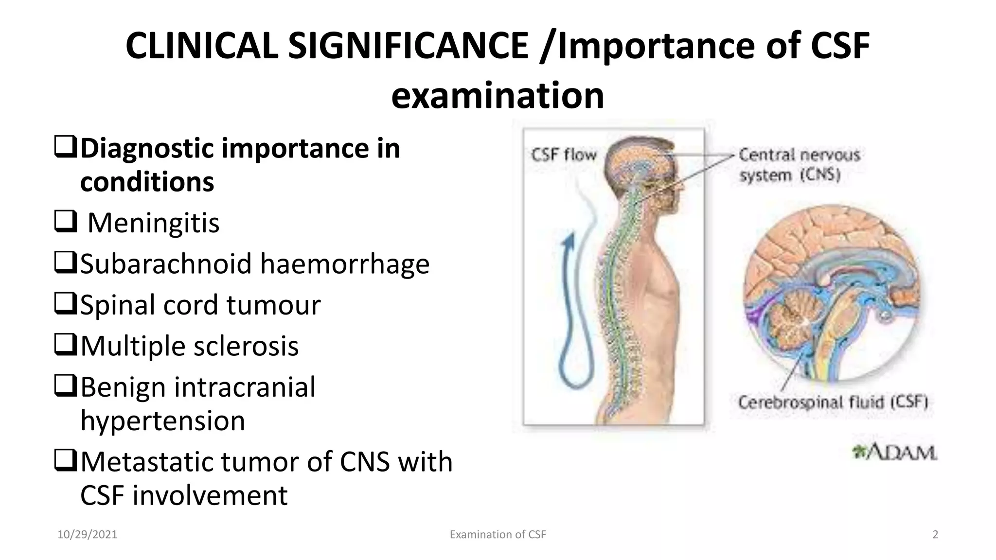
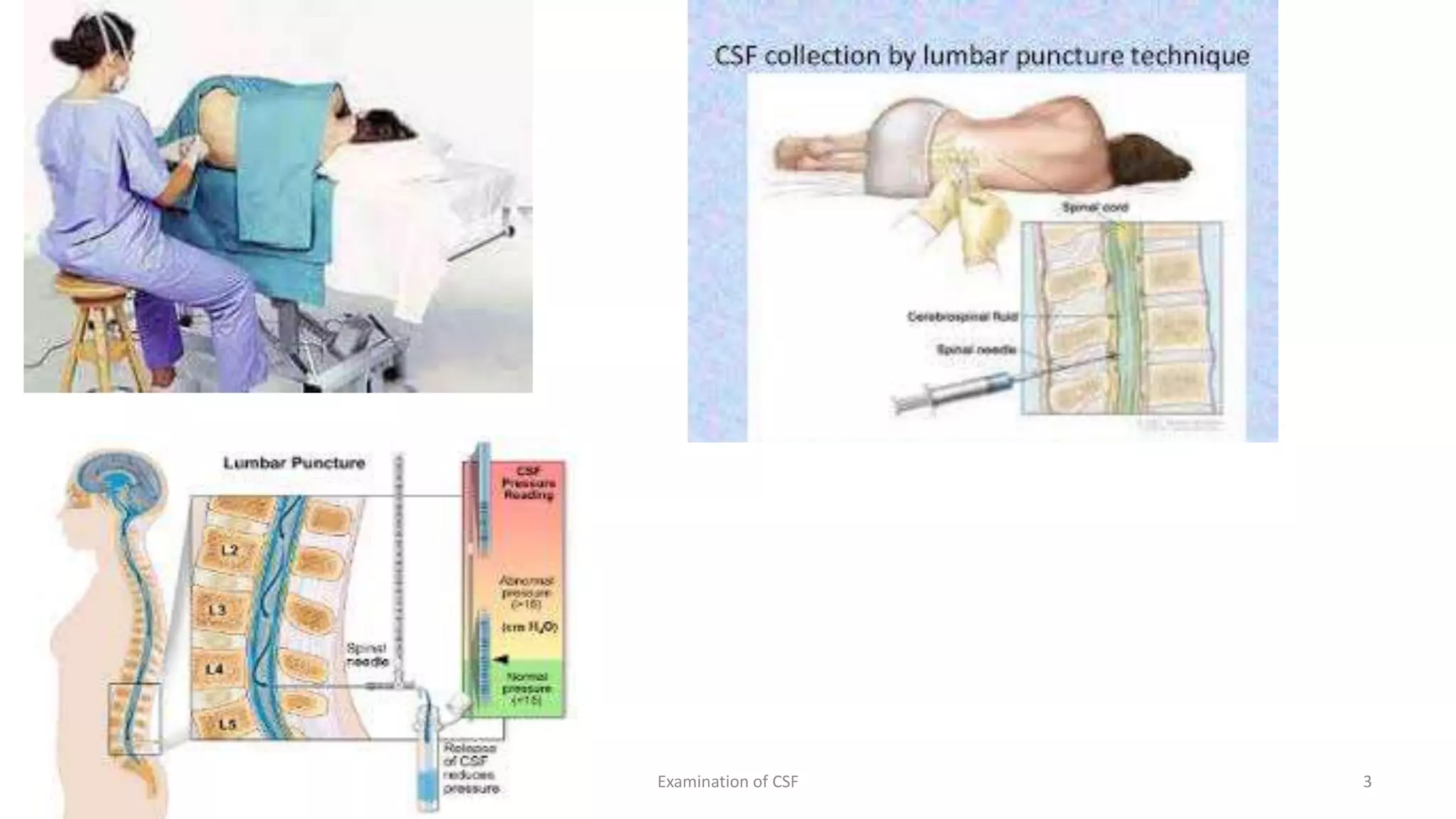
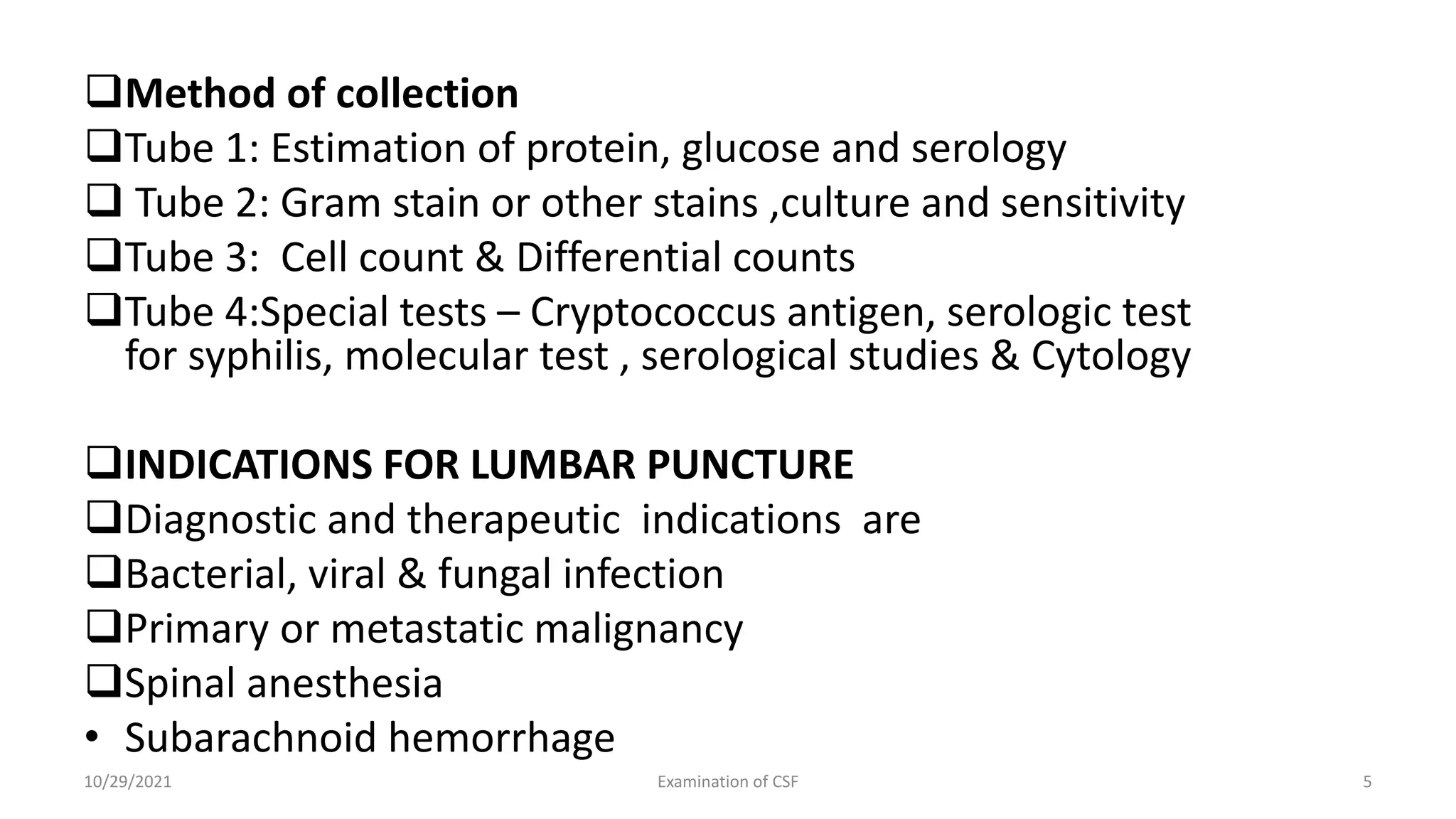
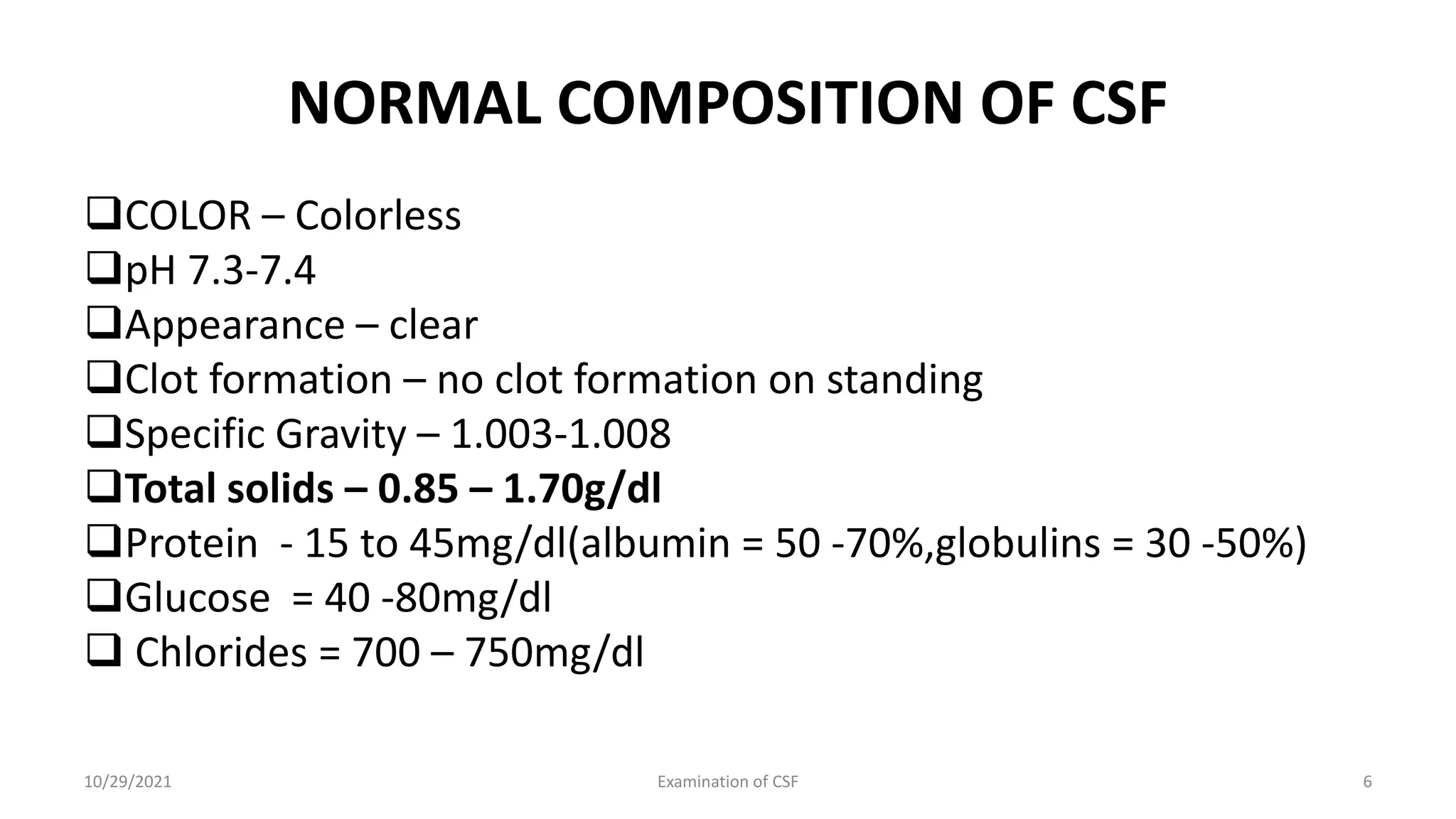
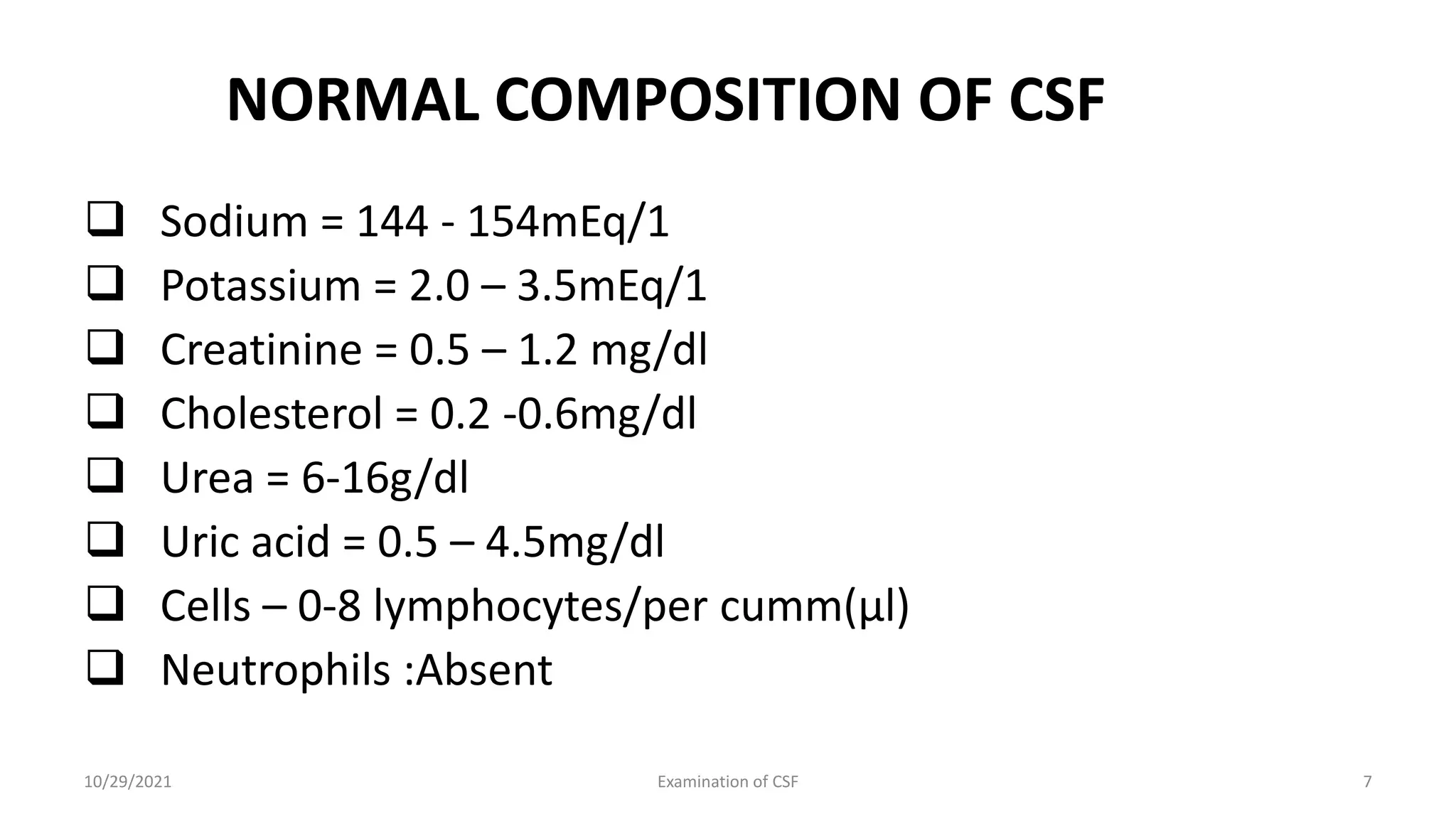
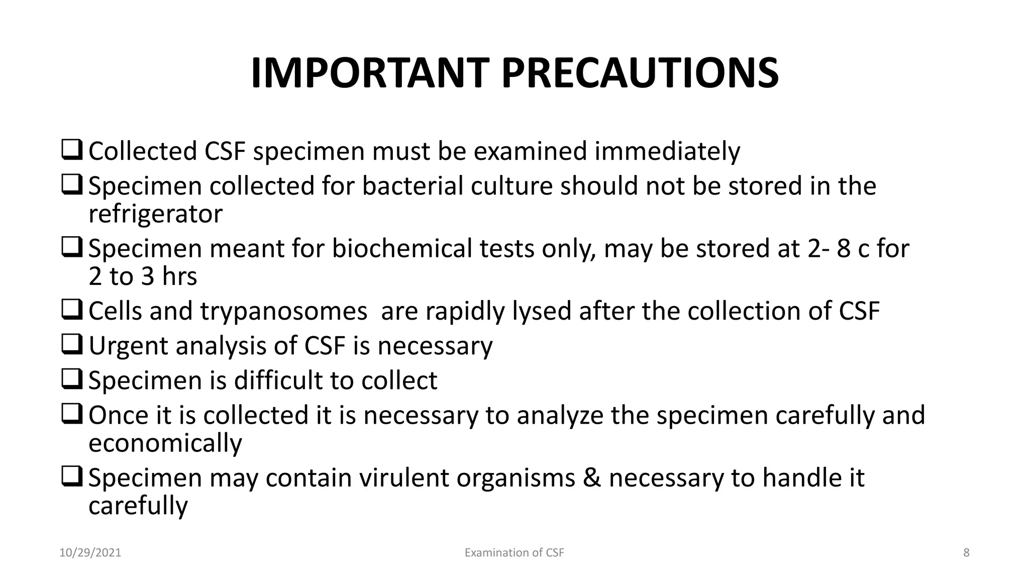
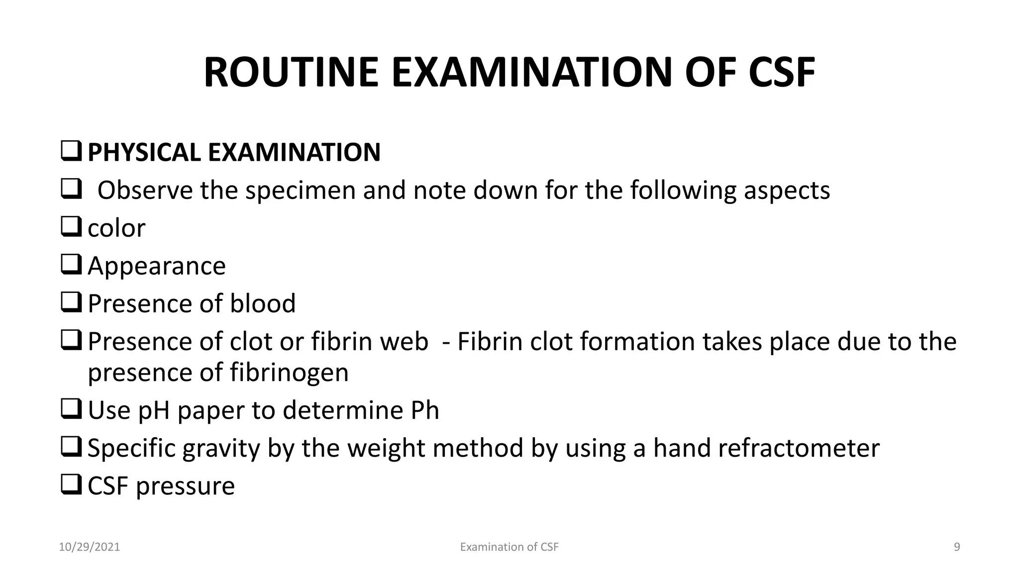
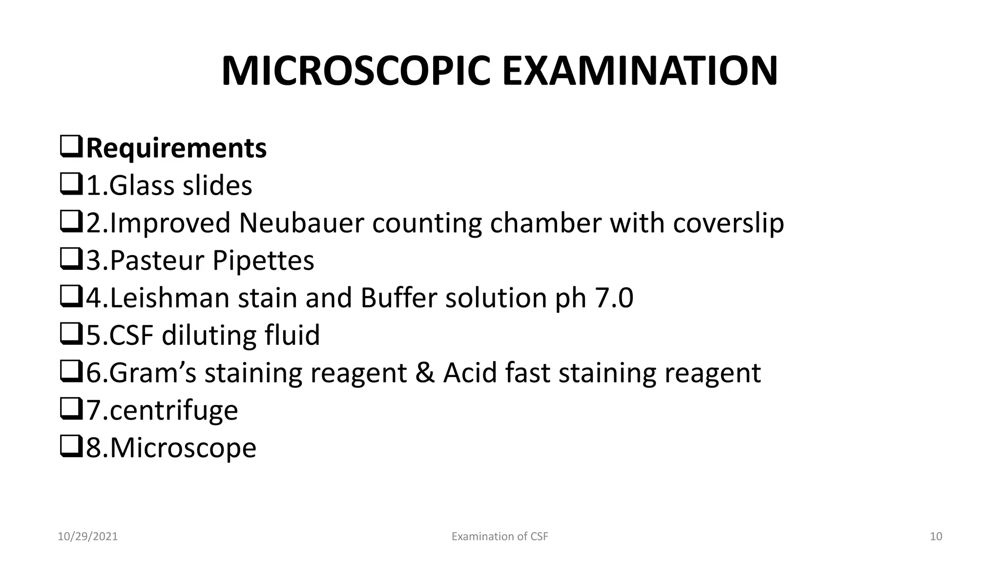
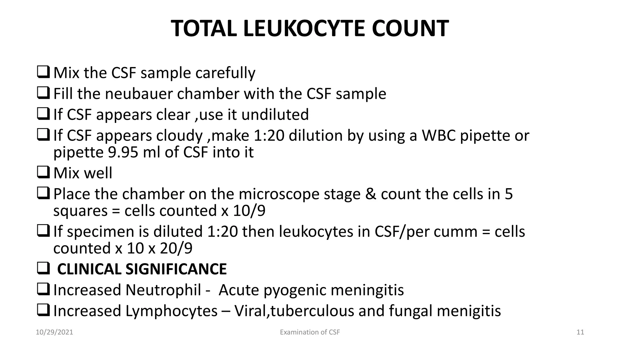
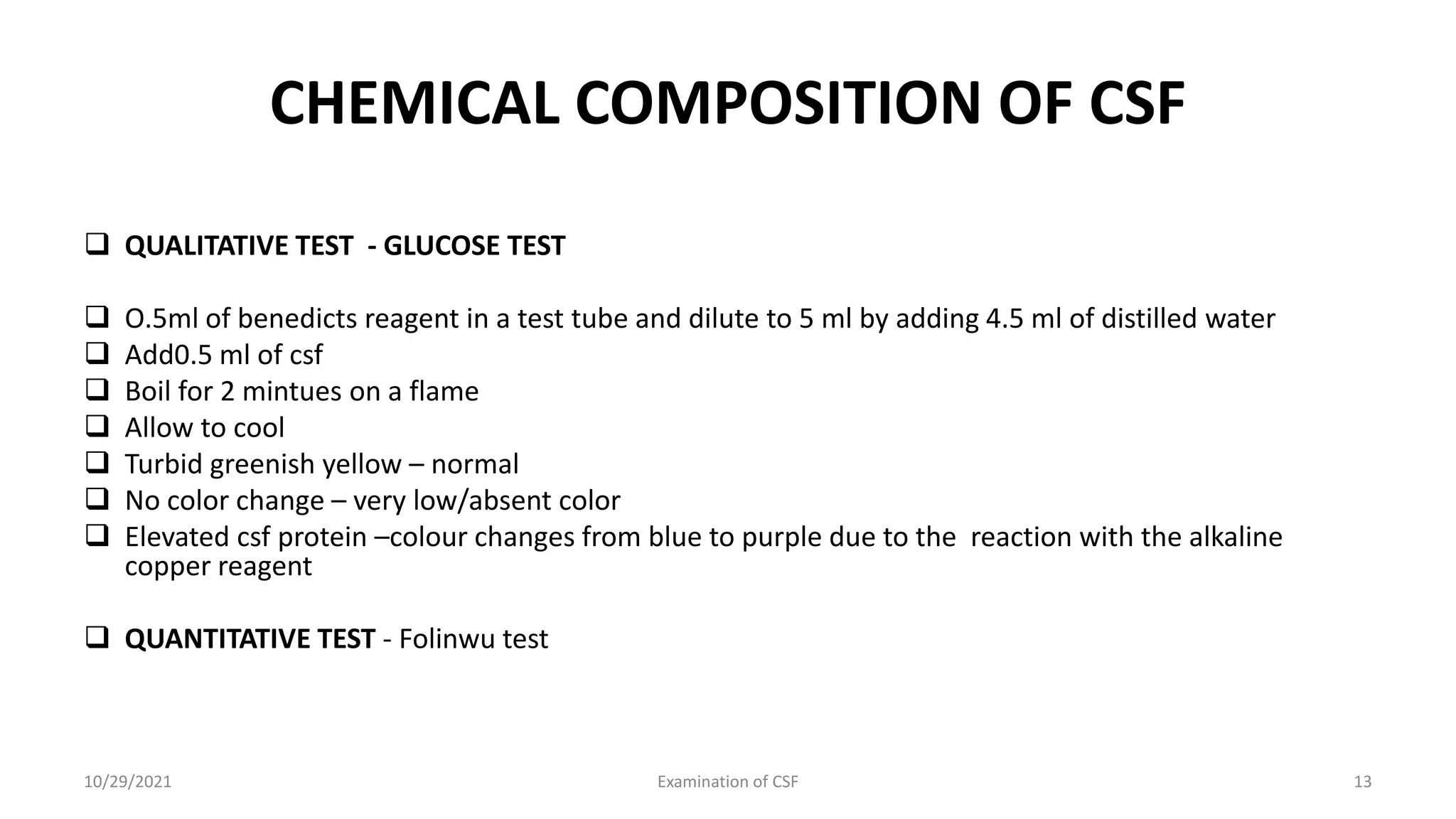
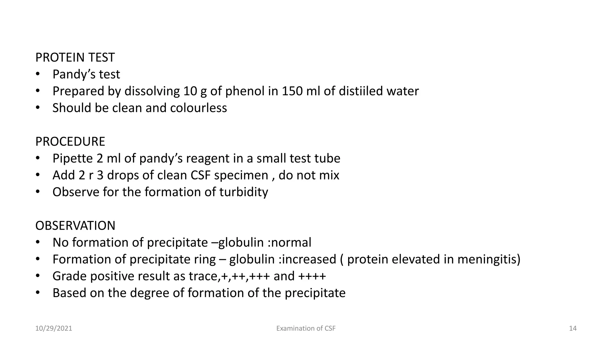
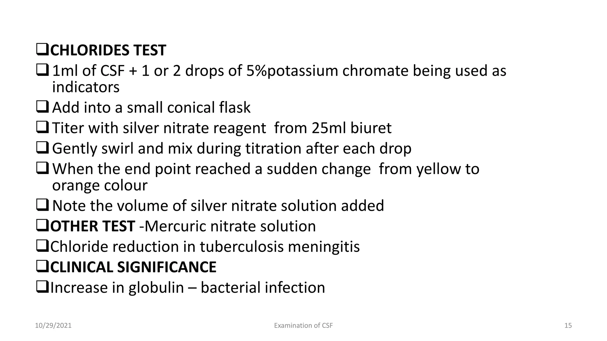
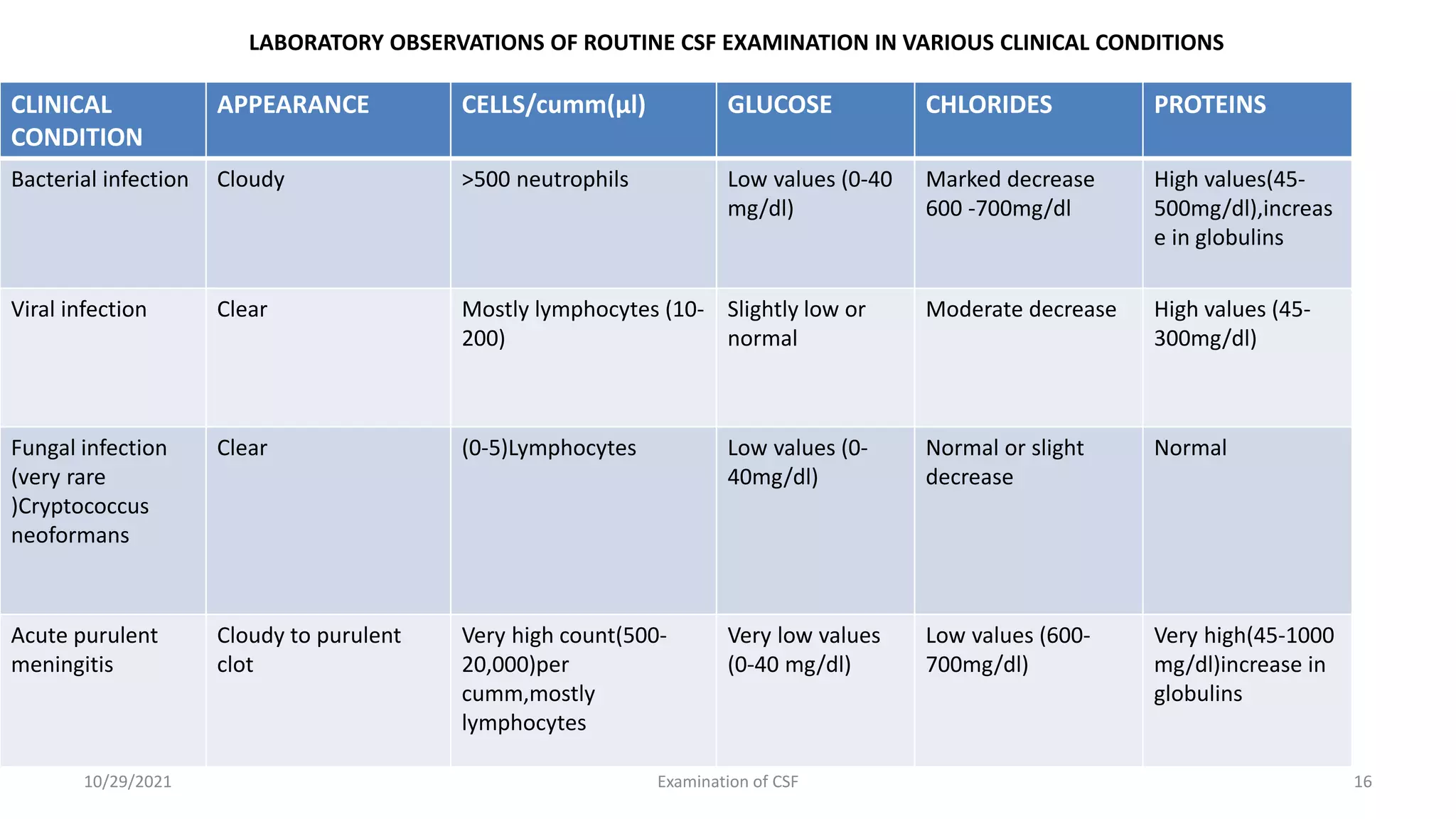
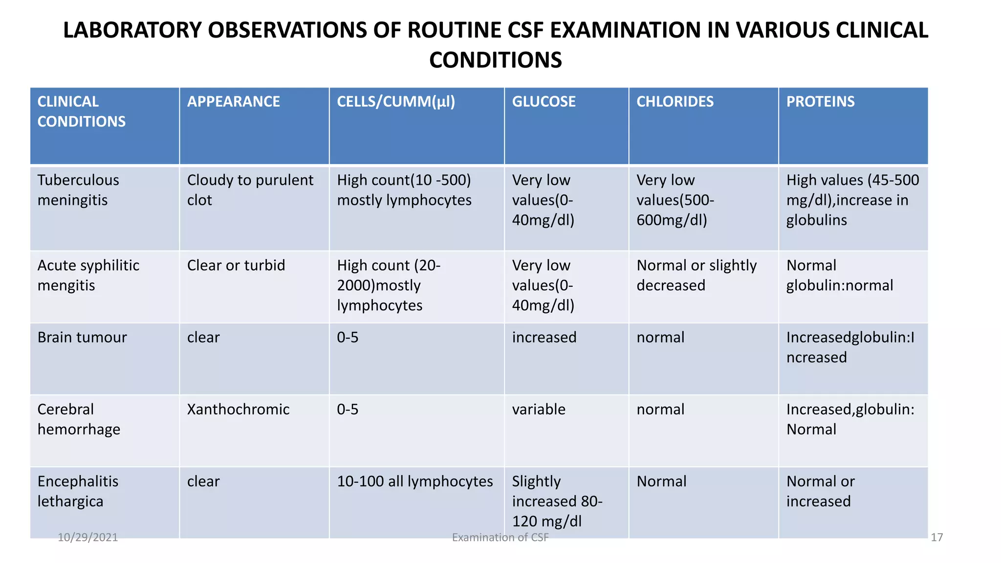
Cerebrospinal fluid (CSF) is formed
within the ventricles and circulates in
the subarachnoid space and ventricles
Total volume of CSF
Adults - 90 to 150 ml
Neonates – 10-60 ml
 Functions of CSF
Protection of brain and spinal cord
from injury (Acts a cushion, lubricant
and shock absorber)
Supply nutrients and removal of
waste products from brain
10/29/2021 Examination of CSF 1
CEREBROSPINAL FLUID
CLINICAL SIGNIFICANCE /Importance of CSF
examination
Diagnostic importance in
conditions
 Meningitis
Subarachnoid haemorrhage
Spinal cord tumour
Multiple sclerosis
Benign intracranial
hypertension
Metastatic tumor of CNS with
CSF involvement
10/29/2021 Examination of CSF 2
10/29/2021 Examination of CSF 3
SPECIMEN COLLECTION
 Specimen should be collected by a physician, a specially trained technician or nurse
 Sterile lumbar puncture needle is inserted between the 4 and 5 lumbar vertebra to a
depth of 4 to 5 cm
 Sites
 Lumbar puncture
 Cisternal puncture
 Ventricular puncture
 CSF Pressure - Normal Range
Adults – 90-100mm of water in lateral positions
Infants & children – 10-100mm of water reaching in adults level by 6-8 Years
 Increased – Meningitis, cerebral edema
Decreased – Dehydration
10/29/2021 Examination of CSF 4
Method of collection
Tube 1: Estimation of protein, glucose and serology
 Tube 2: Gram stain or other stains ,culture and sensitivity
Tube 3: Cell count & Differential counts
Tube 4:Special tests – Cryptococcus antigen, serologic test
for syphilis, molecular test , serological studies & Cytology
INDICATIONS FOR LUMBAR PUNCTURE
Diagnostic and therapeutic indications are
Bacterial, viral & fungal infection
Primary or metastatic malignancy
Spinal anesthesia
• Subarachnoid hemorrhage
10/29/2021 Examination of CSF 5
NORMAL COMPOSITION OF CSF
COLOR – Colorless
pH 7.3-7.4
Appearance – clear
Clot formation – no clot formation on standing
Specific Gravity – 1.003-1.008
Total solids – 0.85 – 1.70g/dl
Protein - 15 to 45mg/dl(albumin = 50 -70%,globulins = 30 -50%)
Glucose = 40 -80mg/dl
 Chlorides = 700 – 750mg/dl
10/29/2021 Examination of CSF 6
NORMAL COMPOSITION OF CSF
 Sodium = 144 - 154mEq/1
 Potassium = 2.0 – 3.5mEq/1
 Creatinine = 0.5 – 1.2 mg/dl
 Cholesterol = 0.2 -0.6mg/dl
 Urea = 6-16g/dl
 Uric acid = 0.5 – 4.5mg/dl
 Cells – 0-8 lymphocytes/per cumm(µl)
 Neutrophils :Absent
10/29/2021 Examination of CSF 7
IMPORTANT PRECAUTIONS
Collected CSF specimen must be examined immediately
Specimen collected for bacterial culture should not be stored in the
refrigerator
Specimen meant for biochemical tests only, may be stored at 2- 8 c for
2 to 3 hrs
Cells and trypanosomes are rapidly lysed after the collection of CSF
Urgent analysis of CSF is necessary
Specimen is difficult to collect
Once it is collected it is necessary to analyze the specimen carefully and
economically
Specimen may contain virulent organisms & necessary to handle it
carefully
10/29/2021 Examination of CSF 8
ROUTINE EXAMINATION OF CSF
PHYSICAL EXAMINATION
 Observe the specimen and note down for the following aspects
color
Appearance
Presence of blood
Presence of clot or fibrin web - Fibrin clot formation takes place due to the
presence of fibrinogen
Use pH paper to determine Ph
Specific gravity by the weight method by using a hand refractometer
CSF pressure
10/29/2021 Examination of CSF 9
MICROSCOPIC EXAMINATION
Requirements
1.Glass slides
2.Improved Neubauer counting chamber with coverslip
3.Pasteur Pipettes
4.Leishman stain and Buffer solution ph 7.0
5.CSF diluting fluid
6.Gram’s staining reagent & Acid fast staining reagent
7.centrifuge
8.Microscope
10/29/2021 Examination of CSF 10
TOTAL LEUKOCYTE COUNT
Mix the CSF sample carefully
Fill the neubauer chamber with the CSF sample
If CSF appears clear ,use it undiluted
If CSF appears cloudy ,make 1:20 dilution by using a WBC pipette or
pipette 9.95 ml of CSF into it
Mix well
Place the chamber on the microscope stage & count the cells in 5
squares = cells counted x 10/9
If specimen is diluted 1:20 then leukocytes in CSF/per cumm = cells
counted x 10 x 20/9
 CLINICAL SIGNIFICANCE
Increased Neutrophil - Acute pyogenic meningitis
Increased Lymphocytes – Viral,tuberculous and fungal menigitis
10/29/2021 Examination of CSF 11
Determination of differential leukocyte count
PROCEDURE
 If the cells count is low centrifuge CSF specimen and prepare a thin smear
of the sediment
 If the cell count is more than 500WBC/cumm
 Prepare a thin smear, of uncenrtifuged CSF
 Add 10 -15 drops of leishman’s stain on the smear
 Wait for 1 minute
 Add equal number of drops of buffer solution pH7.0
 Mix well and keep for 10 minutes
 Wash under running tap water,air dry and observe under oil immersion
CLINICAL CONDITIONS
 Increased lymphocyte count - Viral infection
 Increased Neutrophil count - Bacterial infection
10/29/2021 Examination of CSF 12
CHEMICAL COMPOSITION OF CSF
 QUALITATIVE TEST - GLUCOSE TEST
 O.5ml of benedicts reagent in a test tube and dilute to 5 ml by adding 4.5 ml of distilled water
 Add0.5 ml of csf
 Boil for 2 mintues on a flame
 Allow to cool
 Turbid greenish yellow – normal
 No color change – very low/absent color
 Elevated csf protein –colour changes from blue to purple due to the reaction with the alkaline
copper reagent
 QUANTITATIVE TEST - Folinwu test
10/29/2021 Examination of CSF 13
PROTEIN TEST
• Pandy’s test
• Prepared by dissolving 10 g of phenol in 150 ml of distiiled water
• Should be clean and colourless
PROCEDURE
• Pipette 2 ml of pandy’s reagent in a small test tube
• Add 2 r 3 drops of clean CSF specimen , do not mix
• Observe for the formation of turbidity
OBSERVATION
• No formation of precipitate –globulin :normal
• Formation of precipitate ring – globulin :increased ( protein elevated in meningitis)
• Grade positive result as trace,+,++,+++ and ++++
• Based on the degree of formation of the precipitate
10/29/2021 Examination of CSF 14
CHLORIDES TEST
1ml of CSF + 1 or 2 drops of 5%potassium chromate being used as
indicators
Add into a small conical flask
Titer with silver nitrate reagent from 25ml biuret
Gently swirl and mix during titration after each drop
When the end point reached a sudden change from yellow to
orange colour
Note the volume of silver nitrate solution added
OTHER TEST -Mercuric nitrate solution
Chloride reduction in tuberculosis meningitis
CLINICAL SIGNIFICANCE
Increase in globulin – bacterial infection
10/29/2021 Examination of CSF 15
LABORATORY OBSERVATIONS OF ROUTINE CSF EXAMINATION IN VARIOUS CLINICAL CONDITIONS
CLINICAL
CONDITION
APPEARANCE CELLS/cumm(µl) GLUCOSE CHLORIDES PROTEINS
Bacterial infection Cloudy >500 neutrophils Low values (0-40
mg/dl)
Marked decrease
600 -700mg/dl
High values(45-
500mg/dl),increas
e in globulins
Viral infection Clear Mostly lymphocytes (10-
200)
Slightly low or
normal
Moderate decrease High values (45-
300mg/dl)
Fungal infection
(very rare
)Cryptococcus
neoformans
Clear (0-5)Lymphocytes Low values (0-
40mg/dl)
Normal or slight
decrease
Normal
Acute purulent
meningitis
Cloudy to purulent
clot
Very high count(500-
20,000)per
cumm,mostly
lymphocytes
Very low values
(0-40 mg/dl)
Low values (600-
700mg/dl)
Very high(45-1000
mg/dl)increase in
globulins
10/29/2021 Examination of CSF 16
LABORATORY OBSERVATIONS OF ROUTINE CSF EXAMINATION IN VARIOUS CLINICAL
CONDITIONS
CLINICAL
CONDITIONS
APPEARANCE CELLS/CUMM(µl) GLUCOSE CHLORIDES PROTEINS
Tuberculous
meningitis
Cloudy to purulent
clot
High count(10 -500)
mostly lymphocytes
Very low
values(0-
40mg/dl)
Very low
values(500-
600mg/dl)
High values (45-500
mg/dl),increase in
globulins
Acute syphilitic
mengitis
Clear or turbid High count (20-
2000)mostly
lymphocytes
Very low
values(0-
40mg/dl)
Normal or slightly
decreased
Normal
globulin:normal
Brain tumour clear 0-5 increased normal Increasedglobulin:I
ncreased
Cerebral
hemorrhage
Xanthochromic 0-5 variable normal Increased,globulin:
Normal
Encephalitis
lethargica
clear 10-100 all lymphocytes Slightly
increased 80-
120 mg/dl
Normal Normal or
increased
10/29/2021 Examination of CSF 17
QUESTIONS
1.CSF findings in tuberculosis meningitis( 5 & 10 Marks )
2.CSF findings in pyogenic meningitis ( 5 & 10 Marks )
3.CSF findings in Viral meningitis ( 5 & 10 Marks )
4.Analysis of CSF ( 5 & 10 Marks )
5.Examination of CSF ( 5 & 10 Marks )
6.Indications of Lumbar puncture ( 3 Marks )
10/29/2021 Examination of CSF 18

CSF

  • 1.
    INTRODUCTION Cerebrospinal fluid (CSF)is formed within the ventricles and circulates in the subarachnoid space and ventricles Total volume of CSF Adults - 90 to 150 ml Neonates – 10-60 ml  Functions of CSF Protection of brain and spinal cord from injury (Acts a cushion, lubricant and shock absorber) Supply nutrients and removal of waste products from brain 10/29/2021 Examination of CSF 1 CEREBROSPINAL FLUID
  • 2.
    CLINICAL SIGNIFICANCE /Importanceof CSF examination Diagnostic importance in conditions  Meningitis Subarachnoid haemorrhage Spinal cord tumour Multiple sclerosis Benign intracranial hypertension Metastatic tumor of CNS with CSF involvement 10/29/2021 Examination of CSF 2
  • 3.
  • 4.
    SPECIMEN COLLECTION  Specimenshould be collected by a physician, a specially trained technician or nurse  Sterile lumbar puncture needle is inserted between the 4 and 5 lumbar vertebra to a depth of 4 to 5 cm  Sites  Lumbar puncture  Cisternal puncture  Ventricular puncture  CSF Pressure - Normal Range Adults – 90-100mm of water in lateral positions Infants & children – 10-100mm of water reaching in adults level by 6-8 Years  Increased – Meningitis, cerebral edema Decreased – Dehydration 10/29/2021 Examination of CSF 4
  • 5.
    Method of collection Tube1: Estimation of protein, glucose and serology  Tube 2: Gram stain or other stains ,culture and sensitivity Tube 3: Cell count & Differential counts Tube 4:Special tests – Cryptococcus antigen, serologic test for syphilis, molecular test , serological studies & Cytology INDICATIONS FOR LUMBAR PUNCTURE Diagnostic and therapeutic indications are Bacterial, viral & fungal infection Primary or metastatic malignancy Spinal anesthesia • Subarachnoid hemorrhage 10/29/2021 Examination of CSF 5
  • 6.
    NORMAL COMPOSITION OFCSF COLOR – Colorless pH 7.3-7.4 Appearance – clear Clot formation – no clot formation on standing Specific Gravity – 1.003-1.008 Total solids – 0.85 – 1.70g/dl Protein - 15 to 45mg/dl(albumin = 50 -70%,globulins = 30 -50%) Glucose = 40 -80mg/dl  Chlorides = 700 – 750mg/dl 10/29/2021 Examination of CSF 6
  • 7.
    NORMAL COMPOSITION OFCSF  Sodium = 144 - 154mEq/1  Potassium = 2.0 – 3.5mEq/1  Creatinine = 0.5 – 1.2 mg/dl  Cholesterol = 0.2 -0.6mg/dl  Urea = 6-16g/dl  Uric acid = 0.5 – 4.5mg/dl  Cells – 0-8 lymphocytes/per cumm(µl)  Neutrophils :Absent 10/29/2021 Examination of CSF 7
  • 8.
    IMPORTANT PRECAUTIONS Collected CSFspecimen must be examined immediately Specimen collected for bacterial culture should not be stored in the refrigerator Specimen meant for biochemical tests only, may be stored at 2- 8 c for 2 to 3 hrs Cells and trypanosomes are rapidly lysed after the collection of CSF Urgent analysis of CSF is necessary Specimen is difficult to collect Once it is collected it is necessary to analyze the specimen carefully and economically Specimen may contain virulent organisms & necessary to handle it carefully 10/29/2021 Examination of CSF 8
  • 9.
    ROUTINE EXAMINATION OFCSF PHYSICAL EXAMINATION  Observe the specimen and note down for the following aspects color Appearance Presence of blood Presence of clot or fibrin web - Fibrin clot formation takes place due to the presence of fibrinogen Use pH paper to determine Ph Specific gravity by the weight method by using a hand refractometer CSF pressure 10/29/2021 Examination of CSF 9
  • 10.
    MICROSCOPIC EXAMINATION Requirements 1.Glass slides 2.ImprovedNeubauer counting chamber with coverslip 3.Pasteur Pipettes 4.Leishman stain and Buffer solution ph 7.0 5.CSF diluting fluid 6.Gram’s staining reagent & Acid fast staining reagent 7.centrifuge 8.Microscope 10/29/2021 Examination of CSF 10
  • 11.
    TOTAL LEUKOCYTE COUNT Mixthe CSF sample carefully Fill the neubauer chamber with the CSF sample If CSF appears clear ,use it undiluted If CSF appears cloudy ,make 1:20 dilution by using a WBC pipette or pipette 9.95 ml of CSF into it Mix well Place the chamber on the microscope stage & count the cells in 5 squares = cells counted x 10/9 If specimen is diluted 1:20 then leukocytes in CSF/per cumm = cells counted x 10 x 20/9  CLINICAL SIGNIFICANCE Increased Neutrophil - Acute pyogenic meningitis Increased Lymphocytes – Viral,tuberculous and fungal menigitis 10/29/2021 Examination of CSF 11
  • 12.
    Determination of differentialleukocyte count PROCEDURE  If the cells count is low centrifuge CSF specimen and prepare a thin smear of the sediment  If the cell count is more than 500WBC/cumm  Prepare a thin smear, of uncenrtifuged CSF  Add 10 -15 drops of leishman’s stain on the smear  Wait for 1 minute  Add equal number of drops of buffer solution pH7.0  Mix well and keep for 10 minutes  Wash under running tap water,air dry and observe under oil immersion CLINICAL CONDITIONS  Increased lymphocyte count - Viral infection  Increased Neutrophil count - Bacterial infection 10/29/2021 Examination of CSF 12
  • 13.
    CHEMICAL COMPOSITION OFCSF  QUALITATIVE TEST - GLUCOSE TEST  O.5ml of benedicts reagent in a test tube and dilute to 5 ml by adding 4.5 ml of distilled water  Add0.5 ml of csf  Boil for 2 mintues on a flame  Allow to cool  Turbid greenish yellow – normal  No color change – very low/absent color  Elevated csf protein –colour changes from blue to purple due to the reaction with the alkaline copper reagent  QUANTITATIVE TEST - Folinwu test 10/29/2021 Examination of CSF 13
  • 14.
    PROTEIN TEST • Pandy’stest • Prepared by dissolving 10 g of phenol in 150 ml of distiiled water • Should be clean and colourless PROCEDURE • Pipette 2 ml of pandy’s reagent in a small test tube • Add 2 r 3 drops of clean CSF specimen , do not mix • Observe for the formation of turbidity OBSERVATION • No formation of precipitate –globulin :normal • Formation of precipitate ring – globulin :increased ( protein elevated in meningitis) • Grade positive result as trace,+,++,+++ and ++++ • Based on the degree of formation of the precipitate 10/29/2021 Examination of CSF 14
  • 15.
    CHLORIDES TEST 1ml ofCSF + 1 or 2 drops of 5%potassium chromate being used as indicators Add into a small conical flask Titer with silver nitrate reagent from 25ml biuret Gently swirl and mix during titration after each drop When the end point reached a sudden change from yellow to orange colour Note the volume of silver nitrate solution added OTHER TEST -Mercuric nitrate solution Chloride reduction in tuberculosis meningitis CLINICAL SIGNIFICANCE Increase in globulin – bacterial infection 10/29/2021 Examination of CSF 15
  • 16.
    LABORATORY OBSERVATIONS OFROUTINE CSF EXAMINATION IN VARIOUS CLINICAL CONDITIONS CLINICAL CONDITION APPEARANCE CELLS/cumm(µl) GLUCOSE CHLORIDES PROTEINS Bacterial infection Cloudy >500 neutrophils Low values (0-40 mg/dl) Marked decrease 600 -700mg/dl High values(45- 500mg/dl),increas e in globulins Viral infection Clear Mostly lymphocytes (10- 200) Slightly low or normal Moderate decrease High values (45- 300mg/dl) Fungal infection (very rare )Cryptococcus neoformans Clear (0-5)Lymphocytes Low values (0- 40mg/dl) Normal or slight decrease Normal Acute purulent meningitis Cloudy to purulent clot Very high count(500- 20,000)per cumm,mostly lymphocytes Very low values (0-40 mg/dl) Low values (600- 700mg/dl) Very high(45-1000 mg/dl)increase in globulins 10/29/2021 Examination of CSF 16
  • 17.
    LABORATORY OBSERVATIONS OFROUTINE CSF EXAMINATION IN VARIOUS CLINICAL CONDITIONS CLINICAL CONDITIONS APPEARANCE CELLS/CUMM(µl) GLUCOSE CHLORIDES PROTEINS Tuberculous meningitis Cloudy to purulent clot High count(10 -500) mostly lymphocytes Very low values(0- 40mg/dl) Very low values(500- 600mg/dl) High values (45-500 mg/dl),increase in globulins Acute syphilitic mengitis Clear or turbid High count (20- 2000)mostly lymphocytes Very low values(0- 40mg/dl) Normal or slightly decreased Normal globulin:normal Brain tumour clear 0-5 increased normal Increasedglobulin:I ncreased Cerebral hemorrhage Xanthochromic 0-5 variable normal Increased,globulin: Normal Encephalitis lethargica clear 10-100 all lymphocytes Slightly increased 80- 120 mg/dl Normal Normal or increased 10/29/2021 Examination of CSF 17
  • 18.
    QUESTIONS 1.CSF findings intuberculosis meningitis( 5 & 10 Marks ) 2.CSF findings in pyogenic meningitis ( 5 & 10 Marks ) 3.CSF findings in Viral meningitis ( 5 & 10 Marks ) 4.Analysis of CSF ( 5 & 10 Marks ) 5.Examination of CSF ( 5 & 10 Marks ) 6.Indications of Lumbar puncture ( 3 Marks ) 10/29/2021 Examination of CSF 18