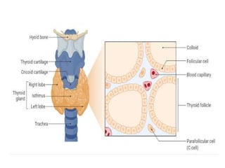
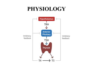
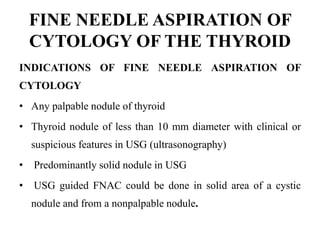
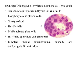
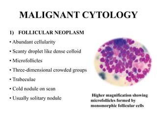
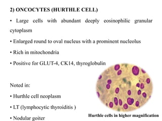
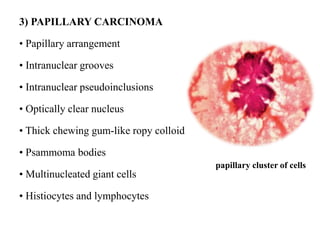
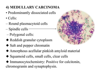
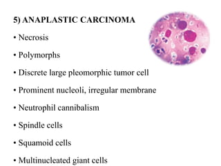
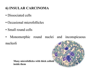
THYROID
ANATOMY AND HISTOLOGY
• Thyroid glands are located in the anterior part of neck
in front of trachea and inferior to larynx.
• The gland is composed of two lobes, which are
connected by central part known as isthmus.
• The thyroid gland is covered by thick fibrous septae.
• The fibroconnective tissue has divided the lobes into
multiple smaller lobules.
• Follicles are the basic structural unit of thyroid gland.
• Each follicle contains homogenous eosinophilic colloid
material.
• Follicles are lined by low cuboidal to columnar cells.
• Thyroid follicular cells liberate tri-iodothyronine (T3 ) and
thyroxine (T4 ) hormone.
• Due to some unknown reason, thyroid follicular cell may
undergo Hurthle cell change.
• These are large cells with abundant eosinophilic cytoplasm
and central round nuclei.
• Parafollicular cells are present in the periphery of the follicles
as small cluster.
• These cells are three to four times larger than the follicular
cells.
• They have pale moderate amount of cytoplasm with central
spherical nuclei.
• Parafollicular cells secrete calcitonin hormone that takes
important role in calcium homeostasis.
• Calcitonin hormone inhibits bone resorption by osteoclasts
and, thereby helps to lower the calcium concentrations in
blood.
PHYSIOLOGY
FINE NEEDLE ASPIRATION OF
CYTOLOGY OF THE THYROID
INDICATIONS OF FINE NEEDLE ASPIRATION OF
CYTOLOGY
• Any palpable nodule of thyroid
• Thyroid nodule of less than 10 mm diameter with clinical or
suspicious features in USG (ultrasonography)
• Predominantly solid nodule in USG
• USG guided FNAC could be done in solid area of a cystic
nodule and from a nonpalpable nodule.
TECHNIQUES
• The patient should be kept in supine position with a small
pillow under his neck to make the thyroid region prominent.
• The technique of FNAC in the thyroid gland is essentially
same.
• After cleaning the area of thyroid, the gland is hold in between
the two fingers so that the thyroid will be prominent.
• Depending on the aspirator’s choice, FNAC or nonsuction fine
needle sampling (FNS) can be done.
• As the thyroid is a vascular organ so FNS is preferable in
small swelling of the thyroid gland.
• However, in case of a cystic lesion, FNAC is preferable as
there is a chance of spillage by FNS technique.
• It is recommended to do three to five passes of needle in each
nodule.
• Multiple smears should be made immediately from the
aspirated material and both air dried smear and alcohol fixed
smears should be kept.
• If necessary, the material should be taken for cell block and
other ancillary investigations.
• The thyroid nodule is hold in between two fingers and gentle
suction is done.
• FNS is preferable in thyroid as the material is free from blood.
• Thyroid swelling is hold tightly in between the two fingers so
that the thyroid will be prominent.
• Now, the needle is moved gently to and fro within the lesion.
• Material comes to the needle hub with the help of capillary
pressure.
• The needle is gently withdrawn and the syringe is attached
with the hub.
• The material in the needle is spread on the slide.
• The FNAC material can also be collected for cell
block and flow cytometry to do ancillary technique.
NORMAL CYTOLOGY
a) Colloid: Gross appearance of colloid is thick to thin
brownish material.
• The MGG stained cytology smear shows a pale blue
acellular material.
pale blue thin colloid material
b) Thyroid follicular cells:
• Follicular cells are present in small clusters and
follicles.
• The cells are round with scanty cytoplasm
and round monomorphic nucleus.
• Nuclear chromatin is homogeneously
spread.
c) Foam cells : The foam cells show abundant foamy
cytoplasm with small monomorphic nucleus.
NON- MALIGNANT
1) COLLOID GOITER –
a) Nodular colloid goiter is the commonest diseases of
thyroid.
Cytology of nodular goiter
• Abundant colloid
• Discrete or flat sheet of thyroid follicular cells
• Macrophages
nodular goiter in a young female
Abundant thick and thin colloid along
with scattered benign thyroid follicular
cells in colloid goiter
b) Adenomatoid hyperplasia or hyperplastic nodules
• Cellular smear
• Dissociated and microfollicular arrangement
• Hurthle cells
• Foamy macrophages
• Scanty colloid
Multiple follicles and thin colloid
c) Diffuse toxic goiter (Grave’s disease)
• Cellular
• Scanty watery colloid
• Enlarged cell with vacuolated cytoplasm
• Prominent nucleoli
• Fire flares appearance
2) THYROIDITIS
a) Acute Thyroiditis- Acute thyroiditis patient presents
with fever, tachycardia, and sudden painful enlargement
of thyroid.
Cytology
• Polymorphs and lymphocytes
• Necrosis
• Degenerated thyroid follicular cells.
b) Subacute Granulomatous Thyroiditis
• Dirty background formed by cellular debris and
scanty colloid
• Epithelioid cell granulomas
• Multinucleated giant cells
• Lymphocytes and plasma cells
• Benign thyroid follicular cells with paravacuolar
granulations
c) Chronic Lymphocytic Thyroiditis (Hashimoto’s Thyroiditis)
• Lymphocytic infiltration in thyroid follicular cells
• Lymphocytes and plasma cells
• Scanty colloid
• Hurthle cells
• Multinucleated giant cells
• Ill-formed epithelioid cell granuloma
• Elevated thyroid antimicrosomal antibody and
antithyroglobulin antibodies.
MALIGNANT CYTOLOGY
1) FOLLICULAR NEOPLASM
• Abundant cellularity
• Scanty droplet like dense colloid
• Microfollicles
• Three-dimensional crowded groups
• Trabeculae
• Cold nodule on scan
• Usually solitary nodule
Higher magnification showing
microfollicles formed by
monomorphic follicular cells
2) ONCOCYTES (HURTHLE CELL)
• Large cells with abundant deeply eosinophilic granular
cytoplasm
• Enlarged round to oval nucleus with a prominent nucleolus
• Rich in mitochondria
• Positive for GLUT-4, CK14, thyroglobulin
Noted in:
• Hurthle cell neoplasm
• LT (lymphocytic thyroiditis )
• Nodular goiter
Hurthle cells in higher magnification
3) PAPILLARY CARCINOMA
• Papillary arrangement
• Intranuclear grooves
• Intranuclear pseudoinclusions
• Optically clear nucleus
• Thick chewing gum-like ropy colloid
• Psammoma bodies
• Multinucleated giant cells
• Histiocytes and lymphocytes
papillary cluster of cells
4) MEDULLARY CARCINOMA
• Predominantly dissociated cells
• Cells:
– Round plasmacytoid cells
– Spindle cells
– Polygonal cells:
◆ Reddish granular cytoplasm
◆ Salt and pepper chromatin
◆ Amorphous acellular pinkish amyloid material
◆ Squamoid cells, small cells, clear cells
◆ Immunocytochemistry: Positive for calcitonin,
chromogranin and synaptophysin.
5) ANAPLASTIC CARCINOMA
• Necrosis
• Polymorphs
• Discrete large pleomorphic tumor cell
• Prominent nucleoli, irregular membrane
• Neutrophil cannibalism
• Spindle cells
• Squamoid cells
• Multinucleated giant cells
6) INSULAR CARCINOMA
• Dissociated cells
• Occasional microfollicles
• Small round cells
• Monomorphic round nuclei and inconspicuous
nucleoli
Many microfollicles with thick colloid
inside them
THYROID - cytology pptx

THYROID - cytology pptx

  • 1.
  • 2.
    ANATOMY AND HISTOLOGY •Thyroid glands are located in the anterior part of neck in front of trachea and inferior to larynx. • The gland is composed of two lobes, which are connected by central part known as isthmus. • The thyroid gland is covered by thick fibrous septae. • The fibroconnective tissue has divided the lobes into multiple smaller lobules.
  • 4.
    • Follicles arethe basic structural unit of thyroid gland. • Each follicle contains homogenous eosinophilic colloid material. • Follicles are lined by low cuboidal to columnar cells. • Thyroid follicular cells liberate tri-iodothyronine (T3 ) and thyroxine (T4 ) hormone. • Due to some unknown reason, thyroid follicular cell may undergo Hurthle cell change. • These are large cells with abundant eosinophilic cytoplasm and central round nuclei.
  • 5.
    • Parafollicular cellsare present in the periphery of the follicles as small cluster. • These cells are three to four times larger than the follicular cells. • They have pale moderate amount of cytoplasm with central spherical nuclei. • Parafollicular cells secrete calcitonin hormone that takes important role in calcium homeostasis. • Calcitonin hormone inhibits bone resorption by osteoclasts and, thereby helps to lower the calcium concentrations in blood.
  • 6.
  • 7.
    FINE NEEDLE ASPIRATIONOF CYTOLOGY OF THE THYROID INDICATIONS OF FINE NEEDLE ASPIRATION OF CYTOLOGY • Any palpable nodule of thyroid • Thyroid nodule of less than 10 mm diameter with clinical or suspicious features in USG (ultrasonography) • Predominantly solid nodule in USG • USG guided FNAC could be done in solid area of a cystic nodule and from a nonpalpable nodule.
  • 8.
    TECHNIQUES • The patientshould be kept in supine position with a small pillow under his neck to make the thyroid region prominent. • The technique of FNAC in the thyroid gland is essentially same. • After cleaning the area of thyroid, the gland is hold in between the two fingers so that the thyroid will be prominent. • Depending on the aspirator’s choice, FNAC or nonsuction fine needle sampling (FNS) can be done.
  • 9.
    • As thethyroid is a vascular organ so FNS is preferable in small swelling of the thyroid gland. • However, in case of a cystic lesion, FNAC is preferable as there is a chance of spillage by FNS technique. • It is recommended to do three to five passes of needle in each nodule. • Multiple smears should be made immediately from the aspirated material and both air dried smear and alcohol fixed smears should be kept. • If necessary, the material should be taken for cell block and other ancillary investigations.
  • 10.
    • The thyroidnodule is hold in between two fingers and gentle suction is done. • FNS is preferable in thyroid as the material is free from blood. • Thyroid swelling is hold tightly in between the two fingers so that the thyroid will be prominent. • Now, the needle is moved gently to and fro within the lesion. • Material comes to the needle hub with the help of capillary pressure. • The needle is gently withdrawn and the syringe is attached with the hub.
  • 11.
    • The materialin the needle is spread on the slide. • The FNAC material can also be collected for cell block and flow cytometry to do ancillary technique.
  • 12.
    NORMAL CYTOLOGY a) Colloid:Gross appearance of colloid is thick to thin brownish material. • The MGG stained cytology smear shows a pale blue acellular material. pale blue thin colloid material
  • 13.
    b) Thyroid follicularcells: • Follicular cells are present in small clusters and follicles. • The cells are round with scanty cytoplasm and round monomorphic nucleus. • Nuclear chromatin is homogeneously spread.
  • 14.
    c) Foam cells: The foam cells show abundant foamy cytoplasm with small monomorphic nucleus.
  • 15.
    NON- MALIGNANT 1) COLLOIDGOITER – a) Nodular colloid goiter is the commonest diseases of thyroid. Cytology of nodular goiter • Abundant colloid • Discrete or flat sheet of thyroid follicular cells • Macrophages
  • 16.
    nodular goiter ina young female Abundant thick and thin colloid along with scattered benign thyroid follicular cells in colloid goiter
  • 17.
    b) Adenomatoid hyperplasiaor hyperplastic nodules • Cellular smear • Dissociated and microfollicular arrangement • Hurthle cells • Foamy macrophages • Scanty colloid Multiple follicles and thin colloid
  • 18.
    c) Diffuse toxicgoiter (Grave’s disease) • Cellular • Scanty watery colloid • Enlarged cell with vacuolated cytoplasm • Prominent nucleoli • Fire flares appearance
  • 19.
    2) THYROIDITIS a) AcuteThyroiditis- Acute thyroiditis patient presents with fever, tachycardia, and sudden painful enlargement of thyroid. Cytology • Polymorphs and lymphocytes • Necrosis • Degenerated thyroid follicular cells.
  • 20.
    b) Subacute GranulomatousThyroiditis • Dirty background formed by cellular debris and scanty colloid • Epithelioid cell granulomas • Multinucleated giant cells • Lymphocytes and plasma cells • Benign thyroid follicular cells with paravacuolar granulations
  • 21.
    c) Chronic LymphocyticThyroiditis (Hashimoto’s Thyroiditis) • Lymphocytic infiltration in thyroid follicular cells • Lymphocytes and plasma cells • Scanty colloid • Hurthle cells • Multinucleated giant cells • Ill-formed epithelioid cell granuloma • Elevated thyroid antimicrosomal antibody and antithyroglobulin antibodies.
  • 22.
    MALIGNANT CYTOLOGY 1) FOLLICULARNEOPLASM • Abundant cellularity • Scanty droplet like dense colloid • Microfollicles • Three-dimensional crowded groups • Trabeculae • Cold nodule on scan • Usually solitary nodule Higher magnification showing microfollicles formed by monomorphic follicular cells
  • 23.
    2) ONCOCYTES (HURTHLECELL) • Large cells with abundant deeply eosinophilic granular cytoplasm • Enlarged round to oval nucleus with a prominent nucleolus • Rich in mitochondria • Positive for GLUT-4, CK14, thyroglobulin Noted in: • Hurthle cell neoplasm • LT (lymphocytic thyroiditis ) • Nodular goiter Hurthle cells in higher magnification
  • 24.
    3) PAPILLARY CARCINOMA •Papillary arrangement • Intranuclear grooves • Intranuclear pseudoinclusions • Optically clear nucleus • Thick chewing gum-like ropy colloid • Psammoma bodies • Multinucleated giant cells • Histiocytes and lymphocytes papillary cluster of cells
  • 25.
    4) MEDULLARY CARCINOMA •Predominantly dissociated cells • Cells: – Round plasmacytoid cells – Spindle cells – Polygonal cells: ◆ Reddish granular cytoplasm ◆ Salt and pepper chromatin ◆ Amorphous acellular pinkish amyloid material ◆ Squamoid cells, small cells, clear cells ◆ Immunocytochemistry: Positive for calcitonin, chromogranin and synaptophysin.
  • 26.
    5) ANAPLASTIC CARCINOMA •Necrosis • Polymorphs • Discrete large pleomorphic tumor cell • Prominent nucleoli, irregular membrane • Neutrophil cannibalism • Spindle cells • Squamoid cells • Multinucleated giant cells
  • 27.
    6) INSULAR CARCINOMA •Dissociated cells • Occasional microfollicles • Small round cells • Monomorphic round nuclei and inconspicuous nucleoli Many microfollicles with thick colloid inside them