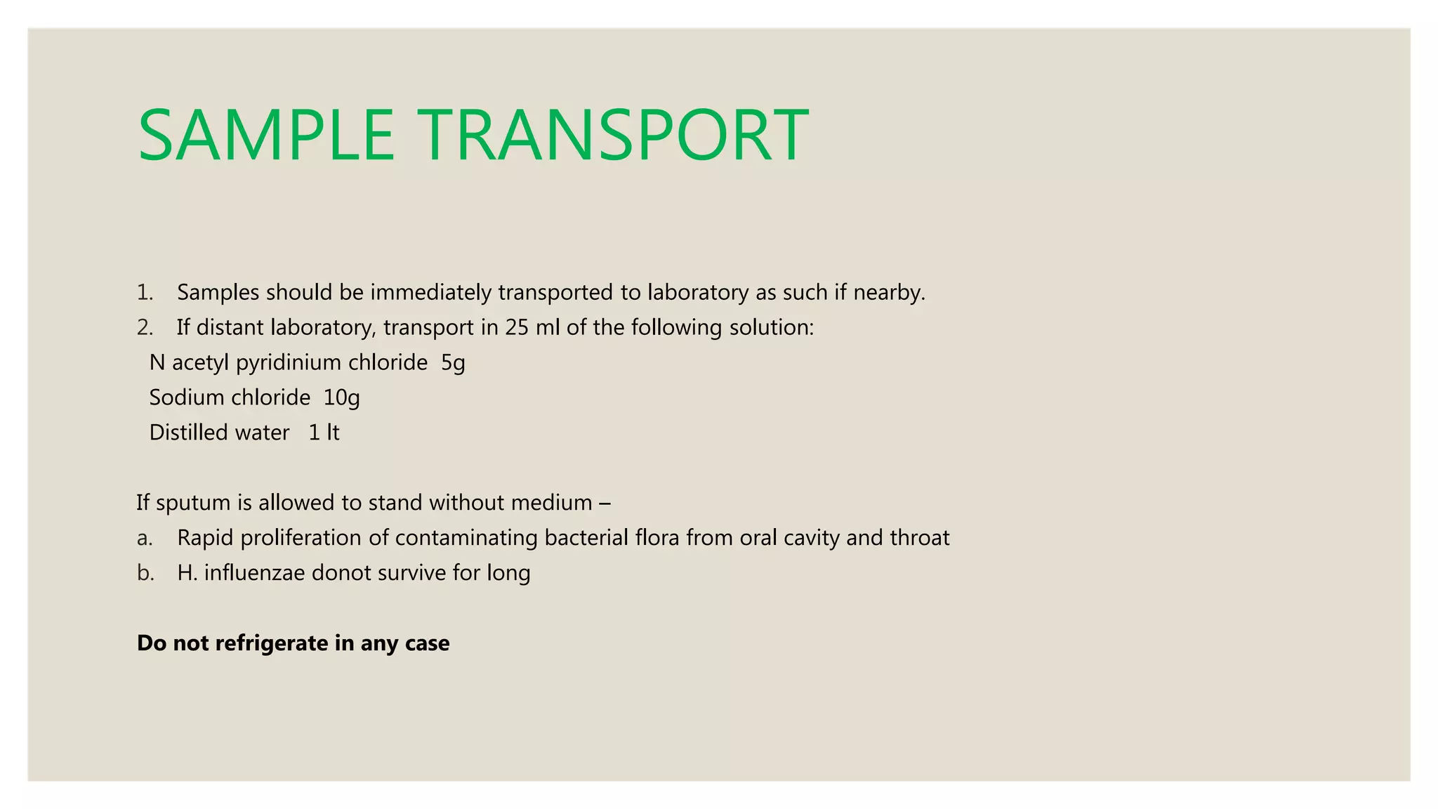
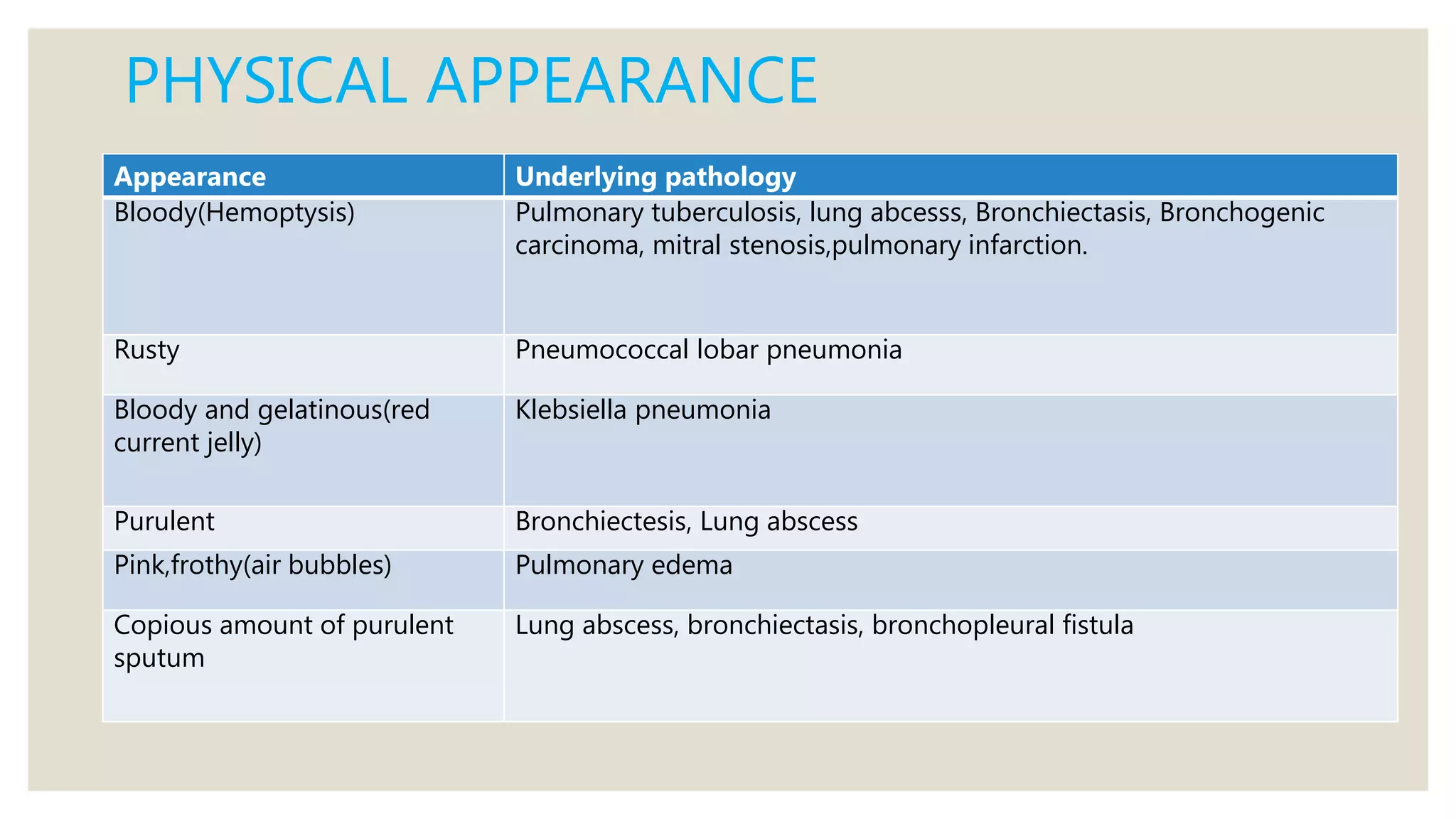
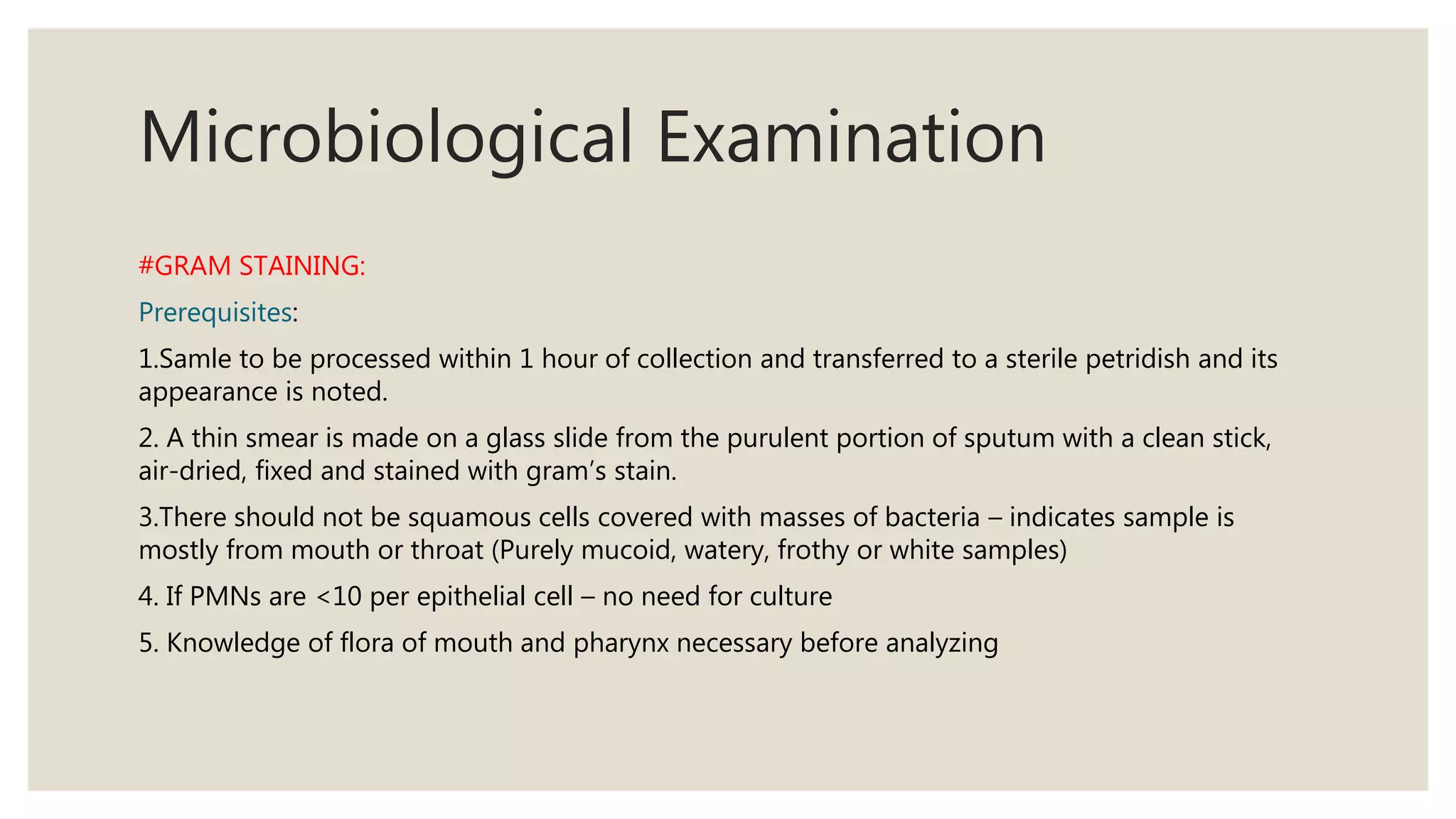
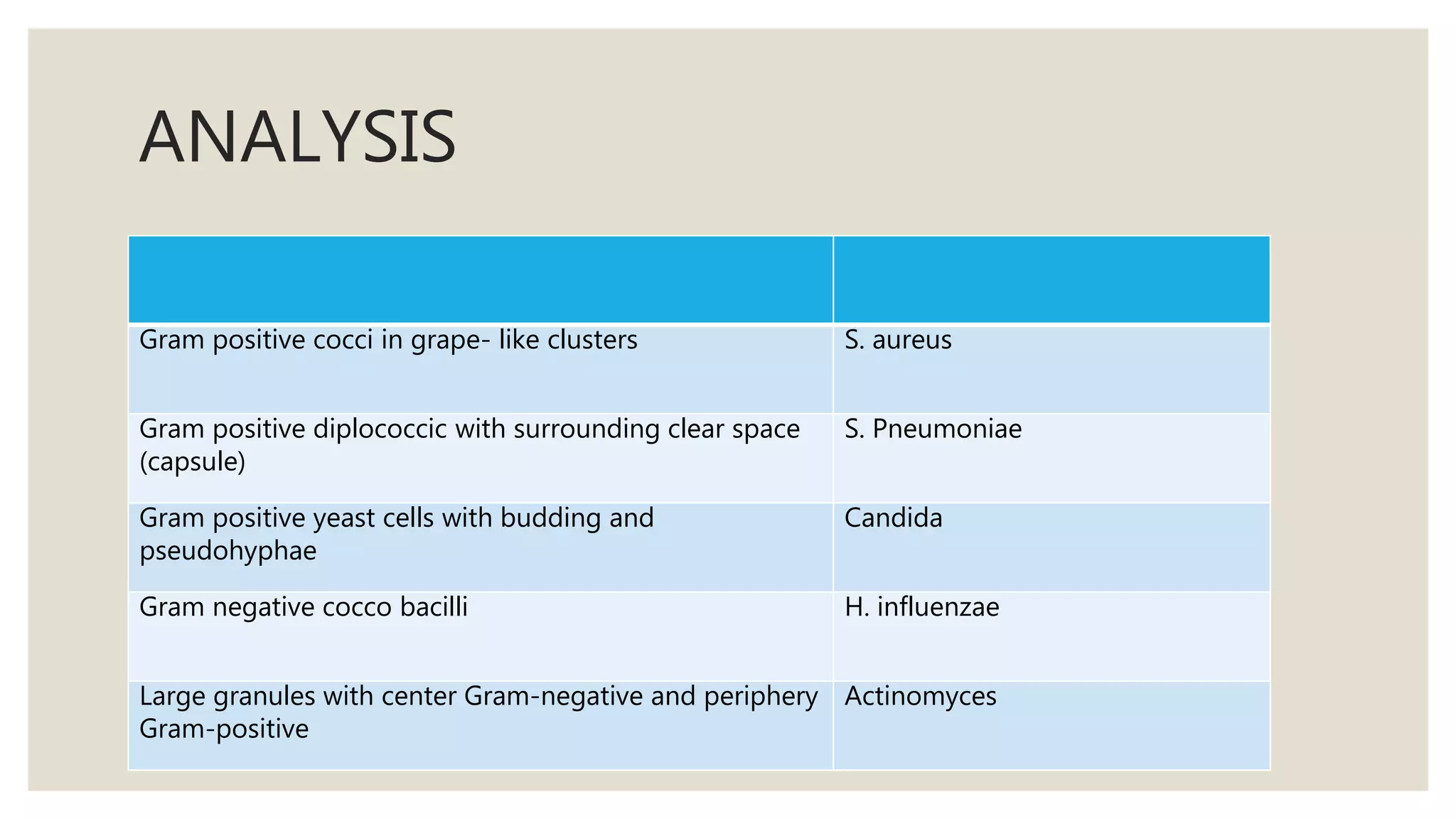
2. ◦ Sputum examination refers to the examination of the material coughed
out from the lungs, bronchi, trachea and larynx.
◦ Normally, sputum is composed predominantly of mucus and also
certain cellular and non-cellular components of host origin.
◦ During expectoration, sputum gets contaminated with cells and normal
bacterial flora from the mouth and pharynx
3. Indications
1. Identification of causative organism in a suspected infection of the
lower respiratory tract- like pneumonia, TB, fungal infection, P. carinii in
HIV, bronchiectasis.
2. Cytological examination
i) For malignant cells
ii) Investigation of viral infections( Viral inclusions in cytomegalo virus
and Herpes simplex infection)
iii) Asbestosis
4. Collection of sputum
1. Early morning deep cough sample is preferred.
2. If unable to cough, induction of sputum can be done by :
a. 15% NaCl aerosol spray & propylene glycol for 20 min
b. Nebulized hypertonic saline or distilled water in association with chest physiotherapy
Collected in:
1. Dry wide mouthed container with 25 ml capacity
2. Leak proof – to prevent aerosols
3. Break resistant – to prevent dessication
5. SAMPLE TRANSPORT
1. Samples should be immediately transported to laboratory as such if nearby.
2. If distant laboratory, transport in 25 ml of the following solution:
N acetyl pyridinium chloride 5g
Sodium chloride 10g
Distilled water 1 lt
If sputum is allowed to stand without medium –
a. Rapid proliferation of contaminating bacterial flora from oral cavity and throat
b. H. influenzae donot survive for long
Do not refrigerate in any case
7. Microbiological Examination
#GRAM STAINING:
Prerequisites:
1.Samle to be processed within 1 hour of collection and transferred to a sterile petridish and its
appearance is noted.
2. A thin smear is made on a glass slide from the purulent portion of sputum with a clean stick,
air-dried, fixed and stained with gram’s stain.
3.There should not be squamous cells covered with masses of bacteria – indicates sample is
mostly from mouth or throat (Purely mucoid, watery, frothy or white samples)
4. If PMNs are <10 per epithelial cell – no need for culture
5. Knowledge of flora of mouth and pharynx necessary before analyzing
9. Normal flora
• Staphylococci(S.aureus,
S.epidermidis)
• Streptococcus(S.viridans, S.
pneumonia)
• Diptheroids, Enterococci,Micrococci
• Yeast(Candida species
Gram
positive
• Neisseria species
• Hemophilus species
• Fusobacteria
• Coliforms
• Moraxella catarrhalis
Gram
Neative
10. ANALYSIS
Gram positive cocci in grape- like clusters S. aureus
Gram positive diplococcic with surrounding clear space
(capsule)
S. Pneumoniae
Gram positive yeast cells with budding and
pseudohyphae
Candida
Gram negative cocco bacilli H. influenzae
Large granules with center Gram-negative and periphery
Gram-positive
Actinomyces
11. BACTERIOLOGICAL CULTURE
◦ Ideal sample for culture
1. Should contain <25 squamous cells per low power filed or
<10 squamous cells per high power field.
2. Sample should contain alveolar macrophages.
3. Neutrophils should be >10 per epithelial cell or >5 per high
power field.
4. Bronchial epithelial cells present.
5. Sample should be washed with normal saline to wash the
saliva.
12. METHOD
Inoculate the sample on blood agar and chocolate agar
Incubate blood agar aerobically and chocolate agar in an atmosphere of extra CO2
Inspect plates after 18 hours.
If growth not satisfactory, incubation for further 24 hrs is indicated
If growth is significant, antibiotic sensitivity testing is carried out
13. EXAMINATION OF SPUTUM FOR MYCOBACTERIUM
TUBERCULOSIS:
1) ZEIHL NEELSON STAINING (AFB stain)
Sample:
1. According to RNTCP guidelines 2 samples are collected, one stat and
one early next morning sample. It should be deep cough sputum
sample.
2. For children, gastric aspirate can be used as they often swallow
sputum.
14. Preparation:
smear is prepared with blood tinged/opaque/grayish/yellowish portion of
the sputum
stained with ZN stain
Examined under the ordinary light microscope. If fluorescent microscope is
available, smear can be examined after staining with a flurochrome
(auramine-rhodamine or auramine O)
17. Reporting guidelines (RNTCP):
1. Mycobacteria appear as bright red, slightly curved or red beaded rods, 2-4 µm in length and
0.2 to 0.5 µm wide, against a blue green background.
2. Atleast 100 fields should be examined before declaring negative.
18. ◦ Drawbacks:
1. sensitivity 60-80%
2. minimum 5000-10000 bacilli / ml should be present for smear to be
positive.
◦ Bleaching technique:
1. A solution of sodium hypochlorite is added to sputum sample – it
leads to liquefaction of mucous and killing of microbes.
2. smears are prepared from sediment and stained with ZN stain.
19. 2. FLUORESCENCE MICROSCOPY
◦ Slides are stained with fluorescent auramine-rhodamine or
auramine O
◦ Observed under fluorescent microscope – mycobacteria appear
bright yellow against green background
◦ Rapid technique and is specially helpful if organisms are few in
number.
◦ It is necessary to confirm a positive smear by ZN stain since false
positive rate is high.
20. 3. CULTURE ON CONVENTIONAL MEDIA
Indications:
1. Drug susceptibility testing
2. Species identification if other than M. tuberculosis suspected
3. Sputum smear negative and strong clinical suspicion
Prerequisites:
1. 4% NaOH should be added before inoculation
2. This is because sputum samples are contaminated with normal flora, which grow and digest the media
before MTB can grow
3. 4% NaOH kills this flora
21. Media used:
1. Solid media – LJ media (egg based) or Middle brook (agar based).
2. Liquid media – middle brook, TH9, TH 12.
Advantage:
1. Sensitivity 80-85% .
2. Can detect as low as 10-100 bacteria/ml.
Drawbacks:
1. Expensive.
2. Requires 6 weeks for results.
22. 4. COMMERCIAL AUTOMATED CULTURE METHODS
(BACTEC)
1.Can give results in 2 weeks.
2. Mycobacteria are inoculated in a broth containing 14C
palmitate.
3. Mycobacteria metabolise 14C palmitate and release 14CO2
which is detected by the instrument.
23. 5. MOLECULAR METHODS (PCR)
◦ Two approaches for molecular diagnosis of tuberculosis in sputum samples:
1.Direct detection of M. tuberculosisin sputum samples.
2.Detection of M. tuberculosis in isolates from culture by nucleic acid probes.
◦ DNA sequences identified in MTB genome by PCR.
◦ Can detect bacteria as low as 10-1000 organisms / ml of sputum.
Disadvantage:
◦ Laboratory cross-contamination is responsible for significant number of false positive results.
◦ Expensive.
◦ Cannot be used to assess the response to therapy as PCR amplifies DNA sequences of both live and dead
bacilli.
28. Cytological Examination
◦ Usually carried out for investigation of bronchogenic carcinoma.
◦ Sometimes it may also be helpful in identification of viral inclusions( CMV and Herpes simplex
virus),fungi,protozoa,and asbestos bodies.
Prerequisites:
1. Fresh morning sample
2. Transport without delay
3. For suspected lung carcinoma collect sample for 5 consecutive days. (because multiple sputum samples
increase the chances of detection of malignant cells).
4. If delay anticipated, prefix with Saccomano’s fixative (50% ethyl alcohol and 2% carbowax)
Method:
1. Smears are made from blood tinged portion or tissue fragments in and stained with pap stain.
2. To be adequate, bronchial epithelial cells and alveolar macrophages must be seen
29. Advantages:
• Easy to collect.
• The wider area of the respiratory tract is covered.
• Simple and non-invasive.
• Good sensitivity for central tumour.
• Best for screening malignancy.
Disadvantage:
• Exact localization of the tumour not possible.
• Degeneration of the cells.
• Not good for peripheral tumour.
• Sensitivity is only 65%
This sensitivity is more if:
1.Smears are examined from multiple samples
2.Lesion is located centrally
3.Larger tumor size
4.histologic type is SCC rather than adenocarcinoma
30.
31. Sputum smear shows discrete malignant cells entangled in
adenocarcinoma of lung(Papanicolaou’s stain)