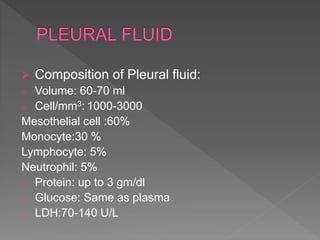
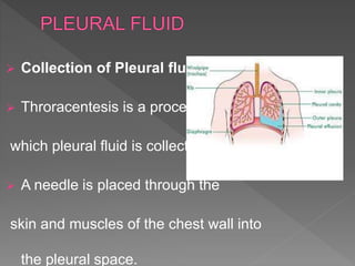
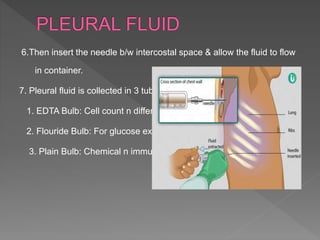
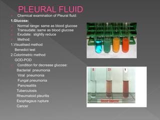
It is fluid which is present in the pleural cavity of
lungs b/w parietal pleura n visceral pleura.
The pleural cavity is a potential space lined by
mesothelium of the visceral n parietal pleura.
Bangalore Call Girl Whatsapp Number 100% Complete Your Sexual Needs
Pleural fluid
1. Ms Ankita R Bhatiya
Assistant Professor
Shree P.M.Patel COLLEGE OF
PARAMEDICAL SCIENCE N
TECHNOLOGY
2. It include:
1.What is Pleural fluid?
2.Function of Pleural fluid.
3.Collection of Pleural fluid.
4. Examination of Pleural fluid.
3. Introduction:
It is fluid which is present in the pleural cavity of
lungs b/w parietal pleura n visceral pleura.
The pleural cavity is a potential space lined by
mesothelium of the visceral n parietal pleura.
Pleural fluid Formation:
Pleural fluid is a selective ultrafiltrate of plasma.
Small amount of the Pleural fluid is also formed
from the cells lining the pleura and other by capillaries.
There is about 60-70 ml of pleural fluid at any one time
and about 125 ml is generated every day.
4. Composition of Pleural fluid:
o Volume: 60-70 ml
o Cell/mm3: 1000-3000
Mesothelial cell :60%
Monocyte:30 %
Lymphocyte: 5%
Neutrophil: 5%
o Protein: up to 3 gm/dl
o Glucose: Same as plasma
o LDH:70-140 U/L
5. Function of Pleural fluid:
Protection: It helps to protect the lungs from the
sudden injury n damaged.
Also acts as a medium for the transfer of
substances from the lung tissue to blood .
Nutrition :
Removal of waste :
Lubrication :
6. Collection of Pleural fluid:
Throracentesis is a process by
which pleural fluid is collected.
A needle is placed through the
skin and muscles of the chest wall into
the pleural space.
8. Procedure:
1.Take consent of a patient.
2.Position of patient: The patient is sitting in an upright
position with arms & head extended to over bed table.
3.Proper aseptic precautions are taken
with the help of spirit –iodine-spirit.
4.Before puncture give injection of atrophin
intramuscularly to prevent vasovagal shock.
5.Give xylocane injection as local anesthesia before puncture.
9. 6.Then insert the needle b/w intercostal space & allow the fluid to flow
in container.
7. Pleural fluid is collected in 3 tubes:
1. EDTA Bulb: Cell count n differential count.
2. Flouride Bulb: For glucose examination.
3. Plain Bulb: Chemical n immunology study
10. Examination of Pleural fluid:
1. Physical examination:
2. Chemical examination:
3. Microscopic examination:
11. Physical examination of Pleural fluid:
1.Volume:
Normally: 60 to 70 ml
Transudates– 100 to 150 ml
Condition: Increase hydrostatic pressure
Decrease plasma oncotic pressure
Congestive heart failure
Hepatic cirrhosis
Hypoproteinemia
Exudates– More than 150 ml
Condition: Pneumonia( Bacterial, Viral,Fungal)
Tuberculosis
Bronchogenic carcinoma, Metastatic carcinoma,
Lymphoma, Mesothelioma
Pulmonary infract
Rheumatoid disease
Systemic lupus erythreomatous
Pancreatitis, Ruptured esophagus,Urinithrorax
12. 2. color:
Normally: colorless
Transudates: Pale yellow or straw clr
Exudates:
Reddish: Presence of blood (Bacterial
pneumonia, Cancer, Pancreatitis.)
Green; Pseudochlyous effusion.
13. 3.Appreance:
Normally: Clear or transparent
Transudate: Clear or transparent
Exudate:
Turbid: Bacterial n viral Pneumonia,
Tuberculosis, Pancreatitis
Cloudy: Bacterial n fungal pneumonia
Purulent: Bacterial Pneumonia
Milky :Chylous effusion