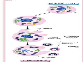
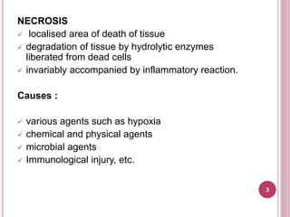
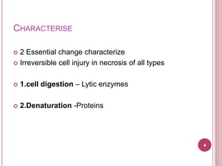
1. Necrosis is the localized death of cells and living tissue. It is caused by factors like hypoxia, chemicals, microbes, or immune reactions. There are two key changes in necrosis - irreversible cell injury and digestion of cells by lytic enzymes.
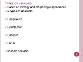
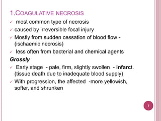
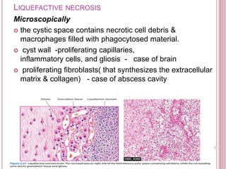
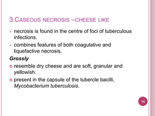
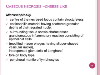
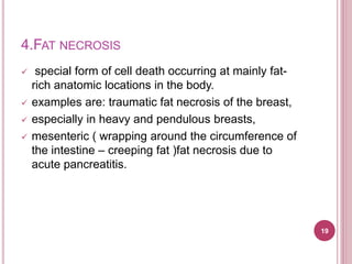
2. There are five main types of necrosis defined by their etiology and appearance: coagulative, liquefactive, caseous, fat, and fibrinoid necrosis. Coagulative necrosis is the most common type seen as pale, swollen areas that become yellow and shrunken over time. Liquefactive necrosis results in a soft, liquefied center containing debris. Caseous necrosis forms a cheese-like focus found in tuberculosis. Fat necrosis