Downloaded 864 times

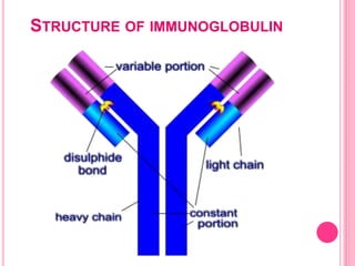
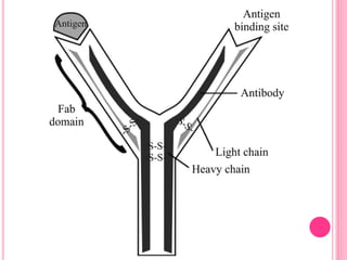
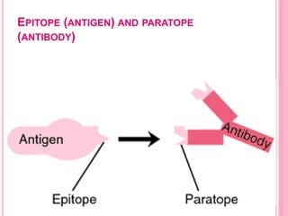
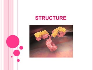


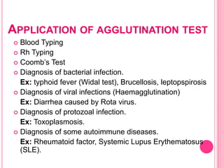
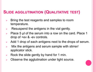
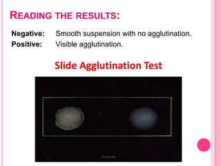
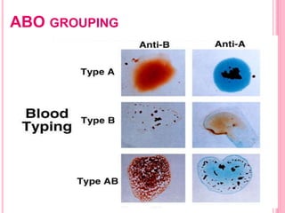
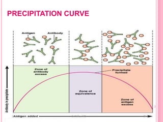
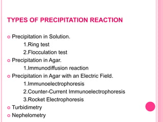
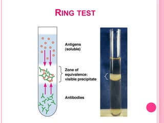
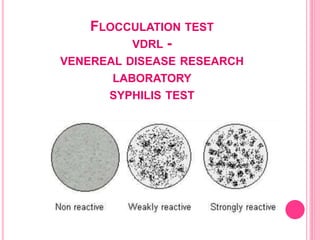
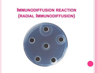
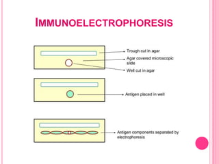
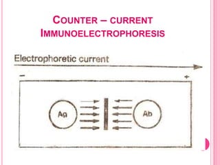
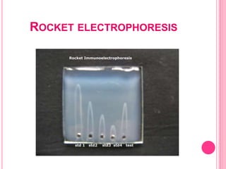


The document provides an overview of antibodies, describing their structure, history, and function in the immune response. It details various types of immunoglobulins, their properties, and the mechanisms by which they interact with antigens, including agglutination and precipitation reactions. Additionally, applications of these reactions in clinical diagnostics are discussed, highlighting their importance in identifying infections and autoimmune diseases.


