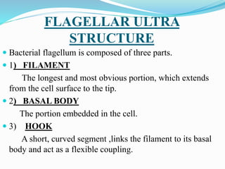
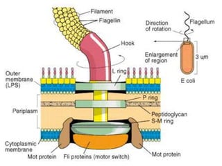
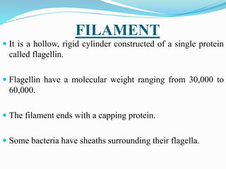
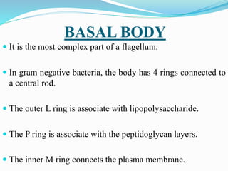
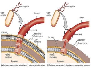
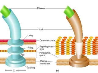
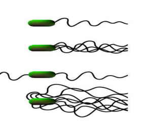
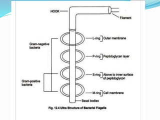
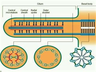
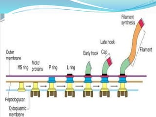
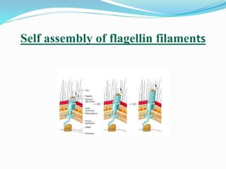
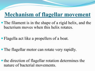
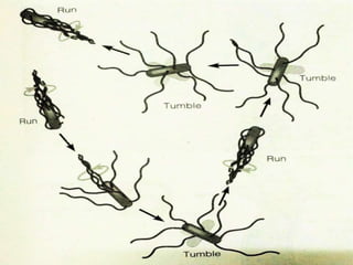
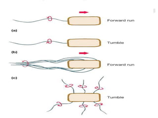
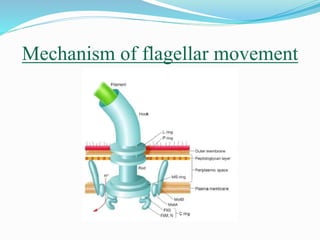
ULTRA STRUCTURE OF
FLAGELLA
SUBMITTED BY
Vivek kumar
MSc . MICROBIOLOGY
Bangalore university
FLAGELLAR ULTRA
STRUCTURE
 Bacterial flagellum is composed of three parts.
 1) FILAMENT
The longest and most obvious portion, which extends
from the cell surface to the tip.
 2) BASAL BODY
The portion embedded in the cell.
 3) HOOK
A short, curved segment ,links the filament to its basal
body and act as a flexible coupling.
FILAMENT
 It is a hollow, rigid cylinder constructed of a single protein
called flagellin.
 Flagellin have a molecular weight ranging from 30,000 to
60,000.
 The filament ends with a capping protein.
 Some bacteria have sheaths surrounding their flagella.
BASAL BODY
 It is the most complex part of a flagellum.
 In gram negative bacteria, the body has 4 rings connected to
a central rod.
 The outer L ring is associate with lipopolysaccharide.
 The P ring is associate with the peptidoglycan layers.
 The inner M ring connects the plasma membrane.
HOOK
 It is quite different from filament.
 It is slightly wider than the filament.
 Hook is made up of different protein subunits.
 Gram positive bacteria have only 2 basal body
rings.
 An inner ring is connected to the plasma
membrane.
 An outer ring probably attached to the
peptidoglycan layer.
 Sometimes the flagellum will have lateral hairs called
flimmer filaments.
 The thicker, stiffer hair are called mastigonemes.
 These filaments change the flagella action.
 so that a wave moving down the filament towards the tip
pulls the cell along instead of pushing it.
 Such a flagellum is called tinsel flagellum.
 The naked flagellum is called whiplash flagellum.
FLAGELLAAND MOTILITY
 Most motile bacteria move by use of flagella.
 Its a thread like locomotor appendages extending outward
from the plasma membrane and the cell wall.
 They are slender, rigid structures, about 20 μm long.
 Flagella can be observed by using special staining
techniques.
FLAGELLAR
ARRANGEMENTS
MONOTRCHOUS FLAGELLA
Having one flagellum. If it is located at an end it is called
polar flagellum.
AMPHITRICHOUS FLAGELLA
Have a single flagellum at each pole.
LOPHOTRICHOUS FLAGELLA
Have a cluster of flagella at one or both ends.
PERITRICHOUS FLAGELLA
Flagella are spread fairly evenly over the whole surface
STRUCTURE OF FLAGELLA
 Flagella are membrane bound cylinders about 0.2 μm in
diameter.
 Located in the matrix of the organelle is a complex, the
axoneme.
 The axoneme consists of 9 pairs of microtubule doublets
arranged in a circle around 2 central tubules.
 This is called 9+2 pattern of microtubules.
 Each doublet also has pair of arms projecting from
subtubule A towards a neighbouring doublet.
 A radial spoke extends from subtubule A towards the
internal pair of microtubule with their central sheath.
 The microtubules are similar to those found in the
cytoplasm.
 It is constructed of α and β tubulins subunits.
 The basal body is a short cylinder with 9 microtubule triplets
around its periphery, a 9+0 pattern.
 Basal body is separated from the rest of the organelle by a basal
plate.
 The doublet arms, about 15nm long, are made of dynein protein.
 Dynein arms interact with the B subtubules of adjacent doublets
to cause the sliding.
 Radial spokes also participate in sliding motion.
 Flagella beats at a rate of about 10 to 40 strokes of
waves per second.
 The common euglenoid flagellate, Euglena gracilis
travels at around 170 μm per second.
 The flagellate Monas stigmatica, swims at a rate of
260 μm / second.
FLAGELLAR SYNTHESIS
 It is a complex process involving at least 20 to 30 genes.
 Besides the gene for flagellin, 10 or more genes code for
hook and basal body proteins.
 Filament synthesis is an example of self-assembly.
 The information required for filament construction is present
in the structure of the flagellin subunit itself.
 The MS ring is synthesised first and inserted into the
cytoplasmic membrane, this is followed by the
formation of other rings, hook and cap.
 These flagellin molecules are then assisted by cap
protein which exist at the tip of a growing flagellum.
 Self-assembly or aggregation of flagellin proteins
lead to the formation of filaments
• The flagellin subunits are transported through the
filament's hollow internal core.
Self assembly of flagellin filaments
Mechanism of flagellar movement
 The filament is in the shape of a rigid helix, and the
bacterium moves when this helix rotates.
 Flagella act like a propellers of a boat.
 The flagellar motor can rotate very rapidly.
 the direction of flagellar rotation determines the
nature of bacterial movements.
 Polar flagella rotate counter clockwise during normal
forward movement.
 The cell itself rotates slowly clockwise.
 The rotating filament thrusts the cell forward with
the flagellum trailing behind.
 Monotrichous flagella stop and tumble randomly by
reversing the direction of flagellar rotation.
 Peritrichous flagella operate in somewhat similar
way.
 To move the flagella rotates counter clockwise.
 They bend as their hook to form a rotating bundle
that propels them forward.
 Clock wise rotation of the flagella disrupts the
bundle and the cell tumbles
 Because bacteria swim through rotation of their rigid
flagella, there must be some sort of motor at the
base.
 The flagellum rotate because of interactions between
the S and M ring, which can rotate freely in the
plasma membrane.
 Torque generated by the motor is transmitted by the
basal body to the hook and the filament
MOTOR
 It is composed of two components
1. THE ROTOR
2. THE STATOR
 It function like an electrical motor, where the rotor turns in
the centre of a ring of electromagnets, the stator.
 In gram negative bacteria, the rotor is composed of the MS
ring and the C ring.
 The flagellar protein FliG is important to interact the rotor
with the stator.
The rotor
 In gram negative bacteria, the rotor is
composed of the MS ring and the C ring.
 The flagellar protein FliG is important to
interact the rotor with the stator.
The stator
 The stator is composed of the proteins MotA
and MotB.
 Both form a channel through the plasma
membrane .
 MotB also anchor MotA to cell wall
peptidoglycan.
 A proton motive force is used to generate torque.
 The channel created by MotA and MotB proteins
allow protons to move across the plasma membrane
from outside to inside.
 They move to the charge and pH gradient.
 This movement releases energy that is used to rotate
the flagellum.
Mechanism of flagellar movement
PERIPLASMIC FLAGELLA
 Certain helical bacteria exhibit swimming motility
particularly in highly viscous media.
 They lack external flagella but possess flagella like
structures located within the cell just beneath the
outer cell envelope.
 These are called periplasmic flagella or endoflagella
or axial flagella.
Periplasmic flagella
THANK YOU
thanks

Flagella

  • 1.
    ULTRA STRUCTURE OF FLAGELLA SUBMITTEDBY Vivek kumar MSc . MICROBIOLOGY Bangalore university
  • 2.
    FLAGELLAR ULTRA STRUCTURE  Bacterialflagellum is composed of three parts.  1) FILAMENT The longest and most obvious portion, which extends from the cell surface to the tip.  2) BASAL BODY The portion embedded in the cell.  3) HOOK A short, curved segment ,links the filament to its basal body and act as a flexible coupling.
  • 4.
    FILAMENT  It isa hollow, rigid cylinder constructed of a single protein called flagellin.  Flagellin have a molecular weight ranging from 30,000 to 60,000.  The filament ends with a capping protein.  Some bacteria have sheaths surrounding their flagella.
  • 5.
    BASAL BODY  Itis the most complex part of a flagellum.  In gram negative bacteria, the body has 4 rings connected to a central rod.  The outer L ring is associate with lipopolysaccharide.  The P ring is associate with the peptidoglycan layers.  The inner M ring connects the plasma membrane.
  • 6.
    HOOK  It isquite different from filament.  It is slightly wider than the filament.  Hook is made up of different protein subunits.
  • 7.
     Gram positivebacteria have only 2 basal body rings.  An inner ring is connected to the plasma membrane.  An outer ring probably attached to the peptidoglycan layer.
  • 9.
     Sometimes theflagellum will have lateral hairs called flimmer filaments.  The thicker, stiffer hair are called mastigonemes.  These filaments change the flagella action.  so that a wave moving down the filament towards the tip pulls the cell along instead of pushing it.  Such a flagellum is called tinsel flagellum.  The naked flagellum is called whiplash flagellum.
  • 11.
    FLAGELLAAND MOTILITY  Mostmotile bacteria move by use of flagella.  Its a thread like locomotor appendages extending outward from the plasma membrane and the cell wall.  They are slender, rigid structures, about 20 μm long.  Flagella can be observed by using special staining techniques.
  • 12.
    FLAGELLAR ARRANGEMENTS MONOTRCHOUS FLAGELLA Having oneflagellum. If it is located at an end it is called polar flagellum. AMPHITRICHOUS FLAGELLA Have a single flagellum at each pole. LOPHOTRICHOUS FLAGELLA Have a cluster of flagella at one or both ends. PERITRICHOUS FLAGELLA Flagella are spread fairly evenly over the whole surface
  • 15.
    STRUCTURE OF FLAGELLA Flagella are membrane bound cylinders about 0.2 μm in diameter.  Located in the matrix of the organelle is a complex, the axoneme.  The axoneme consists of 9 pairs of microtubule doublets arranged in a circle around 2 central tubules.  This is called 9+2 pattern of microtubules.
  • 17.
     Each doubletalso has pair of arms projecting from subtubule A towards a neighbouring doublet.  A radial spoke extends from subtubule A towards the internal pair of microtubule with their central sheath.  The microtubules are similar to those found in the cytoplasm.  It is constructed of α and β tubulins subunits.
  • 19.
     The basalbody is a short cylinder with 9 microtubule triplets around its periphery, a 9+0 pattern.  Basal body is separated from the rest of the organelle by a basal plate.  The doublet arms, about 15nm long, are made of dynein protein.  Dynein arms interact with the B subtubules of adjacent doublets to cause the sliding.  Radial spokes also participate in sliding motion.
  • 21.
     Flagella beatsat a rate of about 10 to 40 strokes of waves per second.  The common euglenoid flagellate, Euglena gracilis travels at around 170 μm per second.  The flagellate Monas stigmatica, swims at a rate of 260 μm / second.
  • 22.
    FLAGELLAR SYNTHESIS  Itis a complex process involving at least 20 to 30 genes.  Besides the gene for flagellin, 10 or more genes code for hook and basal body proteins.  Filament synthesis is an example of self-assembly.  The information required for filament construction is present in the structure of the flagellin subunit itself.
  • 23.
     The MSring is synthesised first and inserted into the cytoplasmic membrane, this is followed by the formation of other rings, hook and cap.  These flagellin molecules are then assisted by cap protein which exist at the tip of a growing flagellum.  Self-assembly or aggregation of flagellin proteins lead to the formation of filaments • The flagellin subunits are transported through the filament's hollow internal core.
  • 25.
    Self assembly offlagellin filaments
  • 26.
    Mechanism of flagellarmovement  The filament is in the shape of a rigid helix, and the bacterium moves when this helix rotates.  Flagella act like a propellers of a boat.  The flagellar motor can rotate very rapidly.  the direction of flagellar rotation determines the nature of bacterial movements.
  • 27.
     Polar flagellarotate counter clockwise during normal forward movement.  The cell itself rotates slowly clockwise.  The rotating filament thrusts the cell forward with the flagellum trailing behind.  Monotrichous flagella stop and tumble randomly by reversing the direction of flagellar rotation.
  • 28.
     Peritrichous flagellaoperate in somewhat similar way.  To move the flagella rotates counter clockwise.  They bend as their hook to form a rotating bundle that propels them forward.  Clock wise rotation of the flagella disrupts the bundle and the cell tumbles
  • 31.
     Because bacteriaswim through rotation of their rigid flagella, there must be some sort of motor at the base.  The flagellum rotate because of interactions between the S and M ring, which can rotate freely in the plasma membrane.  Torque generated by the motor is transmitted by the basal body to the hook and the filament
  • 32.
    MOTOR  It iscomposed of two components 1. THE ROTOR 2. THE STATOR  It function like an electrical motor, where the rotor turns in the centre of a ring of electromagnets, the stator.  In gram negative bacteria, the rotor is composed of the MS ring and the C ring.  The flagellar protein FliG is important to interact the rotor with the stator.
  • 33.
    The rotor  Ingram negative bacteria, the rotor is composed of the MS ring and the C ring.  The flagellar protein FliG is important to interact the rotor with the stator.
  • 34.
    The stator  Thestator is composed of the proteins MotA and MotB.  Both form a channel through the plasma membrane .  MotB also anchor MotA to cell wall peptidoglycan.
  • 35.
     A protonmotive force is used to generate torque.  The channel created by MotA and MotB proteins allow protons to move across the plasma membrane from outside to inside.  They move to the charge and pH gradient.  This movement releases energy that is used to rotate the flagellum.
  • 36.
  • 37.
    PERIPLASMIC FLAGELLA  Certainhelical bacteria exhibit swimming motility particularly in highly viscous media.  They lack external flagella but possess flagella like structures located within the cell just beneath the outer cell envelope.  These are called periplasmic flagella or endoflagella or axial flagella.
  • 38.
  • 39.