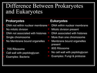
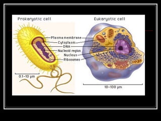
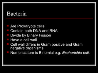
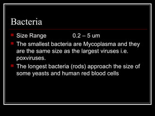
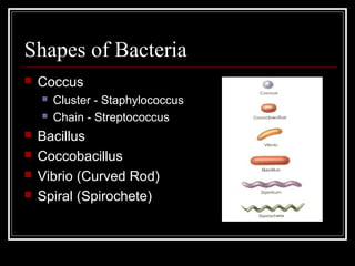
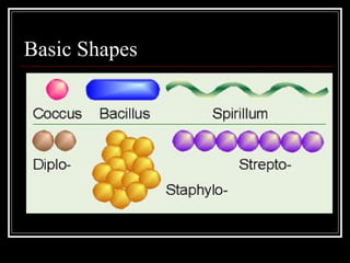
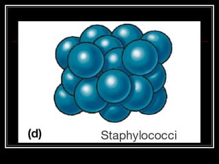
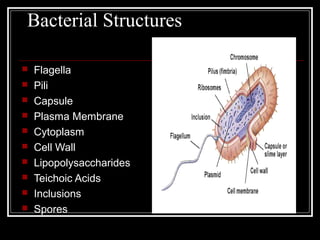
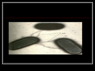
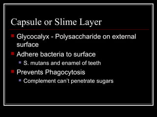
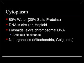
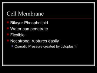
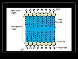
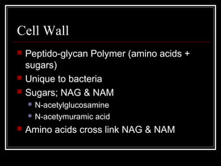
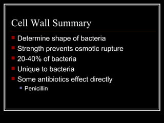
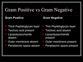
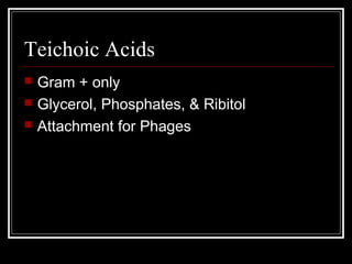
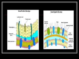
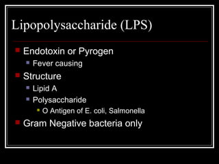
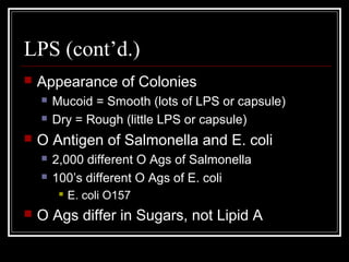
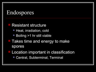
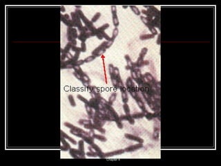
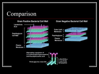
Structure Of
Bacteria
Maj M. Omair Riaz
Assistant Professor of Pathology
QIMS
Difference Between Prokaryotes
and Eukaryotes
Prokaryotes
 DNA not within nuclear membrane
 No mitotic division
 DNA not associated with histones
 Single chromosome
 No Membrane bound organelles
 70S Ribosome
 Cell wall with peptidoglycan
 Examples: Bacteria
Eukaryotes
 DNA within nuclear membrane
 Mitotic division present
 DNA associated with histones
 More than one chromosome
 Membrane bound organelles
present
 80S Ribosome
 No cell wall with peptidoglycan
 Examples: Fungi & protozoa
Bacteria
 Are Prokaryote cells
 Contain both DNA and RNA
 Divide by Binary Fission
 Have a cell wall
 Cell wall differs in Gram positive and Gram
negative organisms
 Nomenclature is Binomial e.g. Escherichia coli.
Bacteria
 Size Range 0.2 – 5 um
 The smallest bacteria are Mycoplasma and they
are the same size as the largest viruses i.e.
poxviruses.
 The longest bacteria (rods) approach the size of
some yeasts and human red blood cells
Shapes of Bacteria
 Coccus
 Cluster - Staphylococcus
 Chain - Streptococcus
 Bacillus
 Coccobacillus
 Vibrio (Curved Rod)
 Spiral (Spirochete)
Basic Shapes
Bacterial Structures
 Flagella
 Pili
 Capsule
 Plasma Membrane
 Cytoplasm
 Cell Wall
 Lipopolysaccharides
 Teichoic Acids
 Inclusions
 Spores
Flagella
 Motility - movement
 Swarming occurs with some bacteria
 Spread across Petri Dish
 Proteus species most evident
 Arrangement basis for classification
 Monotrichous; 1 flagella
 Lophotrichous; tuft at one end
 Amphitrichous; both ends
 Peritrichous; all around bacteria
Pili
 Short protein appendages
 smaller than flagella
 Adhere bacteria to surfaces
 E. coli has numerous types
 F-pilus; used in conjugation
 Exchange of genetic information
 Flotation; increase boyancy
Capsule or Slime Layer
 Glycocalyx - Polysaccharide on external
surface
 Adhere bacteria to surface
 S. mutans and enamel of teeth
 Prevents Phagocytosis
 Complement can’t penetrate sugars
Cytoplasm
 80% Water {20% Salts-Proteins)
 DNA is circular, Haploid
 Plasmids; extra chromosomal DNA
 Antibiotic Resistance
 No organelles (Mitochondria, Golgi, etc.)
Cell Membrane
 Bilayer Phospholipid
 Water can penetrate
 Flexible
 Not strong, ruptures easily
 Osmotic Pressure created by cytoplasm
Cell Wall
 Peptido-glycan Polymer (amino acids +
sugars)
 Unique to bacteria
 Sugars; NAG & NAM
 N-acetylglucosamine
 N-acetymuramic acid
 Amino acids cross link NAG & NAM
Cell Wall Summary
 Determine shape of bacteria
 Strength prevents osmotic rupture
 20-40% of bacteria
 Unique to bacteria
 Some antibiotics effect directly
 Penicillin
Gram Positive vs Gram Negative
Gram Positive
 Thick Peptidoglycan layer
 Teichoic acid present
 Lipopolysaccharide
absent
 Outer membrane absent
 Periplasmic space absent
Gram Negative
 Thin Peptidoglycan layer
 Teichoic acid absent
 Lipopolysaccharide
present
 Outer membrane present
 Periplasmic space present
Teichoic Acids
 Gram + only
 Glycerol, Phosphates, & Ribitol
 Attachment for Phages
Lipopolysaccharide (LPS)
 Endotoxin or Pyrogen
 Fever causing
 Structure
 Lipid A
 Polysaccharide
 O Antigen of E. coli, Salmonella
 Gram Negative bacteria only
LPS (cont’d.)
 Appearance of Colonies
 Mucoid = Smooth (lots of LPS or capsule)
 Dry = Rough (little LPS or capsule)
 O Antigen of Salmonella and E. coli
 2,000 different O Ags of Salmonella
 100’s different O Ags of E. coli
 E. coli O157
 O Ags differ in Sugars, not Lipid A
Endospores
 Resistant structure
 Heat, irradiation, cold
 Boiling >1 hr still viable
 Takes time and energy to make
spores
 Location important in classification
 Central, Subterminal, Terminal
Chapter 4
Comparison

Bacterial anatomy

  • 1.
    Structure Of Bacteria Maj M.Omair Riaz Assistant Professor of Pathology QIMS
  • 2.
    Difference Between Prokaryotes andEukaryotes Prokaryotes  DNA not within nuclear membrane  No mitotic division  DNA not associated with histones  Single chromosome  No Membrane bound organelles  70S Ribosome  Cell wall with peptidoglycan  Examples: Bacteria Eukaryotes  DNA within nuclear membrane  Mitotic division present  DNA associated with histones  More than one chromosome  Membrane bound organelles present  80S Ribosome  No cell wall with peptidoglycan  Examples: Fungi & protozoa
  • 4.
    Bacteria  Are Prokaryotecells  Contain both DNA and RNA  Divide by Binary Fission  Have a cell wall  Cell wall differs in Gram positive and Gram negative organisms  Nomenclature is Binomial e.g. Escherichia coli.
  • 5.
    Bacteria  Size Range0.2 – 5 um  The smallest bacteria are Mycoplasma and they are the same size as the largest viruses i.e. poxviruses.  The longest bacteria (rods) approach the size of some yeasts and human red blood cells
  • 6.
    Shapes of Bacteria Coccus  Cluster - Staphylococcus  Chain - Streptococcus  Bacillus  Coccobacillus  Vibrio (Curved Rod)  Spiral (Spirochete)
  • 7.
  • 9.
    Bacterial Structures  Flagella Pili  Capsule  Plasma Membrane  Cytoplasm  Cell Wall  Lipopolysaccharides  Teichoic Acids  Inclusions  Spores
  • 10.
    Flagella  Motility -movement  Swarming occurs with some bacteria  Spread across Petri Dish  Proteus species most evident  Arrangement basis for classification  Monotrichous; 1 flagella  Lophotrichous; tuft at one end  Amphitrichous; both ends  Peritrichous; all around bacteria
  • 12.
    Pili  Short proteinappendages  smaller than flagella  Adhere bacteria to surfaces  E. coli has numerous types  F-pilus; used in conjugation  Exchange of genetic information  Flotation; increase boyancy
  • 14.
    Capsule or SlimeLayer  Glycocalyx - Polysaccharide on external surface  Adhere bacteria to surface  S. mutans and enamel of teeth  Prevents Phagocytosis  Complement can’t penetrate sugars
  • 15.
    Cytoplasm  80% Water{20% Salts-Proteins)  DNA is circular, Haploid  Plasmids; extra chromosomal DNA  Antibiotic Resistance  No organelles (Mitochondria, Golgi, etc.)
  • 16.
    Cell Membrane  BilayerPhospholipid  Water can penetrate  Flexible  Not strong, ruptures easily  Osmotic Pressure created by cytoplasm
  • 18.
    Cell Wall  Peptido-glycanPolymer (amino acids + sugars)  Unique to bacteria  Sugars; NAG & NAM  N-acetylglucosamine  N-acetymuramic acid  Amino acids cross link NAG & NAM
  • 19.
    Cell Wall Summary Determine shape of bacteria  Strength prevents osmotic rupture  20-40% of bacteria  Unique to bacteria  Some antibiotics effect directly  Penicillin
  • 20.
    Gram Positive vsGram Negative Gram Positive  Thick Peptidoglycan layer  Teichoic acid present  Lipopolysaccharide absent  Outer membrane absent  Periplasmic space absent Gram Negative  Thin Peptidoglycan layer  Teichoic acid absent  Lipopolysaccharide present  Outer membrane present  Periplasmic space present
  • 21.
    Teichoic Acids  Gram+ only  Glycerol, Phosphates, & Ribitol  Attachment for Phages
  • 23.
    Lipopolysaccharide (LPS)  Endotoxinor Pyrogen  Fever causing  Structure  Lipid A  Polysaccharide  O Antigen of E. coli, Salmonella  Gram Negative bacteria only
  • 24.
    LPS (cont’d.)  Appearanceof Colonies  Mucoid = Smooth (lots of LPS or capsule)  Dry = Rough (little LPS or capsule)  O Antigen of Salmonella and E. coli  2,000 different O Ags of Salmonella  100’s different O Ags of E. coli  E. coli O157  O Ags differ in Sugars, not Lipid A
  • 25.
    Endospores  Resistant structure Heat, irradiation, cold  Boiling >1 hr still viable  Takes time and energy to make spores  Location important in classification  Central, Subterminal, Terminal
  • 26.
  • 27.