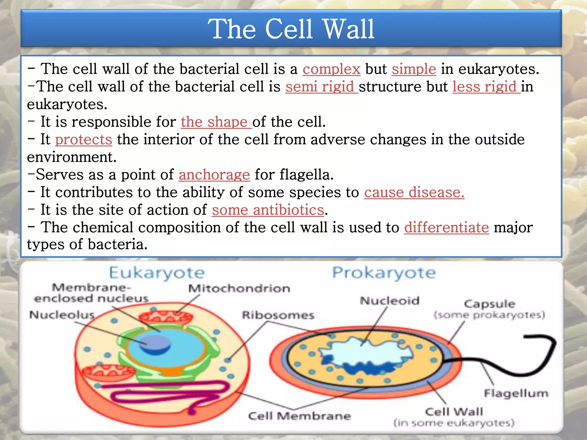
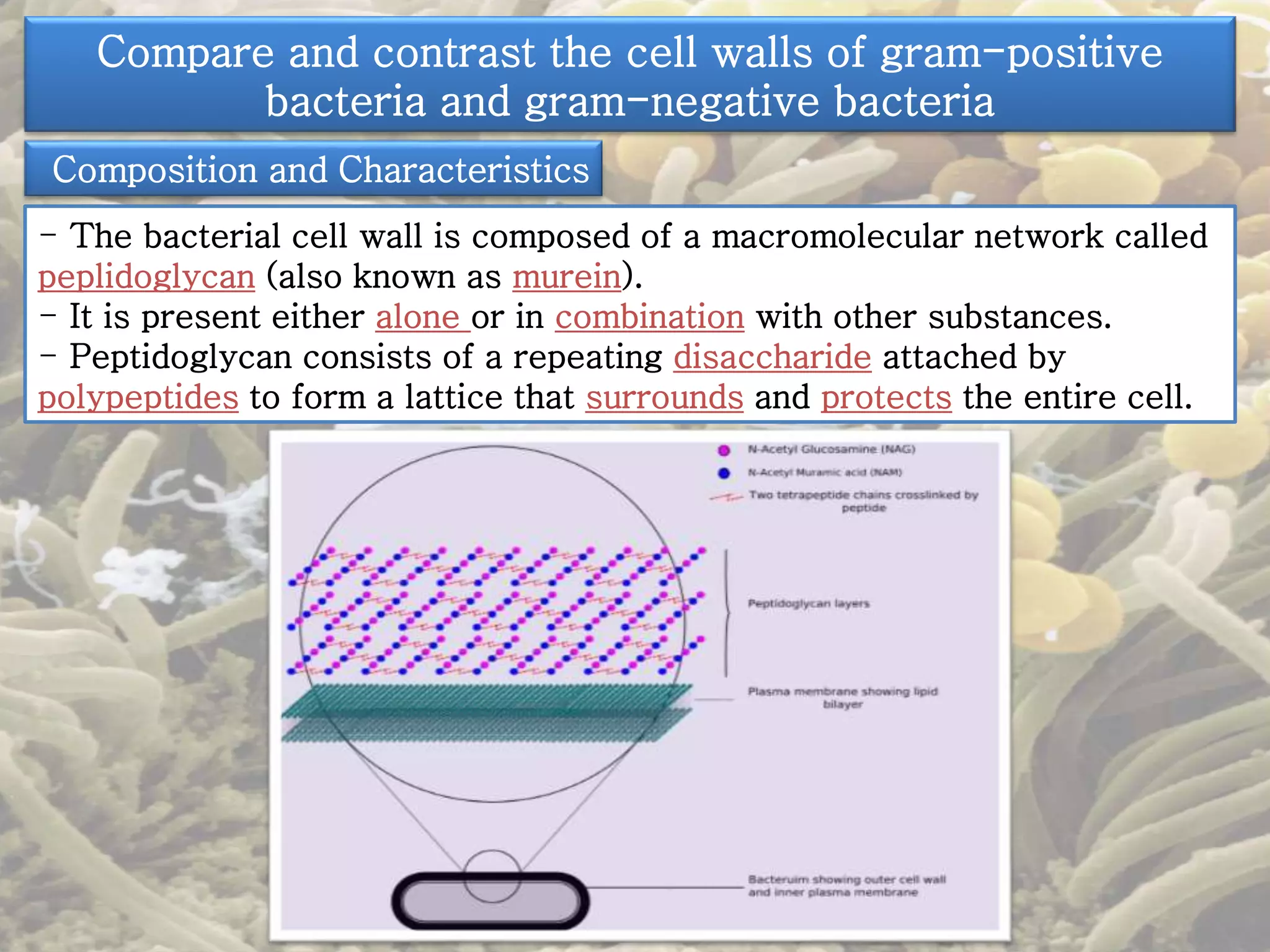
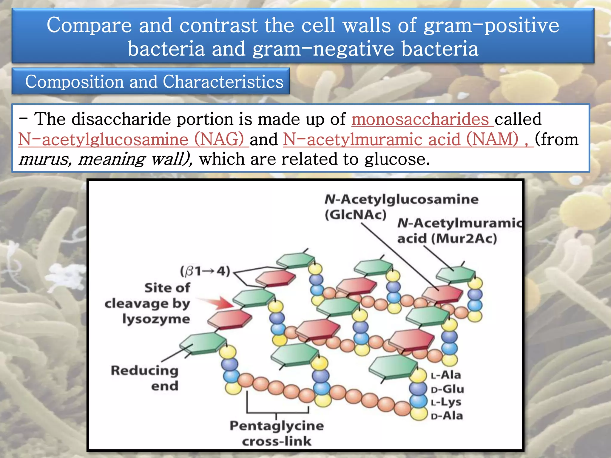
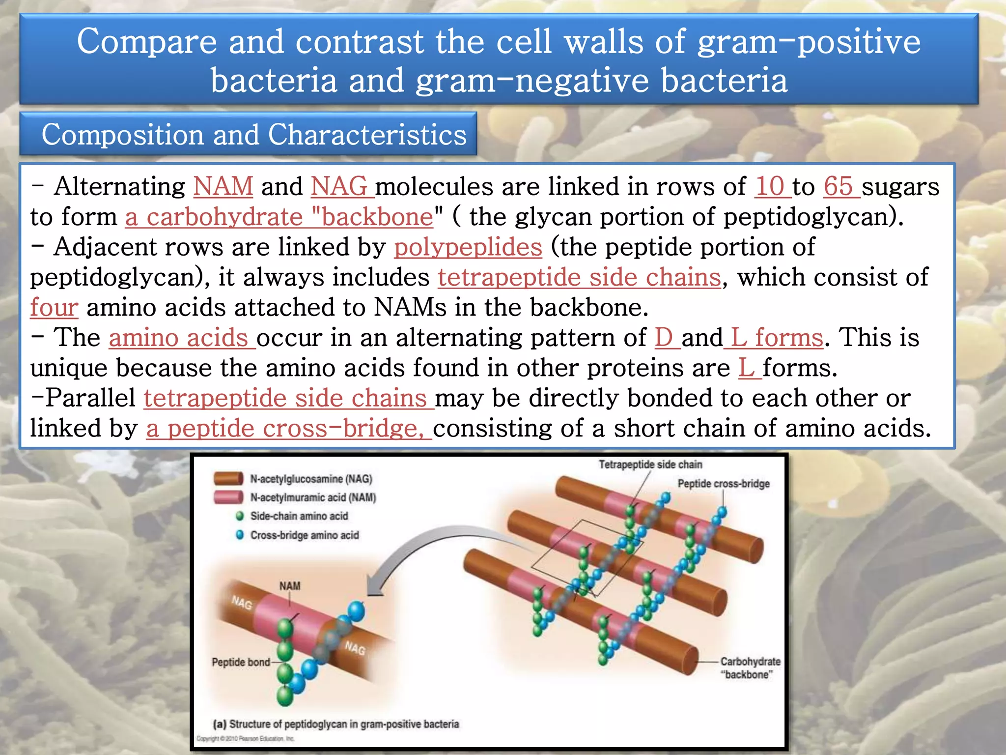
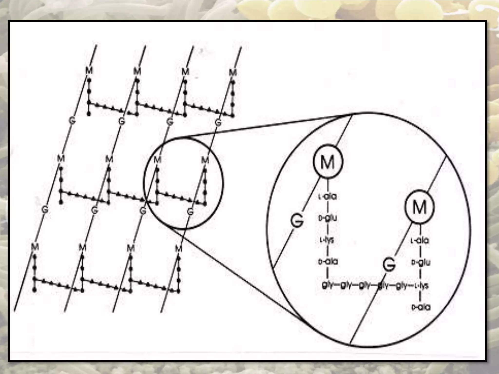
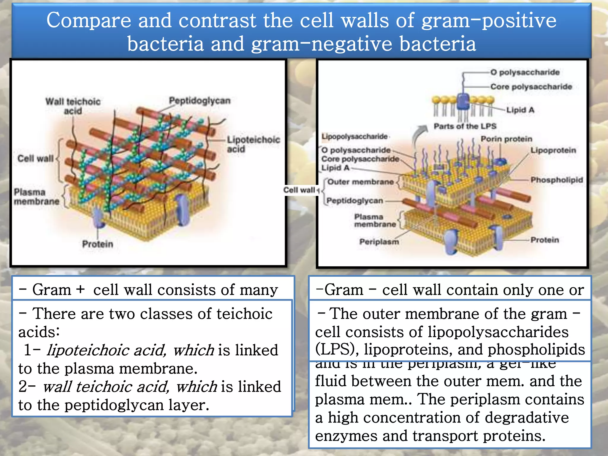
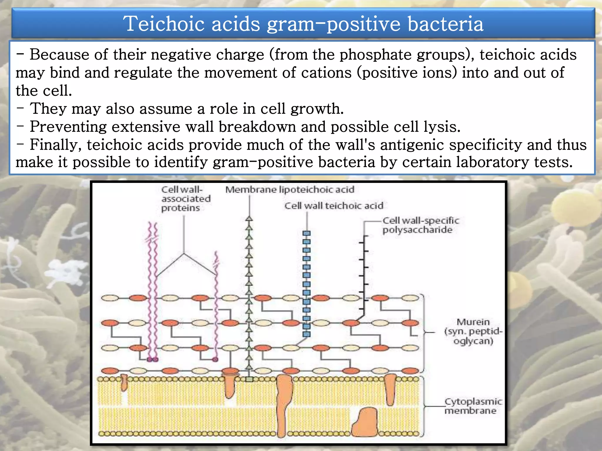
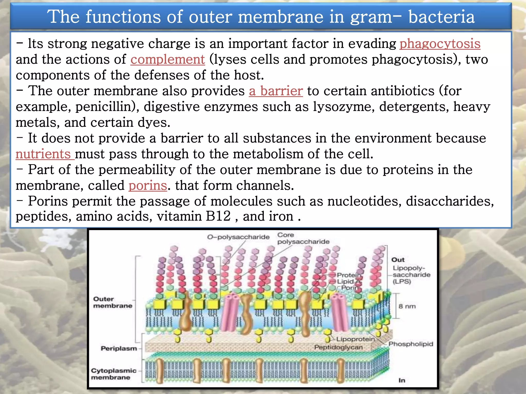
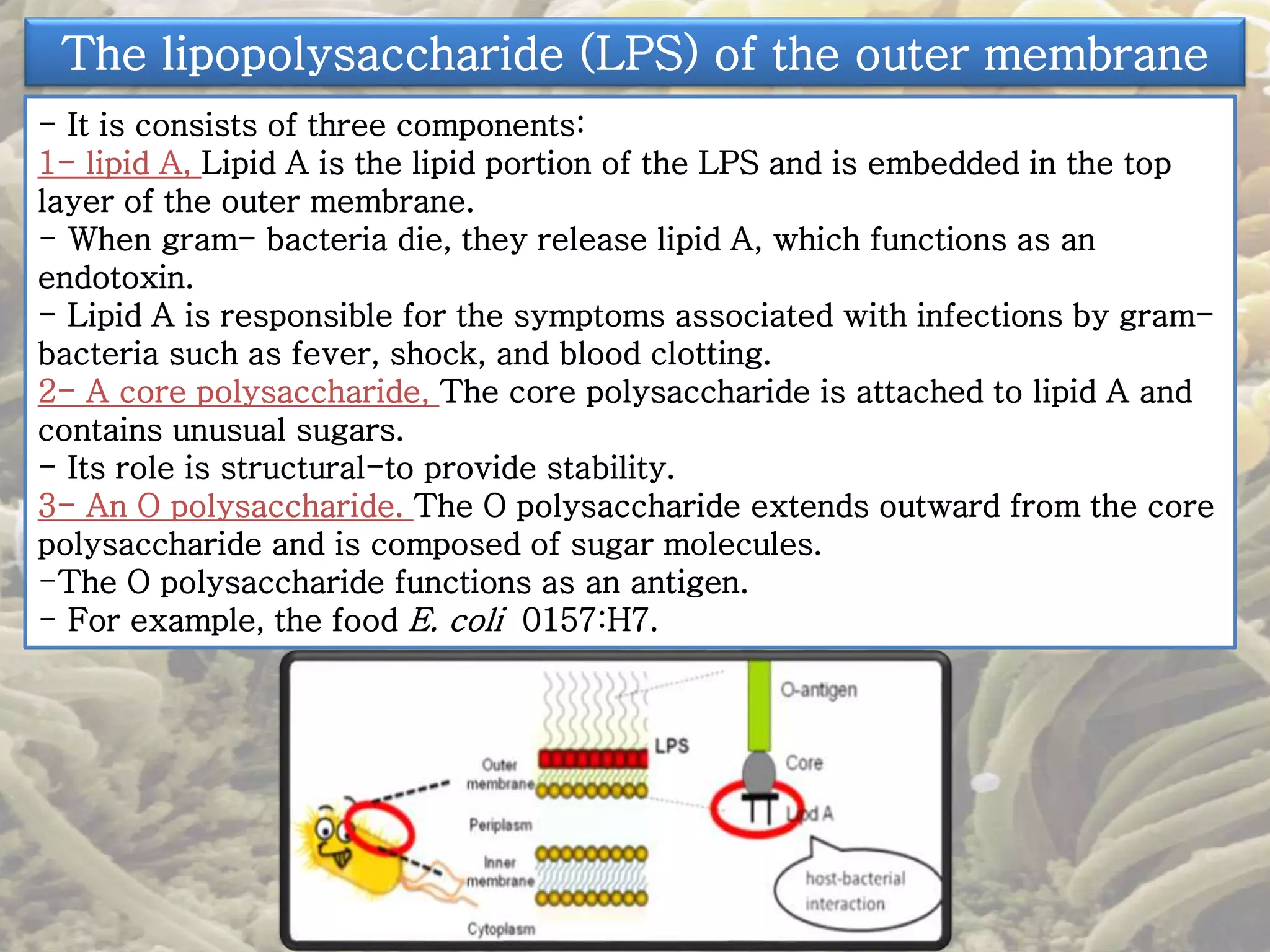
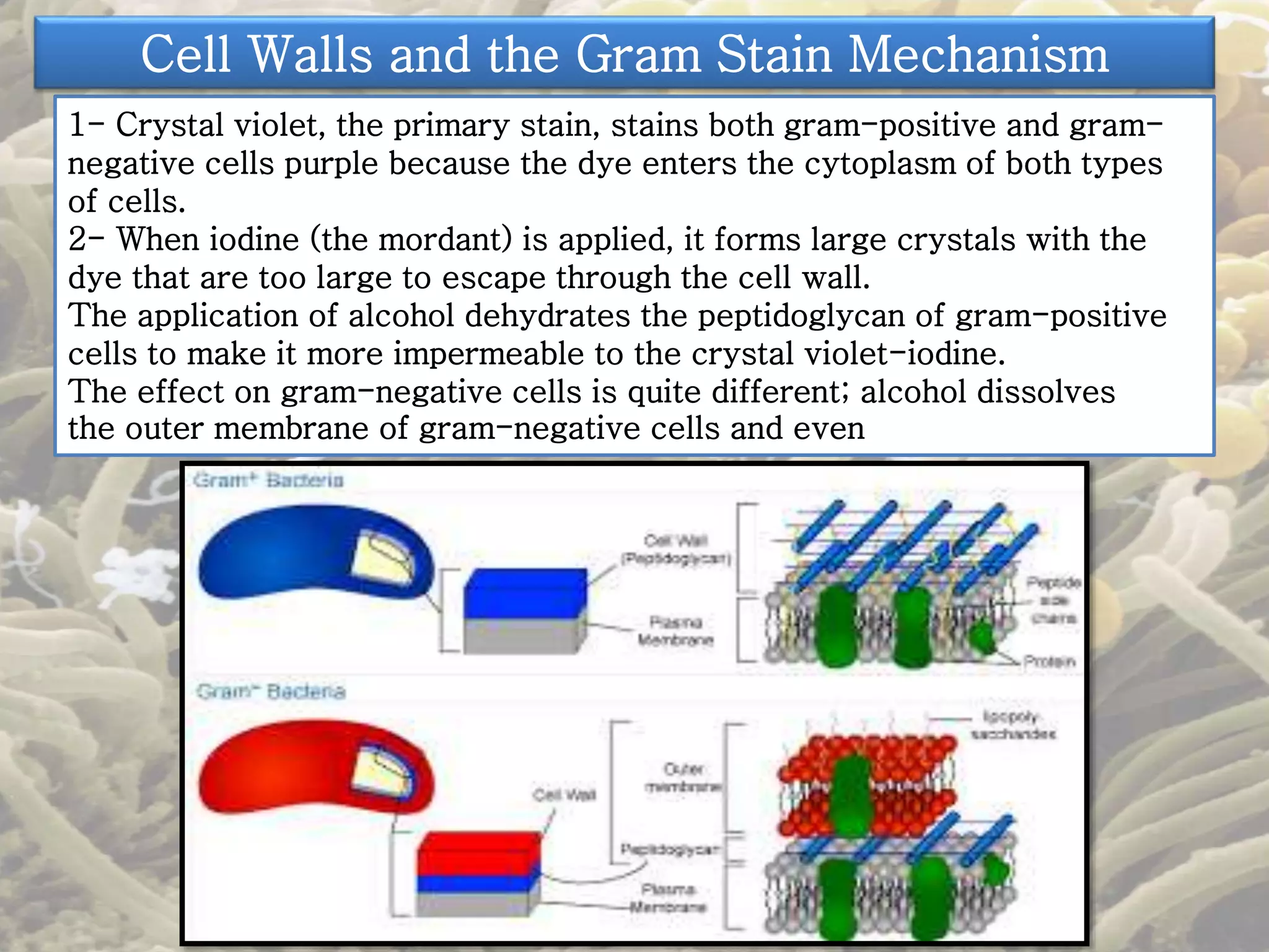
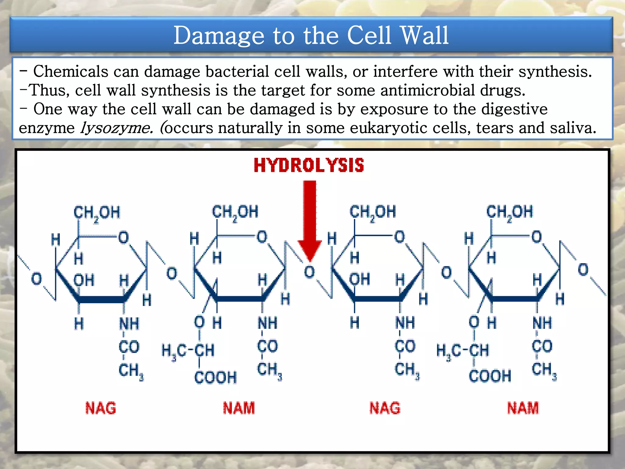
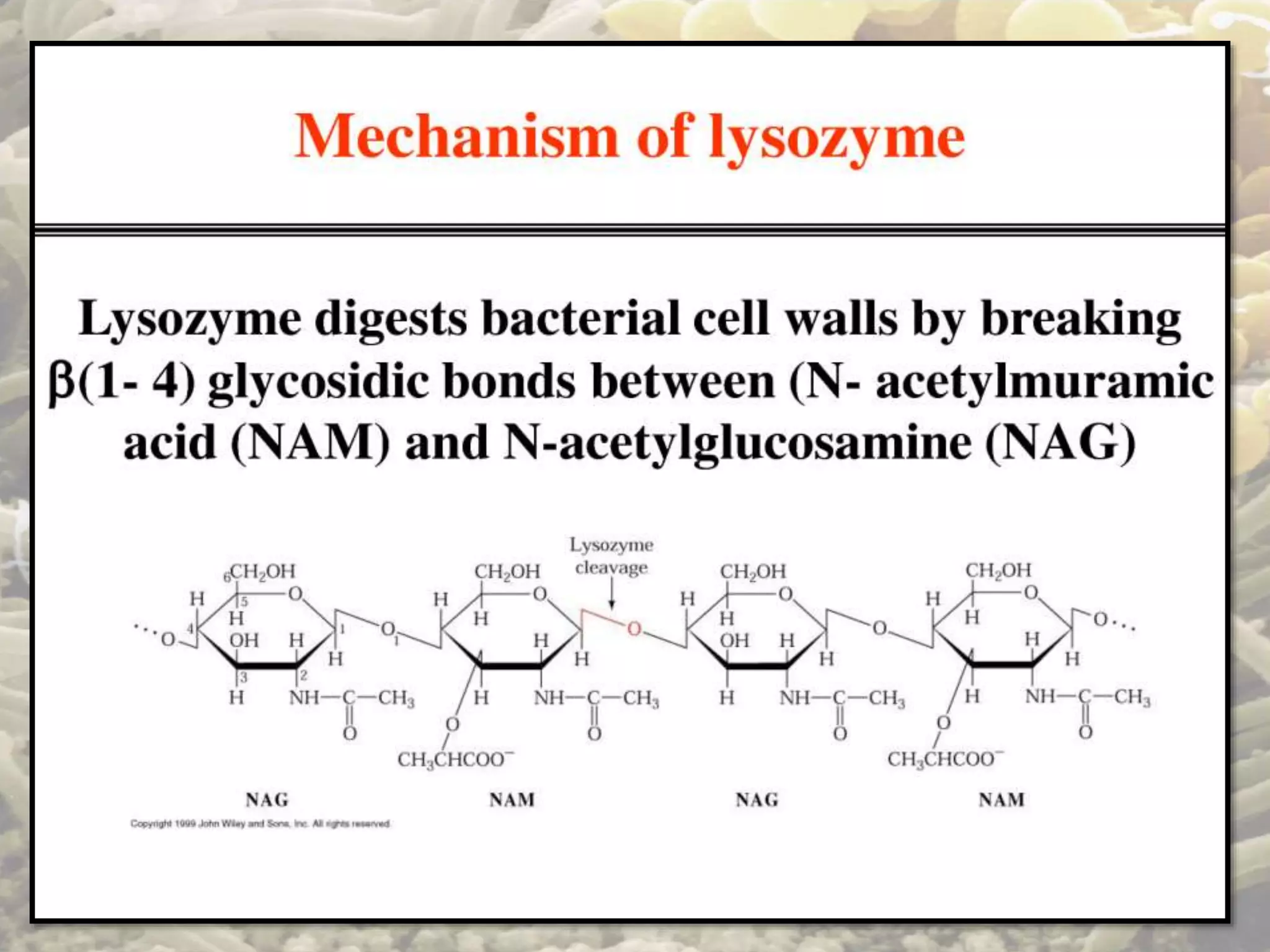
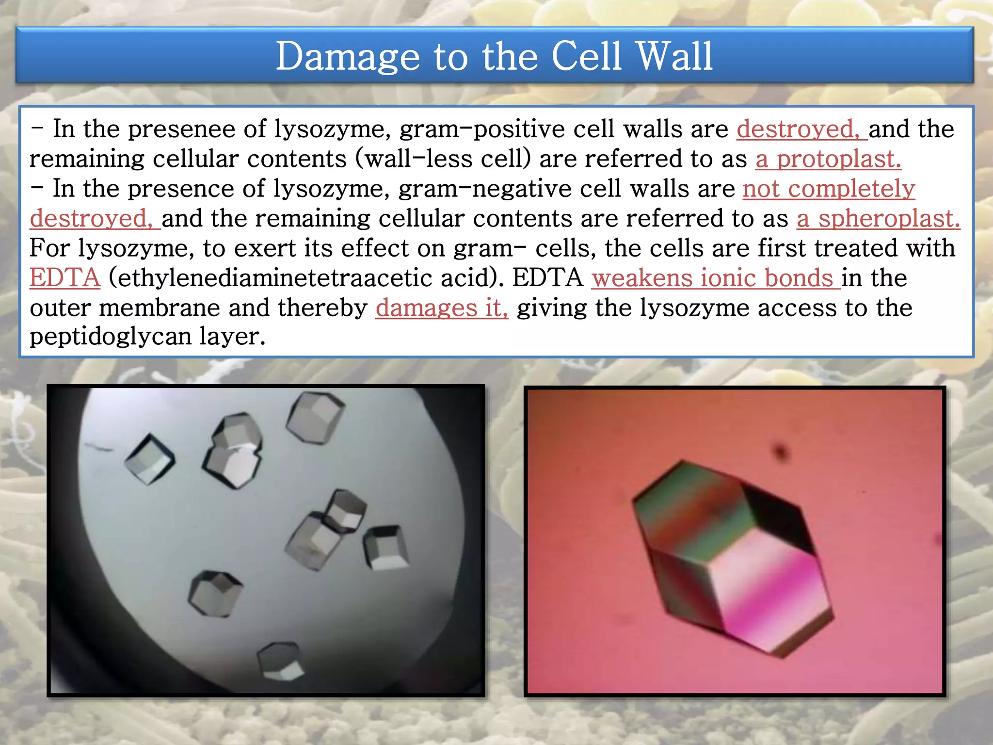
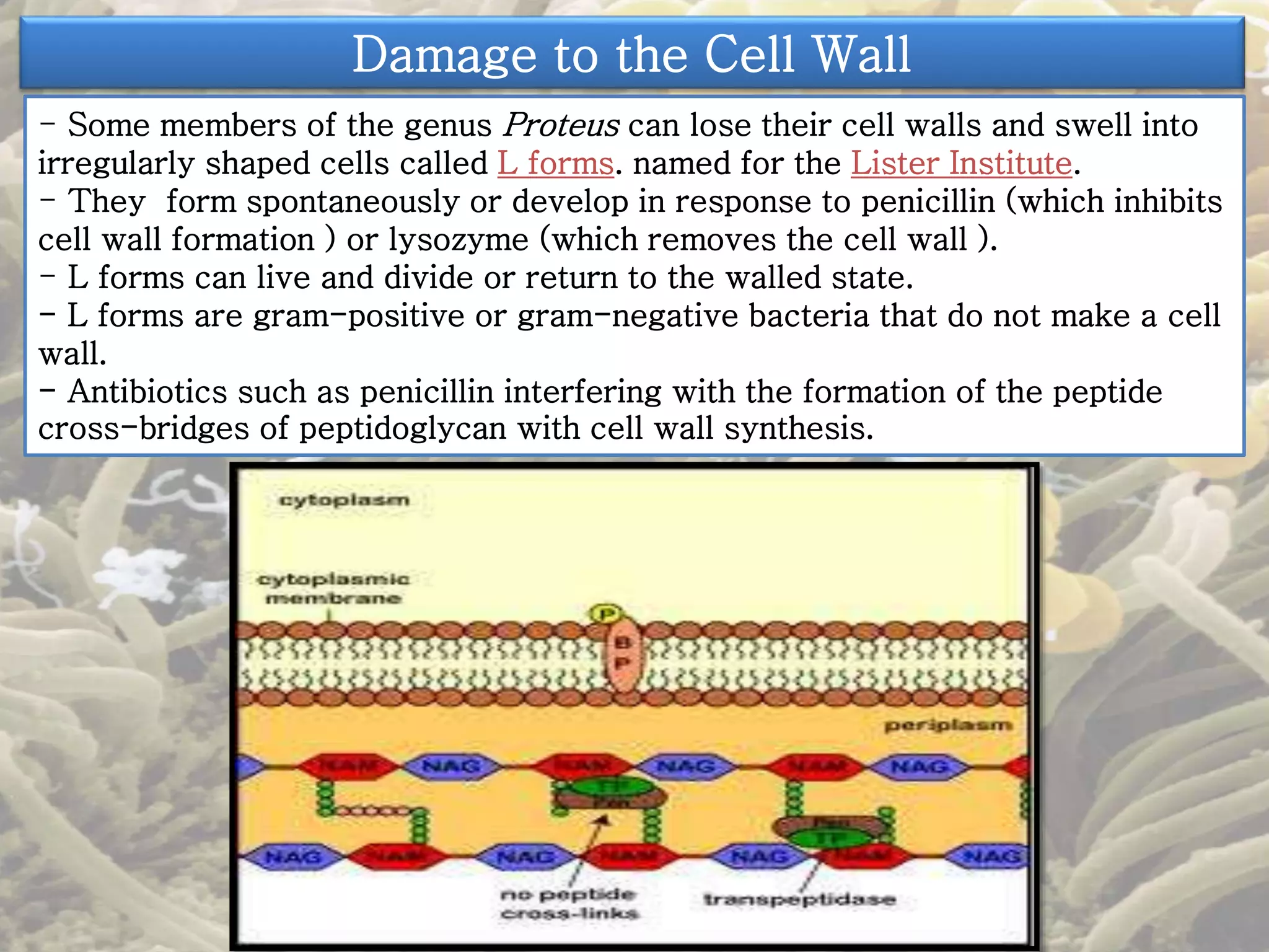
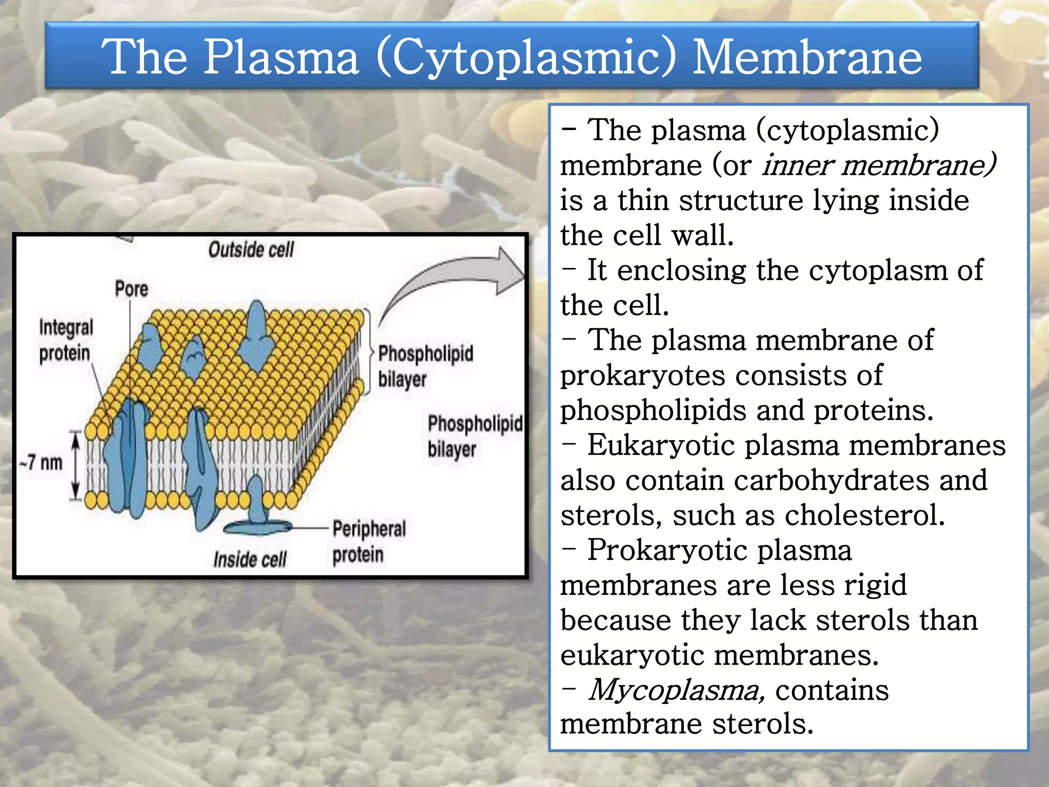
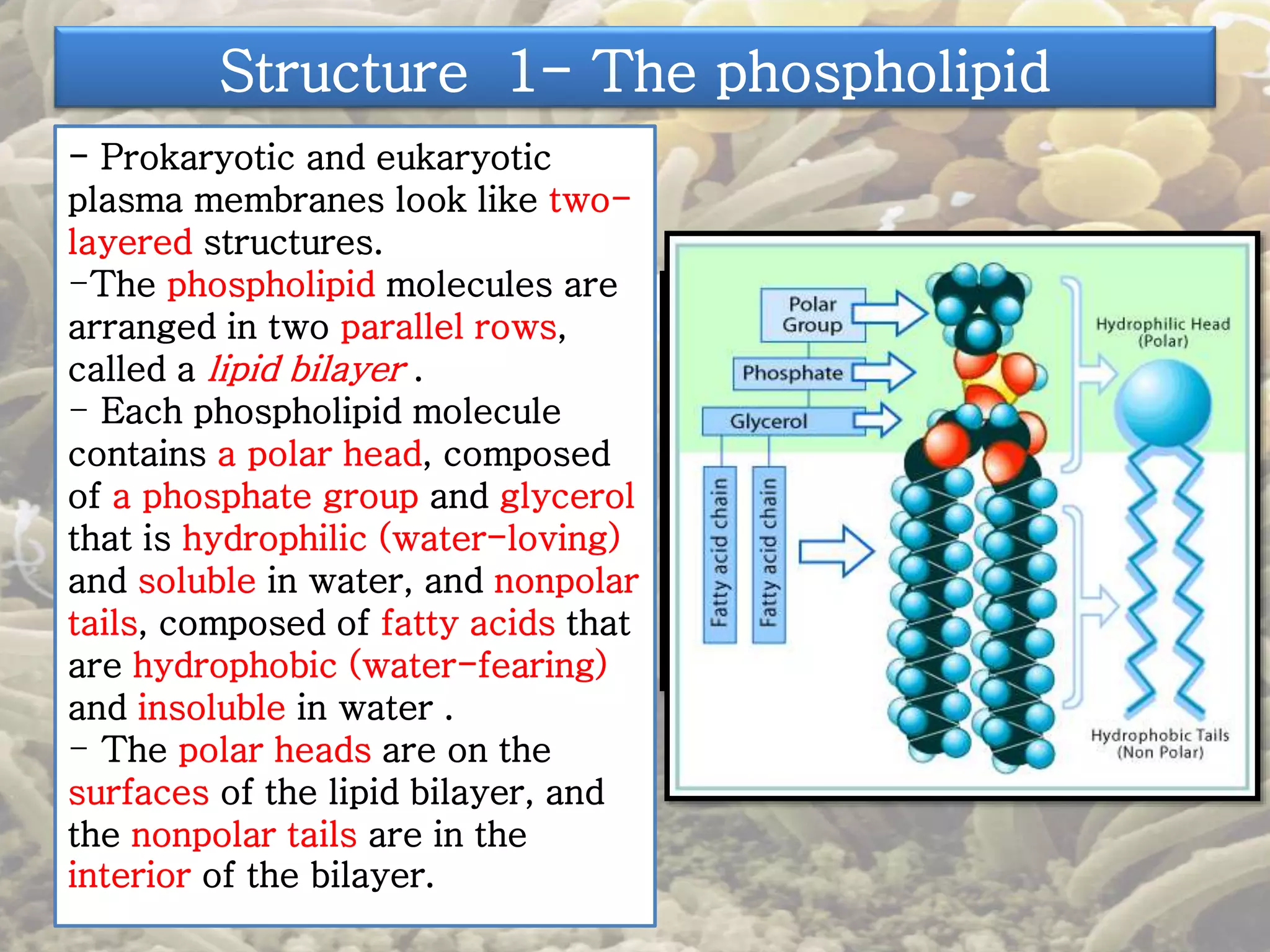
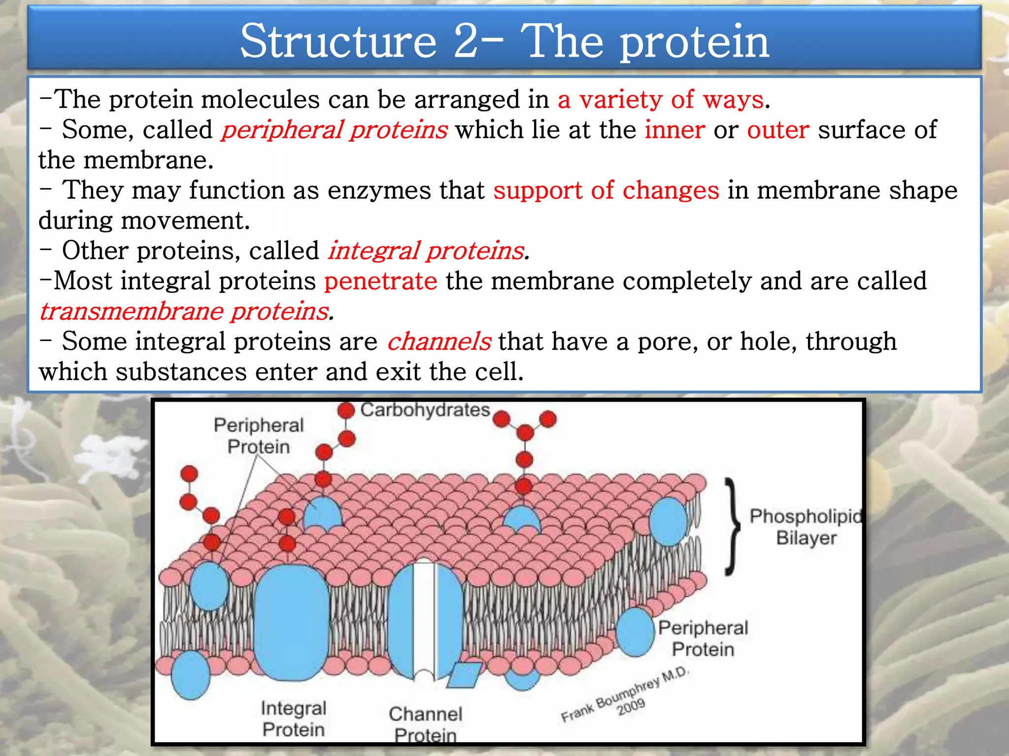
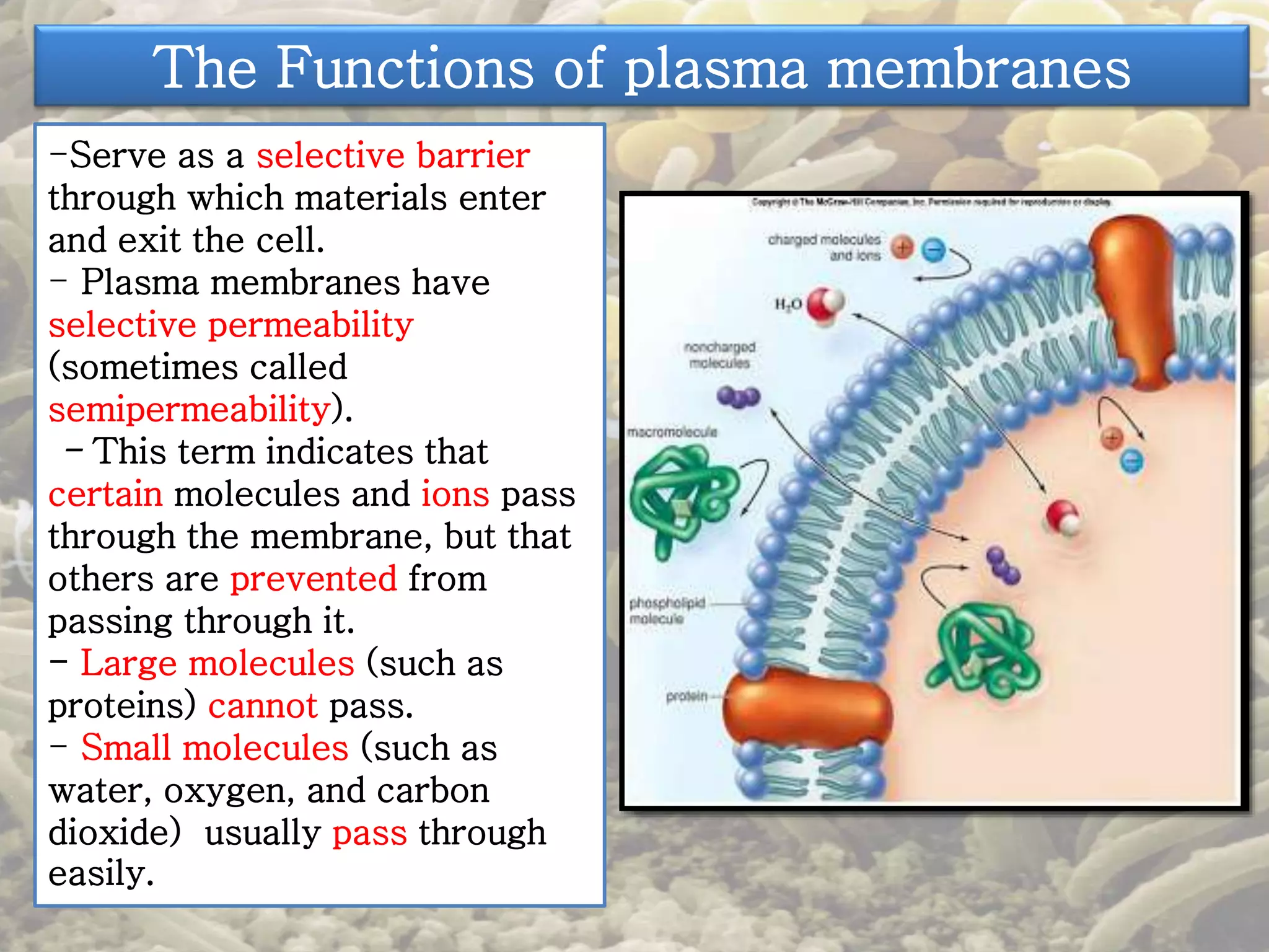
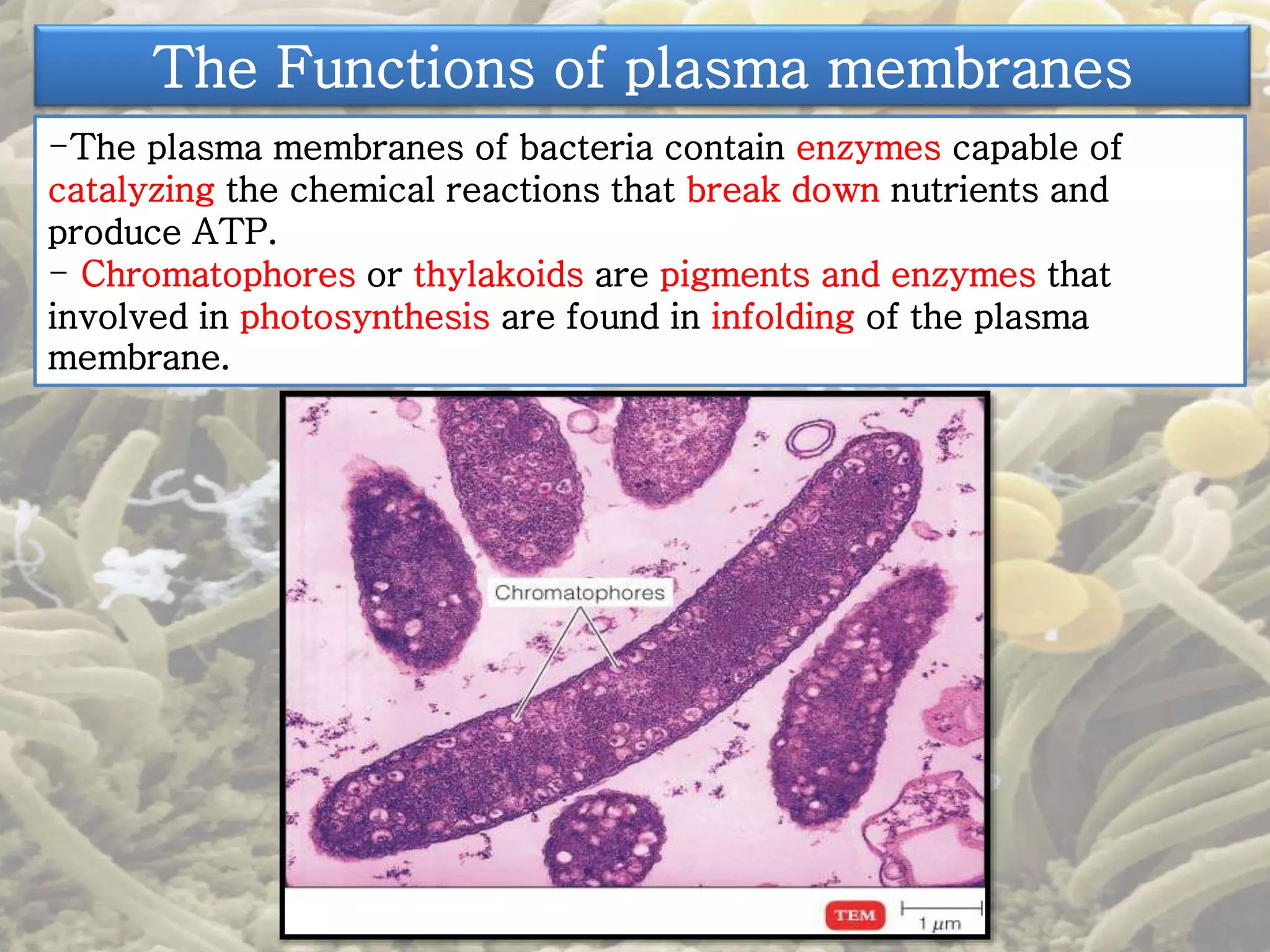
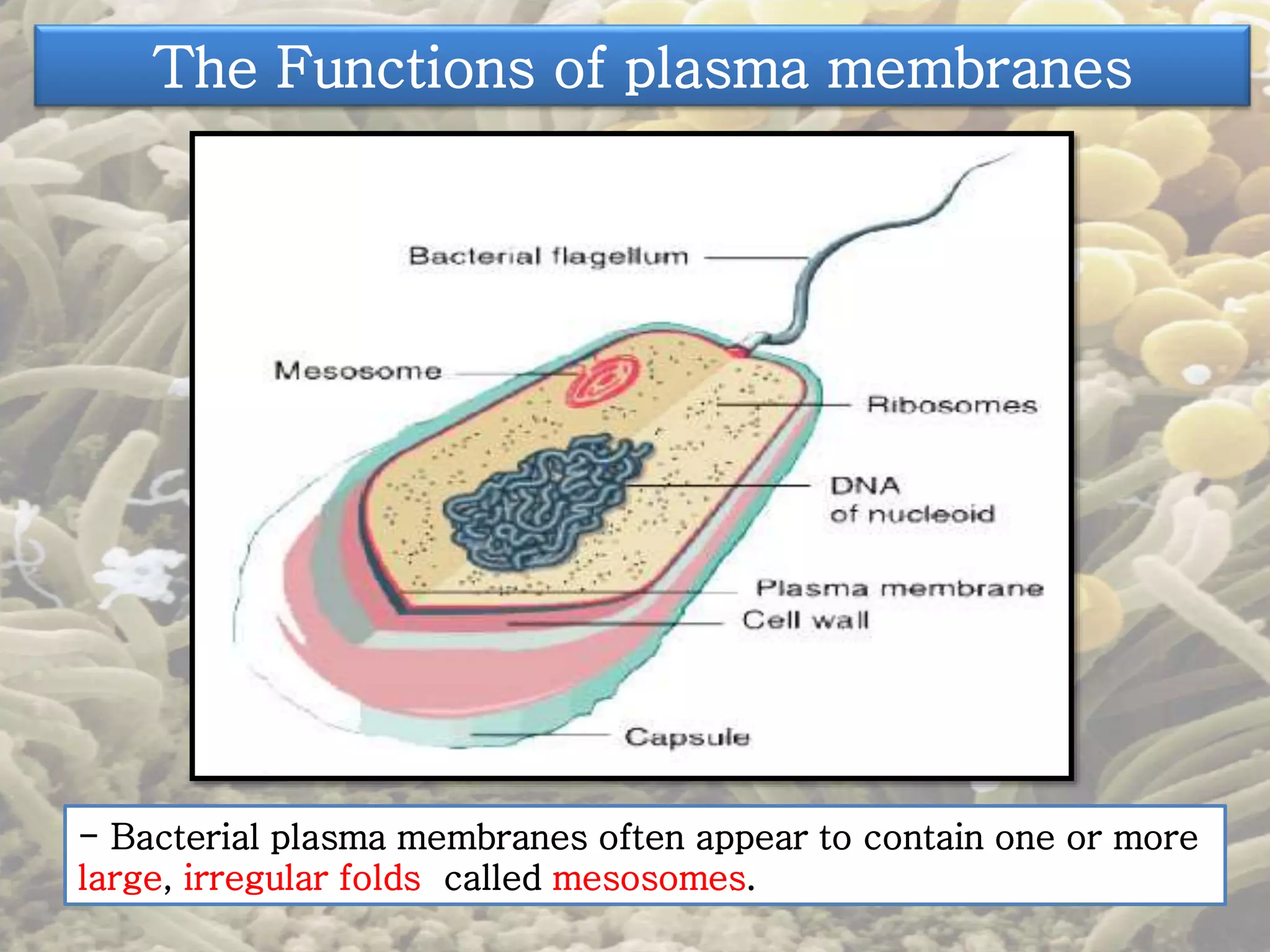
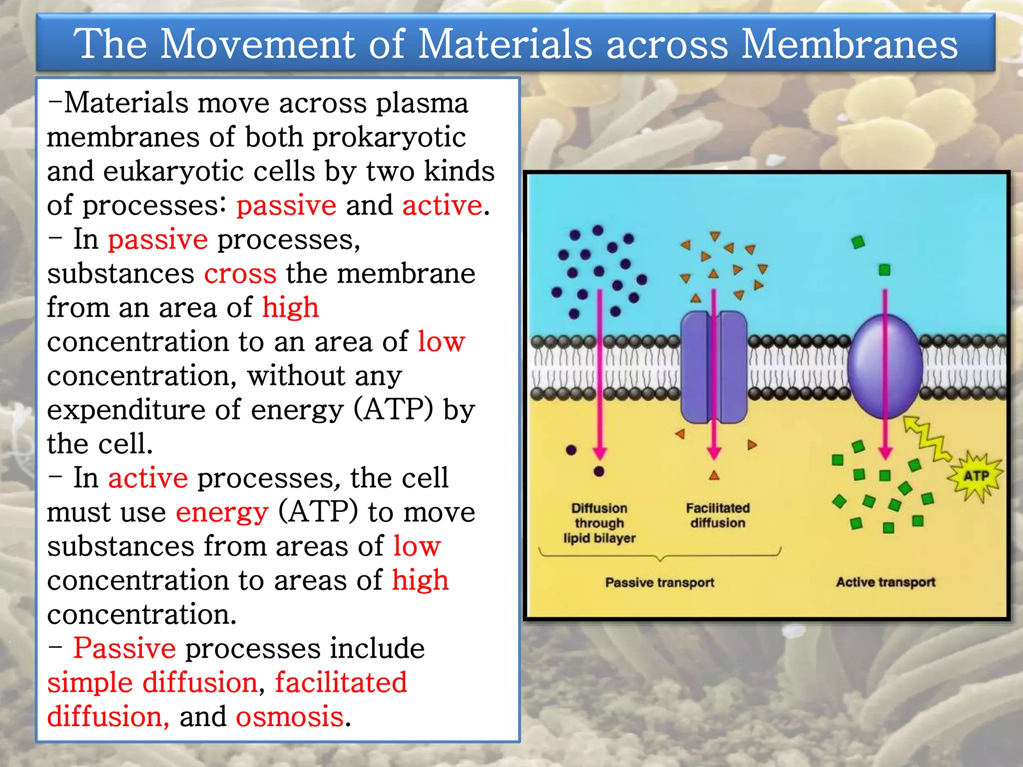
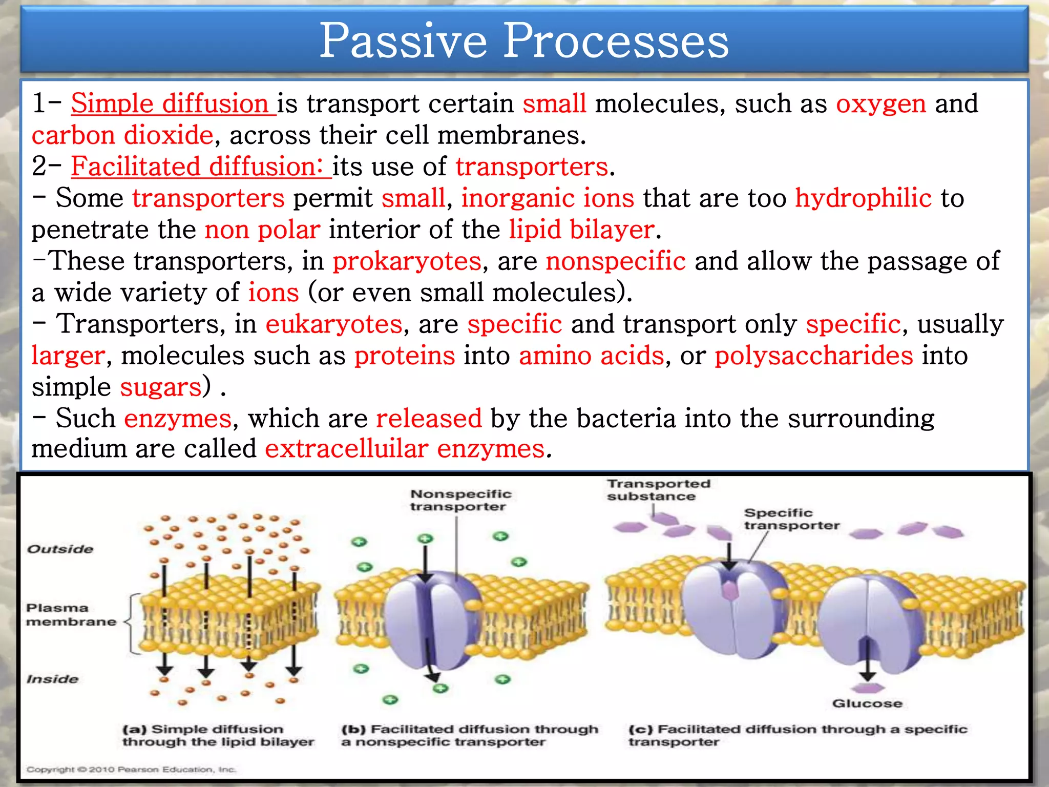
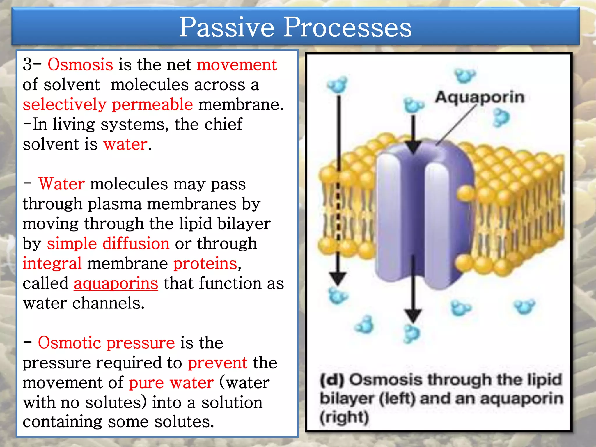
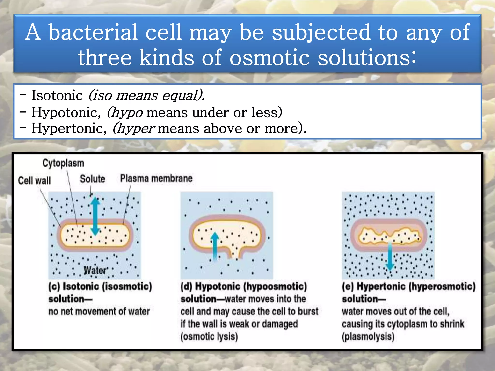
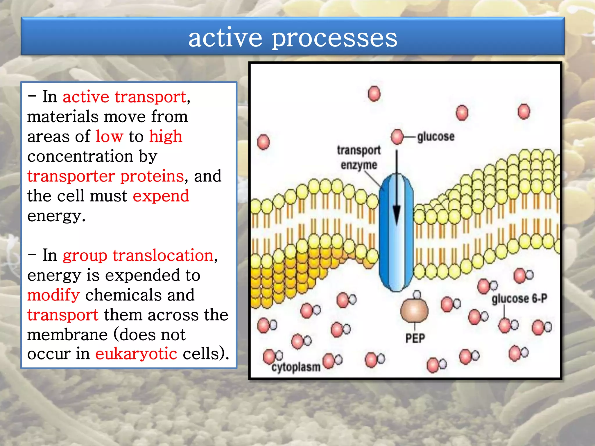
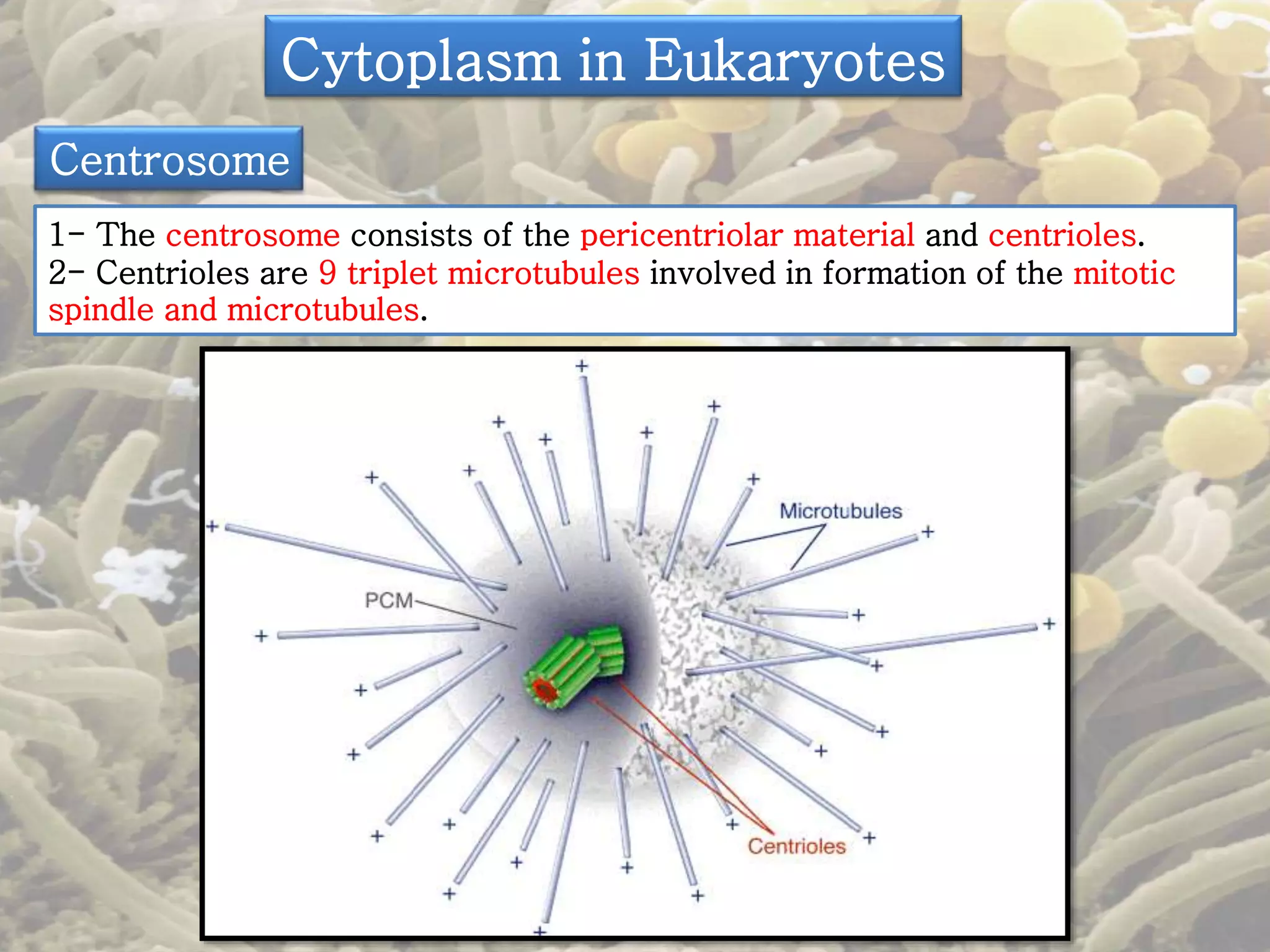
Microbiology 
An 
introduction 
Presented by 
D. Mona 
Othman 
Albureikan 
Lecture (7,8)
The Cell Wall 
- The cell wall of the bacterial cell is a complex but simple in eukaryotes. 
-The cell wall of the bacterial cell is semi rigid structure but less rigid in 
eukaryotes. 
- It is responsible for the shape of the cell. 
- It protects the interior of the cell from adverse changes in the outside 
environment. 
-Serves as a point of anchorage for flagella. 
- It contributes to the ability of some species to cause disease. 
- It is the site of action of some antibiotics. 
- The chemical composition of the cell wall is used to differentiate major 
types of bacteria.
Compare and contrast the cell walls of gram-positive 
bacteria and gram-negative bacteria 
Composition and Characteristics 
- The bacterial cell wall is composed of a macromolecular network called 
peplidoglycan (also known as murein). 
- It is present either alone or in combination with other substances. 
- Peptidoglycan consists of a repeating disaccharide attached by 
polypeptides to form a lattice that surrounds and protects the entire cell.
Compare and contrast the cell walls of gram-positive 
bacteria and gram-negative bacteria 
Composition and Characteristics 
- The disaccharide portion is made up of monosaccharides called 
N-acetylglucosamine (NAG) and N-acetylmuramic acid (NAM) , (from 
murus, meaning wall), which are related to glucose.
Compare and contrast the cell walls of gram-positive 
bacteria and gram-negative bacteria 
Composition and Characteristics 
- Alternating NAM and NAG molecules are linked in rows of 10 to 65 sugars 
to form a carbohydrate "backbone" ( the glycan portion of peptidoglycan). 
- Adjacent rows are linked by polypeplides (the peptide portion of 
peptidoglycan), it always includes tetrapeptide side chains, which consist of 
four amino acids attached to NAMs in the backbone. 
- The amino acids occur in an alternating pattern of D and L forms. This is 
unique because the amino acids found in other proteins are L forms. 
-Parallel tetrapeptide side chains may be directly bonded to each other or 
linked by a peptide cross-bridge, consisting of a short chain of amino acids.
Compare and contrast the cell walls of gram-positive 
bacteria and gram-negative bacteria 
- Gram + cell wall consists of many 
layers of peptidoglycan, forming a 
thick, rigid structure. 
- The cell walls contain teichoic 
acids, which consist of an alcohol 
(such as glycerol or ribitol) and 
phosphate. 
-Gram - cell wall contain only one or 
a very few layers of peptidoglycan. 
- The peptidoglycan is bonded to 
lipoproteins in the outer membrane, 
and is in the periplasm, a gel-like 
fluid between the outer mem. and the 
plasma mem.. The periplasm contains 
a high concentration of degradative 
enzymes and transport proteins. 
- There are two classes of teichoic 
acids: 
1- lipoteichoic acid, which is linked 
to the plasma membrane. 
2- wall teichoic acid, which is linked 
to the peptidoglycan layer. 
- The outer membrane of the gram - 
cell consists of lipopolysaccharides 
(LPS), lipoproteins, and phospholipids
Teichoic acids gram-positive bacteria 
- Because of their negative charge (from the phosphate groups), teichoic acids 
may bind and regulate the movement of cations (positive ions) into and out of 
the cell. 
- They may also assume a role in cell growth. 
- Preventing extensive wall breakdown and possible cell lysis. 
- Finally, teichoic acids provide much of the wall's antigenic specificity and thus 
make it possible to identify gram-positive bacteria by certain laboratory tests.
The functions of outer membrane in gram- bacteria 
- lts strong negative charge is an important factor in evading phagocytosis 
and the actions of complement (lyses cells and promotes phagocytosis), two 
components of the defenses of the host. 
- The outer membrane also provides a barrier to certain antibiotics (for 
example, penicillin), digestive enzymes such as lysozyme, detergents, heavy 
metals, and certain dyes. 
- It does not provide a barrier to all substances in the environment because 
nutrients must pass through to the metabolism of the cell. 
- Part of the permeability of the outer membrane is due to proteins in the 
membrane, called porins. that form channels. 
- Porins permit the passage of molecules such as nucleotides, disaccharides, 
peptides, amino acids, vitamin B12 , and iron .
The lipopolysaccharide (LPS) of the outer membrane 
- It is consists of three components: 
1- lipid A, Lipid A is the lipid portion of the LPS and is embedded in the top 
layer of the outer membrane. 
- When gram- bacteria die, they release lipid A, which functions as an 
endotoxin. 
- Lipid A is responsible for the symptoms associated with infections by gram-bacteria 
such as fever, shock, and blood clotting. 
2- A core polysaccharide, The core polysaccharide is attached to lipid A and 
contains unusual sugars. 
- Its role is structural-to provide stability. 
3- An O polysaccharide. The O polysaccharide extends outward from the core 
polysaccharide and is composed of sugar molecules. 
-The O polysaccharide functions as an antigen. 
- For example, the food E. coli 0157:H7.
Cell Walls and the Gram Stain Mechanism 
1- Crystal violet, the primary stain, stains both gram-positive and gram-negative 
cells purple because the dye enters the cytoplasm of both types 
of cells. 
2- When iodine (the mordant) is applied, it forms large crystals with the 
dye that are too large to escape through the cell wall. 
The application of alcohol dehydrates the peptidoglycan of gram-positive 
cells to make it more impermeable to the crystal violet-iodine. 
The effect on gram-negative cells is quite different; alcohol dissolves 
the outer membrane of gram-negative cells and even
Atypical Cell Walls. 
1- Mycoplasma is a bacterial genus 
that naturally lacks cell walls. 
2- Archaea have pseudomurein; they 
lack peptidoglycan. 
3- Acid-fast cell walls have a layer of 
mycolic acid outside a thin 
peptidoglycan layer.
Damage to the Cell Wall 
- Chemicals can damage bacterial cell walls, or interfere with their synthesis. 
-Thus, cell wall synthesis is the target for some antimicrobial drugs. 
- One way the cell wall can be damaged is by exposure to the digestive 
enzyme lysozyme. (occurs naturally in some eukaryotic cells, tears and saliva.
Damage to the Cell Wall 
- In the presenee of lysozyme, gram-positive cell walls are destroyed, and the 
remaining cellular contents (wall-less cell) are referred to as a protoplast. 
- In the presence of lysozyme, gram-negative cell walls are not completely 
destroyed, and the remaining cellular contents are referred to as a spheroplast. 
For lysozyme, to exert its effect on gram- cells, the cells are first treated with 
EDTA (ethylenediaminetetraacetic acid). EDTA weakens ionic bonds in the 
outer membrane and thereby damages it, giving the lysozyme access to the 
peptidoglycan layer.
Damage to the Cell Wall 
- Some members of the genus Proteus can lose their cell walls and swell into 
irregularly shaped cells called L forms. named for the Lister Institute. 
- They form spontaneously or develop in response to penicillin (which inhibits 
cell wall formation ) or lysozyme (which removes the cell wall ). 
- L forms can live and divide or return to the walled state. 
- L forms are gram-positive or gram-negative bacteria that do not make a cell 
wall. 
- Antibiotics such as penicillin interfering with the formation of the peptide 
cross-bridges of peptidoglycan with cell wall synthesis.
The Plasma (Cytoplasmic) Membrane 
- The plasma (cytoplasmic) 
membrane (or inner membrane) 
is a thin structure lying inside 
the cell wall. 
- It enclosing the cytoplasm of 
the cell. 
- The plasma membrane of 
prokaryotes consists of 
phospholipids and proteins. 
- Eukaryotic plasma membranes 
also contain carbohydrates and 
sterols, such as cholesterol. 
- Prokaryotic plasma 
membranes are less rigid 
because they lack sterols than 
eukaryotic membranes. 
- Mycoplasma, contains 
membrane sterols.
Structure 1- The phospholipid 
- Prokaryotic and eukaryotic 
plasma membranes look like two-layered 
structures. 
-The phospholipid molecules are 
arranged in two parallel rows, 
called a lipid bilayer . 
- Each phospholipid molecule 
contains a polar head, composed 
of a phosphate group and glycerol 
that is hydrophilic (water-loving) 
and soluble in water, and nonpolar 
tails, composed of fatty acids that 
are hydrophobic (water-fearing) 
and insoluble in water . 
- The polar heads are on the 
surfaces of the lipid bilayer, and 
the nonpolar tails are in the 
interior of the bilayer.
Structure 2- The protein 
-The protein molecules can be arranged in a variety of ways. 
- Some, called peripheral proteins which lie at the inner or outer surface of 
the membrane. 
- They may function as enzymes that support of changes in membrane shape 
during movement. 
- Other proteins, called integral proteins. 
-Most integral proteins penetrate the membrane completely and are called 
transmembrane proteins. 
- Some integral proteins are channels that have a pore, or hole, through 
which substances enter and exit the cell.
What is the fluid mosaic model
The Functions of plasma membranes 
-Serve as a selective barrier 
through which materials enter 
and exit the cell. 
- Plasma membranes have 
selective permeability 
(sometimes called 
semipermeability). 
- This term indicates that 
certain molecules and ions pass 
through the membrane, but that 
others are prevented from 
passing through it. 
- Large molecules (such as 
proteins) cannot pass. 
- Small molecules (such as 
water, oxygen, and carbon 
dioxide) usually pass through 
easily.
The Functions of plasma membranes 
-The plasma membranes of bacteria contain enzymes capable of 
catalyzing the chemical reactions that break down nutrients and 
produce ATP. 
- Chromatophores or thylakoids are pigments and enzymes that 
involved in photosynthesis are found in infolding of the plasma 
membrane.
The Functions of plasma membranes 
- Bacterial plasma membranes often appear to contain one or more 
large, irregular folds called mesosomes.
Destruction of the Plasma Membmne by 
Antimicrobial Agents 
- Several antimicrobial agents effects at cell membrance. 
- The chemicals also, damage the cell wall and expose the 
membrane to injury. 
- Certain alcohols and quaternary ammonium compounds are 
disrupting the membrane's phospholipids.
The Movement of Materials across Membranes 
-Materials move across plasma 
membranes of both prokaryotic 
and eukaryotic cells by two kinds 
of processes: passive and active. 
- In passive processes, 
substances cross the membrane 
from an area of high 
concentration to an area of low 
concentration, without any 
expenditure of energy (ATP) by 
the cell. 
- In active processes, the cell 
must use energy (ATP) to move 
substances from areas of low 
concentration to areas of high 
concentration. 
- Passive processes include 
simple diffusion, facilitated 
diffusion, and osmosis.
Passive Processes 
1- Simple diffusion is transport certain small molecules, such as oxygen and 
carbon dioxide, across their cell membranes. 
2- Facilitated diffusion: its use of transporters. 
- Some transporters permit small, inorganic ions that are too hydrophilic to 
penetrate the non polar interior of the lipid bilayer. 
-These transporters, in prokaryotes, are nonspecific and allow the passage of 
a wide variety of ions (or even small molecules). 
- Transporters, in eukaryotes, are specific and transport only specific, usually 
larger, molecules such as proteins into amino acids, or polysaccharides into 
simple sugars) . 
- Such enzymes, which are released by the bacteria into the surrounding 
medium are called extracelluilar enzymes.
Passive Processes 
3- Osmosis is the net movement 
of solvent molecules across a 
selectively permeable membrane. 
-In living systems, the chief 
solvent is water. 
- Water molecules may pass 
through plasma membranes by 
moving through the lipid bilayer 
by simple diffusion or through 
integral membrane proteins, 
called aquaporins that function as 
water channels. 
- Osmotic pressure is the 
pressure required to prevent the 
movement of pure water (water 
with no solutes) into a solution 
containing some solutes.
A bacterial cell may be subjected to any of 
three kinds of osmotic solutions: 
- Isotonic (iso means equal). 
- Hypotonic, (hypo means under or less) 
- Hypertonic, (hyper means above or more).
active processes 
- In active transport, 
materials move from 
areas of low to high 
concentration by 
transporter proteins, and 
the cell must expend 
energy. 
- In group translocation, 
energy is expended to 
modify chemicals and 
transport them across the 
membrane (does not 
occur in eukaryotic cells).
Endocytosis 
- Eukaryotic cells can use a mechanism called endocytosis. 
-This occurs when a segment of the plasma membrane surrounds a particle or 
large molecule, encloses it, and brings it into the cell. 
- Two very important types of endocytosis are phagocytosis and pinocytosis. 
- During phagocytosis, cellular projections called pseudopods engulf particles 
and bring them into the cell. 
- Phagocytosis is used by white blood cells to destroy bacteria and foreign 
substances . 
-In pinocytosis, the plasma membrane folds inward, bringing extracellular fluid 
into the cell, along with whatever substances are dissolved in the fluid . 
- Pinocytosis is one of the ways viruses can enter animal cells.
Cytoplasm in prokaryotes 
1- Cytoplasm is the fluid component inside the plasma membrane. 
2- The cytoplasm is mostly water, with molecules, DNA, ribosomes, 
and inclusions. 
The Nucleoid 
1- The nucleoid contains the DNA 
of the bacterial chromosome. 
2- Bacteria can also contain 
plasmids, which are circular, 
extrachromosomal DNA molecules. 
Ribosomes 
1- The cytoplasm of a prokaryote 
contains numerous 70S ribosomes; 
ribosomes consist of rRNA and 
protein. 
2- Protein synthesis occurs at 
ribosomes; it can be inhibited by 
certain antibiotics.
Cytoplasm in prokaryotes 
Inclusions 
3- Inclusions arc reserve deposits 
found in prokaryotic and eukaryotic 
cells. 
4- Among the inclusions found in 
bacteria are metachromatic granules 
(inorganic phosphate), polysaccharide 
granules (usually glycogen or starch), 
lipid inclusions, sulfur granules, 
magnetosomes , and gas vacuoles. 
Endospores 
1- Endospores are resting structures 
formed by some bacteria; they allow 
survival during adverse environmental 
conditions. 
2- The process of endospore formation 
is called sporulation; the return of an 
endospore to its vegetative state is 
called germination.
Cytoplasm in Eukaryotes 
1- The cytoplasm of eukaryotic cells includes everything inside the plasma 
membrane and external to the nucleus. 
2- Eukaryotic cytoplasm has a cytoskeleton (consisting of exceedingly small 
rods called microfilaments and intermediate filaments and cylinders called 
microtubules and exhibits cytoplasmic streaming. 
The Nucleoid 
- It is usually spherical or oval, is 
frequently the largest structure in the cell, 
and contains chromosomes, Some DNA is 
also found in mitochondria and in the 
chloroplasts of photosynthetic organisms. 
Ribosomes 
1. 80S ribosomes are found in the 
cytoplasm or attached to the rough 
endoplasmic reticulum. 
2. Chloroplasts and mitochondria con 
tain 70S ribosomes. 
3. Free ribosomes, membrane bound 
ribosome, and polyribosome
Cytoplasm in Eukaryotes 
Endoplasmic Reticulum 
1- The nuclear envelope is connected to a system of membranes in the 
cytoplasm called the Endoplasmic Reticulum (ER). 
2- The ER provides a surface for chemical reactions, serves as a transporting 
network, and stores synthesized molecules. 
3- Protein synthesis and transport on rough ER. 
4- Lipid synthesis occurs on smooth ER.
Cytoplasm in Eukaryotes 
Golgi Complex 
1- The Golgi complex consists of flattened sacs called cisterns. 
2- It functions in membrane formation and protein secretion. 
3- Lysosomes are formed from Golgi complexes, they store digestive 
enzymes (40). 
4- Vacuoles are membrane-enclosed cavities derived from the Golgi complex 
or endocytosis. 
5- They are usually found in plant cells that store various substances, 
increase cell size, and provide rigidity to leaves.
Cytoplasm in Eukaryotes 
Mitochondria 
1- Spherical or rod-shaped organelles. 
2- Mitochondria are the primary sites of 
ATP 
production. 
3- They contain 70S ribosomes and 
DNA, and they multiply by binary fission. 
Chloroplasts 
1- Chloroplasts contain 
chlorophyll and enzymes for 
photosynthesis. 
2- Like mitochondria, they 
contain 70S ribosomes and 
DNA and multiply by binary 
fission.
Cytoplasm in Eukaryotes 
Peroxisomes 
1- Peroxisomes contain one or more enzymes that can oxidize various 
organic substances. 
2- Peroxisomes contain the enzyme catalase, which decomposes hydrogen 
peroxide (toxic compound). 
3- Organelles similar In structure to Iysosomes, but smaller.
Cytoplasm in Eukaryotes 
Centrosome 
1- The centrosome consists of the pericentriolar material and centrioles. 
2- Centrioles are 9 triplet microtubules involved in formation of the mitotic 
spindle and microtubules.
References 
• Microbiology: An Introduction Plus 
MasteringMicrobiology with eText - Access Card 
Package (11th Edition) Hardcover – January 2, 2012 
by Gerard J. Tortora, Berdell R. Funke, Christine L. 
Case. 
• Some pictures from different sits.

Presentation 4

  • 1.
    Microbiology An introduction Presented by D. Mona Othman Albureikan Lecture (7,8)
  • 2.
    The Cell Wall - The cell wall of the bacterial cell is a complex but simple in eukaryotes. -The cell wall of the bacterial cell is semi rigid structure but less rigid in eukaryotes. - It is responsible for the shape of the cell. - It protects the interior of the cell from adverse changes in the outside environment. -Serves as a point of anchorage for flagella. - It contributes to the ability of some species to cause disease. - It is the site of action of some antibiotics. - The chemical composition of the cell wall is used to differentiate major types of bacteria.
  • 3.
    Compare and contrastthe cell walls of gram-positive bacteria and gram-negative bacteria Composition and Characteristics - The bacterial cell wall is composed of a macromolecular network called peplidoglycan (also known as murein). - It is present either alone or in combination with other substances. - Peptidoglycan consists of a repeating disaccharide attached by polypeptides to form a lattice that surrounds and protects the entire cell.
  • 4.
    Compare and contrastthe cell walls of gram-positive bacteria and gram-negative bacteria Composition and Characteristics - The disaccharide portion is made up of monosaccharides called N-acetylglucosamine (NAG) and N-acetylmuramic acid (NAM) , (from murus, meaning wall), which are related to glucose.
  • 5.
    Compare and contrastthe cell walls of gram-positive bacteria and gram-negative bacteria Composition and Characteristics - Alternating NAM and NAG molecules are linked in rows of 10 to 65 sugars to form a carbohydrate "backbone" ( the glycan portion of peptidoglycan). - Adjacent rows are linked by polypeplides (the peptide portion of peptidoglycan), it always includes tetrapeptide side chains, which consist of four amino acids attached to NAMs in the backbone. - The amino acids occur in an alternating pattern of D and L forms. This is unique because the amino acids found in other proteins are L forms. -Parallel tetrapeptide side chains may be directly bonded to each other or linked by a peptide cross-bridge, consisting of a short chain of amino acids.
  • 7.
    Compare and contrastthe cell walls of gram-positive bacteria and gram-negative bacteria - Gram + cell wall consists of many layers of peptidoglycan, forming a thick, rigid structure. - The cell walls contain teichoic acids, which consist of an alcohol (such as glycerol or ribitol) and phosphate. -Gram - cell wall contain only one or a very few layers of peptidoglycan. - The peptidoglycan is bonded to lipoproteins in the outer membrane, and is in the periplasm, a gel-like fluid between the outer mem. and the plasma mem.. The periplasm contains a high concentration of degradative enzymes and transport proteins. - There are two classes of teichoic acids: 1- lipoteichoic acid, which is linked to the plasma membrane. 2- wall teichoic acid, which is linked to the peptidoglycan layer. - The outer membrane of the gram - cell consists of lipopolysaccharides (LPS), lipoproteins, and phospholipids
  • 8.
    Teichoic acids gram-positivebacteria - Because of their negative charge (from the phosphate groups), teichoic acids may bind and regulate the movement of cations (positive ions) into and out of the cell. - They may also assume a role in cell growth. - Preventing extensive wall breakdown and possible cell lysis. - Finally, teichoic acids provide much of the wall's antigenic specificity and thus make it possible to identify gram-positive bacteria by certain laboratory tests.
  • 9.
    The functions ofouter membrane in gram- bacteria - lts strong negative charge is an important factor in evading phagocytosis and the actions of complement (lyses cells and promotes phagocytosis), two components of the defenses of the host. - The outer membrane also provides a barrier to certain antibiotics (for example, penicillin), digestive enzymes such as lysozyme, detergents, heavy metals, and certain dyes. - It does not provide a barrier to all substances in the environment because nutrients must pass through to the metabolism of the cell. - Part of the permeability of the outer membrane is due to proteins in the membrane, called porins. that form channels. - Porins permit the passage of molecules such as nucleotides, disaccharides, peptides, amino acids, vitamin B12 , and iron .
  • 10.
    The lipopolysaccharide (LPS)of the outer membrane - It is consists of three components: 1- lipid A, Lipid A is the lipid portion of the LPS and is embedded in the top layer of the outer membrane. - When gram- bacteria die, they release lipid A, which functions as an endotoxin. - Lipid A is responsible for the symptoms associated with infections by gram-bacteria such as fever, shock, and blood clotting. 2- A core polysaccharide, The core polysaccharide is attached to lipid A and contains unusual sugars. - Its role is structural-to provide stability. 3- An O polysaccharide. The O polysaccharide extends outward from the core polysaccharide and is composed of sugar molecules. -The O polysaccharide functions as an antigen. - For example, the food E. coli 0157:H7.
  • 11.
    Cell Walls andthe Gram Stain Mechanism 1- Crystal violet, the primary stain, stains both gram-positive and gram-negative cells purple because the dye enters the cytoplasm of both types of cells. 2- When iodine (the mordant) is applied, it forms large crystals with the dye that are too large to escape through the cell wall. The application of alcohol dehydrates the peptidoglycan of gram-positive cells to make it more impermeable to the crystal violet-iodine. The effect on gram-negative cells is quite different; alcohol dissolves the outer membrane of gram-negative cells and even
  • 12.
    Atypical Cell Walls. 1- Mycoplasma is a bacterial genus that naturally lacks cell walls. 2- Archaea have pseudomurein; they lack peptidoglycan. 3- Acid-fast cell walls have a layer of mycolic acid outside a thin peptidoglycan layer.
  • 13.
    Damage to theCell Wall - Chemicals can damage bacterial cell walls, or interfere with their synthesis. -Thus, cell wall synthesis is the target for some antimicrobial drugs. - One way the cell wall can be damaged is by exposure to the digestive enzyme lysozyme. (occurs naturally in some eukaryotic cells, tears and saliva.
  • 15.
    Damage to theCell Wall - In the presenee of lysozyme, gram-positive cell walls are destroyed, and the remaining cellular contents (wall-less cell) are referred to as a protoplast. - In the presence of lysozyme, gram-negative cell walls are not completely destroyed, and the remaining cellular contents are referred to as a spheroplast. For lysozyme, to exert its effect on gram- cells, the cells are first treated with EDTA (ethylenediaminetetraacetic acid). EDTA weakens ionic bonds in the outer membrane and thereby damages it, giving the lysozyme access to the peptidoglycan layer.
  • 16.
    Damage to theCell Wall - Some members of the genus Proteus can lose their cell walls and swell into irregularly shaped cells called L forms. named for the Lister Institute. - They form spontaneously or develop in response to penicillin (which inhibits cell wall formation ) or lysozyme (which removes the cell wall ). - L forms can live and divide or return to the walled state. - L forms are gram-positive or gram-negative bacteria that do not make a cell wall. - Antibiotics such as penicillin interfering with the formation of the peptide cross-bridges of peptidoglycan with cell wall synthesis.
  • 17.
    The Plasma (Cytoplasmic)Membrane - The plasma (cytoplasmic) membrane (or inner membrane) is a thin structure lying inside the cell wall. - It enclosing the cytoplasm of the cell. - The plasma membrane of prokaryotes consists of phospholipids and proteins. - Eukaryotic plasma membranes also contain carbohydrates and sterols, such as cholesterol. - Prokaryotic plasma membranes are less rigid because they lack sterols than eukaryotic membranes. - Mycoplasma, contains membrane sterols.
  • 18.
    Structure 1- Thephospholipid - Prokaryotic and eukaryotic plasma membranes look like two-layered structures. -The phospholipid molecules are arranged in two parallel rows, called a lipid bilayer . - Each phospholipid molecule contains a polar head, composed of a phosphate group and glycerol that is hydrophilic (water-loving) and soluble in water, and nonpolar tails, composed of fatty acids that are hydrophobic (water-fearing) and insoluble in water . - The polar heads are on the surfaces of the lipid bilayer, and the nonpolar tails are in the interior of the bilayer.
  • 19.
    Structure 2- Theprotein -The protein molecules can be arranged in a variety of ways. - Some, called peripheral proteins which lie at the inner or outer surface of the membrane. - They may function as enzymes that support of changes in membrane shape during movement. - Other proteins, called integral proteins. -Most integral proteins penetrate the membrane completely and are called transmembrane proteins. - Some integral proteins are channels that have a pore, or hole, through which substances enter and exit the cell.
  • 20.
    What is thefluid mosaic model
  • 21.
    The Functions ofplasma membranes -Serve as a selective barrier through which materials enter and exit the cell. - Plasma membranes have selective permeability (sometimes called semipermeability). - This term indicates that certain molecules and ions pass through the membrane, but that others are prevented from passing through it. - Large molecules (such as proteins) cannot pass. - Small molecules (such as water, oxygen, and carbon dioxide) usually pass through easily.
  • 22.
    The Functions ofplasma membranes -The plasma membranes of bacteria contain enzymes capable of catalyzing the chemical reactions that break down nutrients and produce ATP. - Chromatophores or thylakoids are pigments and enzymes that involved in photosynthesis are found in infolding of the plasma membrane.
  • 23.
    The Functions ofplasma membranes - Bacterial plasma membranes often appear to contain one or more large, irregular folds called mesosomes.
  • 24.
    Destruction of thePlasma Membmne by Antimicrobial Agents - Several antimicrobial agents effects at cell membrance. - The chemicals also, damage the cell wall and expose the membrane to injury. - Certain alcohols and quaternary ammonium compounds are disrupting the membrane's phospholipids.
  • 25.
    The Movement ofMaterials across Membranes -Materials move across plasma membranes of both prokaryotic and eukaryotic cells by two kinds of processes: passive and active. - In passive processes, substances cross the membrane from an area of high concentration to an area of low concentration, without any expenditure of energy (ATP) by the cell. - In active processes, the cell must use energy (ATP) to move substances from areas of low concentration to areas of high concentration. - Passive processes include simple diffusion, facilitated diffusion, and osmosis.
  • 26.
    Passive Processes 1-Simple diffusion is transport certain small molecules, such as oxygen and carbon dioxide, across their cell membranes. 2- Facilitated diffusion: its use of transporters. - Some transporters permit small, inorganic ions that are too hydrophilic to penetrate the non polar interior of the lipid bilayer. -These transporters, in prokaryotes, are nonspecific and allow the passage of a wide variety of ions (or even small molecules). - Transporters, in eukaryotes, are specific and transport only specific, usually larger, molecules such as proteins into amino acids, or polysaccharides into simple sugars) . - Such enzymes, which are released by the bacteria into the surrounding medium are called extracelluilar enzymes.
  • 27.
    Passive Processes 3-Osmosis is the net movement of solvent molecules across a selectively permeable membrane. -In living systems, the chief solvent is water. - Water molecules may pass through plasma membranes by moving through the lipid bilayer by simple diffusion or through integral membrane proteins, called aquaporins that function as water channels. - Osmotic pressure is the pressure required to prevent the movement of pure water (water with no solutes) into a solution containing some solutes.
  • 28.
    A bacterial cellmay be subjected to any of three kinds of osmotic solutions: - Isotonic (iso means equal). - Hypotonic, (hypo means under or less) - Hypertonic, (hyper means above or more).
  • 29.
    active processes -In active transport, materials move from areas of low to high concentration by transporter proteins, and the cell must expend energy. - In group translocation, energy is expended to modify chemicals and transport them across the membrane (does not occur in eukaryotic cells).
  • 30.
    Endocytosis - Eukaryoticcells can use a mechanism called endocytosis. -This occurs when a segment of the plasma membrane surrounds a particle or large molecule, encloses it, and brings it into the cell. - Two very important types of endocytosis are phagocytosis and pinocytosis. - During phagocytosis, cellular projections called pseudopods engulf particles and bring them into the cell. - Phagocytosis is used by white blood cells to destroy bacteria and foreign substances . -In pinocytosis, the plasma membrane folds inward, bringing extracellular fluid into the cell, along with whatever substances are dissolved in the fluid . - Pinocytosis is one of the ways viruses can enter animal cells.
  • 31.
    Cytoplasm in prokaryotes 1- Cytoplasm is the fluid component inside the plasma membrane. 2- The cytoplasm is mostly water, with molecules, DNA, ribosomes, and inclusions. The Nucleoid 1- The nucleoid contains the DNA of the bacterial chromosome. 2- Bacteria can also contain plasmids, which are circular, extrachromosomal DNA molecules. Ribosomes 1- The cytoplasm of a prokaryote contains numerous 70S ribosomes; ribosomes consist of rRNA and protein. 2- Protein synthesis occurs at ribosomes; it can be inhibited by certain antibiotics.
  • 32.
    Cytoplasm in prokaryotes Inclusions 3- Inclusions arc reserve deposits found in prokaryotic and eukaryotic cells. 4- Among the inclusions found in bacteria are metachromatic granules (inorganic phosphate), polysaccharide granules (usually glycogen or starch), lipid inclusions, sulfur granules, magnetosomes , and gas vacuoles. Endospores 1- Endospores are resting structures formed by some bacteria; they allow survival during adverse environmental conditions. 2- The process of endospore formation is called sporulation; the return of an endospore to its vegetative state is called germination.
  • 33.
    Cytoplasm in Eukaryotes 1- The cytoplasm of eukaryotic cells includes everything inside the plasma membrane and external to the nucleus. 2- Eukaryotic cytoplasm has a cytoskeleton (consisting of exceedingly small rods called microfilaments and intermediate filaments and cylinders called microtubules and exhibits cytoplasmic streaming. The Nucleoid - It is usually spherical or oval, is frequently the largest structure in the cell, and contains chromosomes, Some DNA is also found in mitochondria and in the chloroplasts of photosynthetic organisms. Ribosomes 1. 80S ribosomes are found in the cytoplasm or attached to the rough endoplasmic reticulum. 2. Chloroplasts and mitochondria con tain 70S ribosomes. 3. Free ribosomes, membrane bound ribosome, and polyribosome
  • 34.
    Cytoplasm in Eukaryotes Endoplasmic Reticulum 1- The nuclear envelope is connected to a system of membranes in the cytoplasm called the Endoplasmic Reticulum (ER). 2- The ER provides a surface for chemical reactions, serves as a transporting network, and stores synthesized molecules. 3- Protein synthesis and transport on rough ER. 4- Lipid synthesis occurs on smooth ER.
  • 35.
    Cytoplasm in Eukaryotes Golgi Complex 1- The Golgi complex consists of flattened sacs called cisterns. 2- It functions in membrane formation and protein secretion. 3- Lysosomes are formed from Golgi complexes, they store digestive enzymes (40). 4- Vacuoles are membrane-enclosed cavities derived from the Golgi complex or endocytosis. 5- They are usually found in plant cells that store various substances, increase cell size, and provide rigidity to leaves.
  • 36.
    Cytoplasm in Eukaryotes Mitochondria 1- Spherical or rod-shaped organelles. 2- Mitochondria are the primary sites of ATP production. 3- They contain 70S ribosomes and DNA, and they multiply by binary fission. Chloroplasts 1- Chloroplasts contain chlorophyll and enzymes for photosynthesis. 2- Like mitochondria, they contain 70S ribosomes and DNA and multiply by binary fission.
  • 37.
    Cytoplasm in Eukaryotes Peroxisomes 1- Peroxisomes contain one or more enzymes that can oxidize various organic substances. 2- Peroxisomes contain the enzyme catalase, which decomposes hydrogen peroxide (toxic compound). 3- Organelles similar In structure to Iysosomes, but smaller.
  • 38.
    Cytoplasm in Eukaryotes Centrosome 1- The centrosome consists of the pericentriolar material and centrioles. 2- Centrioles are 9 triplet microtubules involved in formation of the mitotic spindle and microtubules.
  • 39.
    References • Microbiology:An Introduction Plus MasteringMicrobiology with eText - Access Card Package (11th Edition) Hardcover – January 2, 2012 by Gerard J. Tortora, Berdell R. Funke, Christine L. Case. • Some pictures from different sits.