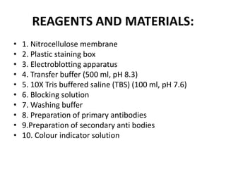
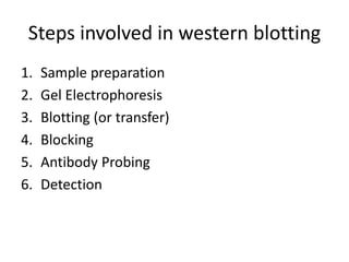
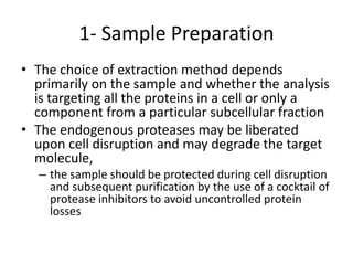
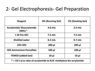
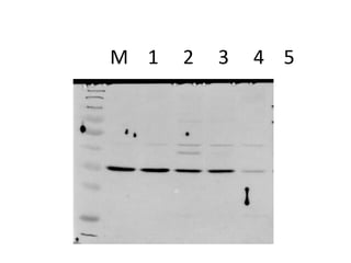
Principle
• Basic principle is protein protein interaction.
• Or antigen/antibody interaction.
Introduction
• The western blot (sometimes called
the protein blotting or immunoblotting) is a
widely used analytical technique used
in molecular biology, immunogenetics and
other molecular biology disciplines to detect
specific proteins in a sample of tissue
homogenate or extract.
• Allow to identify a particular protein of
interest among many proteins in a sample.
Contin….
• Western Blotting was performed by the rapid
method of Towbin et al., (1979) to detect the
expression pattern of a protein. To detect the
antigens blotted on a nitrocellulose
membrane with the use of an antibody.
• By using a western blot, researchers are able
to identify specific proteins from a complex
mixture of proteins extracted from cells. The
technique uses three elements to accomplish
this task: (1) separation by size, (2) transfer to
a solid support, and (3) marking target protein
using a proper primary and secondary
antibody to visualize.
Why do a western blot ?
• To see if specific protein of interest is present
in a sample.
• To compare the amounts of a protein of
interest among different samples.
• To see if the state of a protein changes under
different biological condition (e.g in
disease,stress etc).
REAGENTS AND MATERIALS:
• 1. Nitrocellulose membrane
• 2. Plastic staining box
• 3. Electroblotting apparatus
• 4. Transfer buffer (500 ml, pH 8.3)
• 5. 10X Tris buffered saline (TBS) (100 ml, pH 7.6)
• 6. Blocking solution
• 7. Washing buffer
• 8. Preparation of primary antibodies
• 9.Preparation of secondary anti bodies
• 10. Colour indicator solution
Steps involved in western blotting
1. Sample preparation
2. Gel Electrophoresis
3. Blotting (or transfer)
4. Blocking
5. Antibody Probing
6. Detection
1- Sample Preparation
• All sources of protein, from single cells to whole
tissues, biological fluids and proteins secreted in
vitro, are open to analysis by Western blotting
• In most cases, the cells are harvested, washed,
and lysed to release the target protein
• For best results, all these steps should be carried
out on ice
• This will minimize proteolysis,
dephosphorylation, and denaturation, since all
begin to occur once the cells are disrupted
1- Sample Preparation
• The choice of extraction method depends
primarily on the sample and whether the analysis
is targeting all the proteins in a cell or only a
component from a particular subcellular fraction
• The endogenous proteases may be liberated
upon cell disruption and may degrade the target
molecule,
– the sample should be protected during cell disruption
and subsequent purification by the use of a cocktail of
protease inhibitors to avoid uncontrolled protein
losses
1- Sample Preparation
• Numerous methods are available for disrupting cells and
preparing their contents for analysis by Western blotting
Detergent lysis The membranes are solubilized, lysing cells and liberating
their contents
Ultrasonication The sound waves generate a region of low pressure, causing
disruption of the membranes of cells
Freeze/thaw lysis Cells are disrupted by the repeated formation of
ice crystals and the method is usually combined with
enzymatic lysis
Enzymatic digestion The enzymes dissolve cell walls, coats, capsules, capsids, or
other structures
• To ensure that samples are in the proper range of detection for the
assay, and so they can be compared on an equivalent basis, it is
important to know the concentration of total protein in each sample
2- Gel Electrophoresis- Gel Preparation
Reagent 8% (Running Gel) 5% (Stacking Gel)
Acrylamide/ Bisacrylamide
(40%) *
4.0 mls 2.5 mls
1 M Tris-HCl 7.5 mls 7.5 mls
Distilled water 8.2 mls 9.7 mls
10% SDS 200 µl 200 µl
10% Ammonium Persulfate 100 µl 100 µl
TEMED (added last) 10 µl 10 µl
* = 19:1 w:w ratio of acrylamide to N,N'-methylene bis-acrylamide
2- Gel Electrophoresis- Gel Preparation
• Mix ingredients in the order
shown above, ensuring no air
bubbles form
• Pour the separating gel into glass
plate assembly
• Overlay gel with water to ensure a
flat surface and to exclude air
• Leave to polymerize for ~ 20
minutes
• Then prepare the stacking gel and
pour on the running gel, insert
comb and leave for 20 min
2- Gel Electrophoresis- Sample Buffer
• The end result has two important features:
1.All proteins contain only primary structure and
2.All proteins have a large negative charge which
means they will all migrate towards the positive
pole when placed in an electric field.
• They migrate through a gel towards the
positive pole at a rate proportional to their
linear size
3- Blotting
• Following gel electrophoresis, the separated
protein mixtures are transferred to a solid
support for further analysis
3- Blotting- Blotting Membranes
• The solid support onto which the separated
proteins are transferred is usually of two
types, both of which bind proteins with high
affinity:
– Nitrocellulose membrane
• has excellent protein binding and retention capabilities
• is brittle and thus it is usually less effective when blots need
to be reused
– Polyvinylidene fluoride (PVDF) membrane
• PVDF demonstrates superior mechanical strength making it
suitable for stripping/reprobing
3- Blotting- Visualization of proteins in
membranes: Ponceau Red stain
• Ponceau Red is a reversible stain with poor
sensitivity
• Ponceau S is compatible with both
nitrocellulose and PVDF membranes
• This is a quick and easy way to visualize
proteins transferred to membranes
• Ponceau S is easily removed with water and
is regarded as a “gentle” treatment that
does not interfere with subsequent
immunological detection steps
4- Blocking
• Blocking is a very important step in the
immunodetection phase of Western blotting
because it prevents non-specific binding of
antibody to the blotting membrane
• The most commonly used blocking solutions
contain 3-5% BSA or non-fat dried milk in a
solution of PBS (phosphate buffered saline) or
TBS (tris buffered saline)
• Often, a small amount of Tween 20 detergent is
added to blocking and washing solutions to
reduce background staining
5- Antibody Probing
• Once the protein samples are separated and transferred onto a
membrane, the protein of interest is detected and localized using a
specific antibody
• The blot will be incubated in a dilute solution of antibody, usually
for a few hours at room temperature or overnight at 4°C
• The antibody is diluted in wash buffer or in the blocking solution,
the choice depends upon the antibody
• Since antibody preparations vary in their levels of purity and
specific binding properties, there will be differences in the level of
dilution required
• The manufacturer’s datasheet should provide dilution
recommendations for a particular preparation
5- Antibody Probing
• Usually, Western blotting protocols utilize a non-labeled
primary antibody directed against the target protein
• Wash the membrane several times in TBST while agitating,
5 minutes or more per wash, to remove residual primary
antibody
• A species-specific, labeled secondary antibody directed
against the constant region of the primary antibody is then
used
• The secondary antibody serves not only as a carrier of the
label but is also a mechanism to amplify the emitted
signals, as many secondary antibodies can theoretically
bind simultaneously to the primary antibody
• Secondary Ab is also diluted according to the
manufacturer’s recommendations and incubated for 1 hour
at RT
6- Detection with Substrate
• The most common antibody label used in
Western blots is HRP, a small, stable enzyme
with high specificity and rapid turnover
• The signal is detected when HRP is exposed to
a substrate solution in the final step of the
immunodetection procedure
• Substrate solutions for Western blotting are
chemical reagents that are acted upon by the
enzyme to yield a signal that can be easily
measured
• HRP label is typically detected with either
colorimetric or chemiluminescent substrates
6- Detection with Substrate
• Colorimetric substrates for HRP {eg.
Tetramethylbenzidine(TMB)} produce
purple/black bands directly on the surface of
the blot
• These substrates are very easy to use and take
from a few minutes to a few hours to produce
visible bands
• Detection limits for colorimetric substrates are
in the low nanogram range
6- Detection with Substrate
• More routinely, HRP is used with ECL (enhanced
chemiluminescence) detection
• For ECL detection, the substrate is luminol which is oxidized by
HRP in the presence of H2O2 to produce light
• The emitted light is detected by exposing the Western blot to
X-ray film, or by using a CCD camera for light capture
• The emitted light forms a band on the film, or on the screen
of the imaging system, indicating where the HRP-labeled
antibody has bound to the target protein
• ECL detection of HRP is extraordinarily sensitive, allowing for
the visualization of picogram to femtogram amounts of target
protein
145
M 1 2 3 4 5
Western Blot Applications for Medical
Diagnosis
• For HIV
• Western blot is applied in a confirmatory HIV-test to
detect anti-HIV antibody in a human serum sample.
Proteins like gp41, gp120, from known HIV-infected
cells are separated and blotted on a membrane. Then,
in the primary antibody incubation step, the serum to
be tested is applied; free antibody is washed away, and
a secondary anti-human antibody conjugated with an
enzyme signal is added. Then the stained bands will
indicate whether the patient’s serum contains anti-HIV
antibody. This is the main principle of western blot
medical diagnosis assay for HIV infection.
Application
• For HBV
– confirmatory test for Hepatitis B infection
• For Herps
– detection of HSV infections
• Under appropriate conditions, the western
immunoblotting technique is quantitative.
Sodium dodecyl sulfate polyacrylamide gel
electrophoresis (SDS-PAGE) separated viral
proteins ..
Application…
• Also, in veterinary medical field, western blot
is sometimes applicated in the medical
diagnosis for FIV in cats. The initial test was
the ELISA. However, in ELISA, both anti-FIV
and anti-FeLV antibodies can be tested at the
same time, so there can be false positives with
the ELISA test. An initial positive for FIV is
followed up by a laboratory test, such as
western blot test, which confirms that anti-FIV
antibodies are present in the serum.
Application
• Western blotting is also applied in some forms
of Lyme disease diagnosis test. For medical
tests like Lyme disease, western blotting is
used in combination with another technique
of ELISA (enzyme-linked immunosorbent
assay). Since ELISA might sometimes yield
false-positive results, western blotting works
as a confirmation tool for the test result of
ELISA.
Advantages
• While ELISA being a non specific test, Western
blotting is a more specific test for detection of
HIV.
• It can detect one protein in a mixture of proteins
while giving information about the size of the
protein and so is more specific.
• Western blot test is referred to as the 'Gold
Standard'
• It also tells you how much protein has
accumulated in cells
Western blotting

Western blotting

  • 2.
    Principle • Basic principleis protein protein interaction. • Or antigen/antibody interaction.
  • 3.
    Introduction • The westernblot (sometimes called the protein blotting or immunoblotting) is a widely used analytical technique used in molecular biology, immunogenetics and other molecular biology disciplines to detect specific proteins in a sample of tissue homogenate or extract. • Allow to identify a particular protein of interest among many proteins in a sample.
  • 4.
    Contin…. • Western Blottingwas performed by the rapid method of Towbin et al., (1979) to detect the expression pattern of a protein. To detect the antigens blotted on a nitrocellulose membrane with the use of an antibody.
  • 5.
    • By usinga western blot, researchers are able to identify specific proteins from a complex mixture of proteins extracted from cells. The technique uses three elements to accomplish this task: (1) separation by size, (2) transfer to a solid support, and (3) marking target protein using a proper primary and secondary antibody to visualize.
  • 6.
    Why do awestern blot ? • To see if specific protein of interest is present in a sample. • To compare the amounts of a protein of interest among different samples. • To see if the state of a protein changes under different biological condition (e.g in disease,stress etc).
  • 7.
    REAGENTS AND MATERIALS: •1. Nitrocellulose membrane • 2. Plastic staining box • 3. Electroblotting apparatus • 4. Transfer buffer (500 ml, pH 8.3) • 5. 10X Tris buffered saline (TBS) (100 ml, pH 7.6) • 6. Blocking solution • 7. Washing buffer • 8. Preparation of primary antibodies • 9.Preparation of secondary anti bodies • 10. Colour indicator solution
  • 8.
    Steps involved inwestern blotting 1. Sample preparation 2. Gel Electrophoresis 3. Blotting (or transfer) 4. Blocking 5. Antibody Probing 6. Detection
  • 9.
    1- Sample Preparation •All sources of protein, from single cells to whole tissues, biological fluids and proteins secreted in vitro, are open to analysis by Western blotting • In most cases, the cells are harvested, washed, and lysed to release the target protein • For best results, all these steps should be carried out on ice • This will minimize proteolysis, dephosphorylation, and denaturation, since all begin to occur once the cells are disrupted
  • 10.
    1- Sample Preparation •The choice of extraction method depends primarily on the sample and whether the analysis is targeting all the proteins in a cell or only a component from a particular subcellular fraction • The endogenous proteases may be liberated upon cell disruption and may degrade the target molecule, – the sample should be protected during cell disruption and subsequent purification by the use of a cocktail of protease inhibitors to avoid uncontrolled protein losses
  • 11.
    1- Sample Preparation •Numerous methods are available for disrupting cells and preparing their contents for analysis by Western blotting Detergent lysis The membranes are solubilized, lysing cells and liberating their contents Ultrasonication The sound waves generate a region of low pressure, causing disruption of the membranes of cells Freeze/thaw lysis Cells are disrupted by the repeated formation of ice crystals and the method is usually combined with enzymatic lysis Enzymatic digestion The enzymes dissolve cell walls, coats, capsules, capsids, or other structures • To ensure that samples are in the proper range of detection for the assay, and so they can be compared on an equivalent basis, it is important to know the concentration of total protein in each sample
  • 12.
    2- Gel Electrophoresis-Gel Preparation Reagent 8% (Running Gel) 5% (Stacking Gel) Acrylamide/ Bisacrylamide (40%) * 4.0 mls 2.5 mls 1 M Tris-HCl 7.5 mls 7.5 mls Distilled water 8.2 mls 9.7 mls 10% SDS 200 µl 200 µl 10% Ammonium Persulfate 100 µl 100 µl TEMED (added last) 10 µl 10 µl * = 19:1 w:w ratio of acrylamide to N,N'-methylene bis-acrylamide
  • 13.
    2- Gel Electrophoresis-Gel Preparation • Mix ingredients in the order shown above, ensuring no air bubbles form • Pour the separating gel into glass plate assembly • Overlay gel with water to ensure a flat surface and to exclude air • Leave to polymerize for ~ 20 minutes • Then prepare the stacking gel and pour on the running gel, insert comb and leave for 20 min
  • 14.
    2- Gel Electrophoresis-Sample Buffer • The end result has two important features: 1.All proteins contain only primary structure and 2.All proteins have a large negative charge which means they will all migrate towards the positive pole when placed in an electric field. • They migrate through a gel towards the positive pole at a rate proportional to their linear size
  • 15.
    3- Blotting • Followinggel electrophoresis, the separated protein mixtures are transferred to a solid support for further analysis
  • 16.
    3- Blotting- BlottingMembranes • The solid support onto which the separated proteins are transferred is usually of two types, both of which bind proteins with high affinity: – Nitrocellulose membrane • has excellent protein binding and retention capabilities • is brittle and thus it is usually less effective when blots need to be reused – Polyvinylidene fluoride (PVDF) membrane • PVDF demonstrates superior mechanical strength making it suitable for stripping/reprobing
  • 17.
    3- Blotting- Visualizationof proteins in membranes: Ponceau Red stain • Ponceau Red is a reversible stain with poor sensitivity • Ponceau S is compatible with both nitrocellulose and PVDF membranes • This is a quick and easy way to visualize proteins transferred to membranes • Ponceau S is easily removed with water and is regarded as a “gentle” treatment that does not interfere with subsequent immunological detection steps
  • 18.
    4- Blocking • Blockingis a very important step in the immunodetection phase of Western blotting because it prevents non-specific binding of antibody to the blotting membrane • The most commonly used blocking solutions contain 3-5% BSA or non-fat dried milk in a solution of PBS (phosphate buffered saline) or TBS (tris buffered saline) • Often, a small amount of Tween 20 detergent is added to blocking and washing solutions to reduce background staining
  • 19.
    5- Antibody Probing •Once the protein samples are separated and transferred onto a membrane, the protein of interest is detected and localized using a specific antibody • The blot will be incubated in a dilute solution of antibody, usually for a few hours at room temperature or overnight at 4°C • The antibody is diluted in wash buffer or in the blocking solution, the choice depends upon the antibody • Since antibody preparations vary in their levels of purity and specific binding properties, there will be differences in the level of dilution required • The manufacturer’s datasheet should provide dilution recommendations for a particular preparation
  • 20.
    5- Antibody Probing •Usually, Western blotting protocols utilize a non-labeled primary antibody directed against the target protein • Wash the membrane several times in TBST while agitating, 5 minutes or more per wash, to remove residual primary antibody • A species-specific, labeled secondary antibody directed against the constant region of the primary antibody is then used • The secondary antibody serves not only as a carrier of the label but is also a mechanism to amplify the emitted signals, as many secondary antibodies can theoretically bind simultaneously to the primary antibody • Secondary Ab is also diluted according to the manufacturer’s recommendations and incubated for 1 hour at RT
  • 21.
    6- Detection withSubstrate • The most common antibody label used in Western blots is HRP, a small, stable enzyme with high specificity and rapid turnover • The signal is detected when HRP is exposed to a substrate solution in the final step of the immunodetection procedure • Substrate solutions for Western blotting are chemical reagents that are acted upon by the enzyme to yield a signal that can be easily measured • HRP label is typically detected with either colorimetric or chemiluminescent substrates
  • 22.
    6- Detection withSubstrate • Colorimetric substrates for HRP {eg. Tetramethylbenzidine(TMB)} produce purple/black bands directly on the surface of the blot • These substrates are very easy to use and take from a few minutes to a few hours to produce visible bands • Detection limits for colorimetric substrates are in the low nanogram range
  • 23.
    6- Detection withSubstrate • More routinely, HRP is used with ECL (enhanced chemiluminescence) detection • For ECL detection, the substrate is luminol which is oxidized by HRP in the presence of H2O2 to produce light • The emitted light is detected by exposing the Western blot to X-ray film, or by using a CCD camera for light capture • The emitted light forms a band on the film, or on the screen of the imaging system, indicating where the HRP-labeled antibody has bound to the target protein • ECL detection of HRP is extraordinarily sensitive, allowing for the visualization of picogram to femtogram amounts of target protein
  • 24.
  • 25.
    M 1 23 4 5
  • 26.
    Western Blot Applicationsfor Medical Diagnosis • For HIV • Western blot is applied in a confirmatory HIV-test to detect anti-HIV antibody in a human serum sample. Proteins like gp41, gp120, from known HIV-infected cells are separated and blotted on a membrane. Then, in the primary antibody incubation step, the serum to be tested is applied; free antibody is washed away, and a secondary anti-human antibody conjugated with an enzyme signal is added. Then the stained bands will indicate whether the patient’s serum contains anti-HIV antibody. This is the main principle of western blot medical diagnosis assay for HIV infection.
  • 27.
    Application • For HBV –confirmatory test for Hepatitis B infection • For Herps – detection of HSV infections • Under appropriate conditions, the western immunoblotting technique is quantitative. Sodium dodecyl sulfate polyacrylamide gel electrophoresis (SDS-PAGE) separated viral proteins ..
  • 28.
    Application… • Also, inveterinary medical field, western blot is sometimes applicated in the medical diagnosis for FIV in cats. The initial test was the ELISA. However, in ELISA, both anti-FIV and anti-FeLV antibodies can be tested at the same time, so there can be false positives with the ELISA test. An initial positive for FIV is followed up by a laboratory test, such as western blot test, which confirms that anti-FIV antibodies are present in the serum.
  • 29.
    Application • Western blottingis also applied in some forms of Lyme disease diagnosis test. For medical tests like Lyme disease, western blotting is used in combination with another technique of ELISA (enzyme-linked immunosorbent assay). Since ELISA might sometimes yield false-positive results, western blotting works as a confirmation tool for the test result of ELISA.
  • 30.
    Advantages • While ELISAbeing a non specific test, Western blotting is a more specific test for detection of HIV. • It can detect one protein in a mixture of proteins while giving information about the size of the protein and so is more specific. • Western blot test is referred to as the 'Gold Standard' • It also tells you how much protein has accumulated in cells