- The Gram staining technique was developed in 1884 by Hans Christian Gram as a way to classify bacteria.
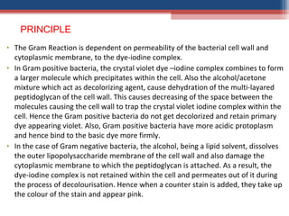
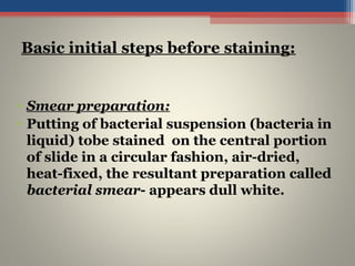
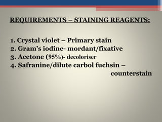
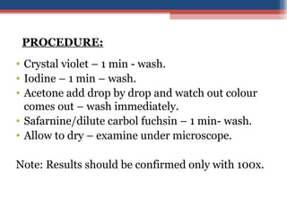
- Gram staining involves staining a bacterial smear with crystal violet dye followed by iodine to form a crystal violet-iodine complex. Bacteria are then decolorized with alcohol or acetone and counterstained with safranin.
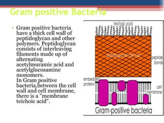
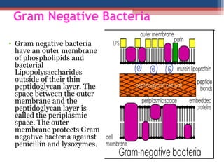
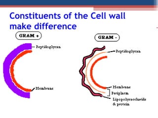
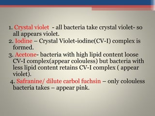
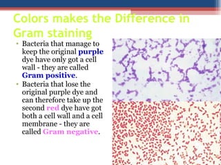
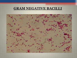
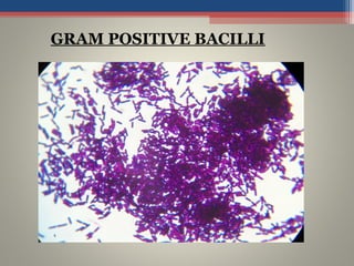
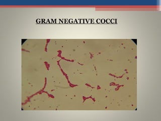
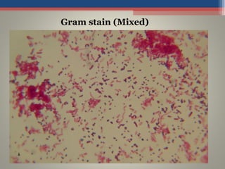
- Based on whether they retain the crystal violet dye after decolorization, bacteria are classified as either Gram-positive or Gram-negative. Gram-positive bacteria retain the crystal violet due to their thick peptidoglycan cell wall, appearing purple under the microscope, while Gram-negative bacteria do not retain the dye due to their thinner cell wall
Capsule is an layer around the bacteria cell which gives bacteria the protection and pathogenicity. Staining such an layer is difficult with the normal stains so it is necessary to stain the background and the cell itself which makes the capsule appear colourless.
Acid fast staining is differential staining technique which differentiate bacteria into two group- acid fast bacteria and non acid bacteria. It used to identify acid-fast organisms such as members of the genus Mycobacterium .
HShjaja jumla ta matokeo ambayo yamepatikan yanaonekana kuwa na faida ku wa saan kwenye jamii kwaiyo tunashukuruni saan kwa michango yeenu kwetu mola tu tunawaombea kuwa awajaalir
this ppt well describes the principle, procedure, modification, usage and limitations of gram's staining.
it is a differential staining method used in bacteriology laboratory
Gram stain is technique used to differntiate gram positive and gram negative bacteria.
If you like this slideshare then please do share and follow me.
You can visit my blogs:pranav2705.blogspot.com
identification of bacteria- lecture 7.pptxOsmanAli92
he culture media are classified in many different ways: Based on the physical state Liquid media Solid media Semisolid media Based on the presence or absence of oxygen Anaerobic media Aerobic media Based on nutritional factors Simple media Synthetic media Complex
micro teaching on communication m.sc nursing.pdfAnurag Sharma
Microteaching is a unique model of practice teaching. It is a viable instrument for the. desired change in the teaching behavior or the behavior potential which, in specified types of real. classroom situations, tends to facilitate the achievement of specified types of objectives.
MANAGEMENT OF ATRIOVENTRICULAR CONDUCTION BLOCK.pdfJim Jacob Roy
Cardiac conduction defects can occur due to various causes.
Atrioventricular conduction blocks ( AV blocks ) are classified into 3 types.
This document describes the acute management of AV block.
TEST BANK for Operations Management, 14th Edition by William J. Stevenson, Ve...kevinkariuki227
TEST BANK for Operations Management, 14th Edition by William J. Stevenson, Verified Chapters 1 - 19, Complete Newest Version.pdf
Anti ulcer drugs and their Advance pharmacology ||
Anti-ulcer drugs are medications used to prevent and treat ulcers in the stomach and upper part of the small intestine (duodenal ulcers). These ulcers are often caused by an imbalance between stomach acid and the mucosal lining, which protects the stomach lining.
||Scope: Overview of various classes of anti-ulcer drugs, their mechanisms of action, indications, side effects, and clinical considerations.
These lecture slides, by Dr Sidra Arshad, offer a quick overview of physiological basis of a normal electrocardiogram.
Learning objectives:
1. Define an electrocardiogram (ECG) and electrocardiography
2. Describe how dipoles generated by the heart produce the waveforms of the ECG
3. Describe the components of a normal electrocardiogram of a typical bipolar leads (limb II)
4. Differentiate between intervals and segments
5. Enlist some common indications for obtaining an ECG
Study Resources:
1. Chapter 11, Guyton and Hall Textbook of Medical Physiology, 14th edition
2. Chapter 9, Human Physiology - From Cells to Systems, Lauralee Sherwood, 9th edition
3. Chapter 29, Ganong’s Review of Medical Physiology, 26th edition
4. Electrocardiogram, StatPearls - https://www.ncbi.nlm.nih.gov/books/NBK549803/
5. ECG in Medical Practice by ABM Abdullah, 4th edition
6. ECG Basics, http://www.nataliescasebook.com/tag/e-c-g-basics
Couples presenting to the infertility clinic- Do they really have infertility...Sujoy Dasgupta
Dr Sujoy Dasgupta presented the study on "Couples presenting to the infertility clinic- Do they really have infertility? – The unexplored stories of non-consummation" in the 13th Congress of the Asia Pacific Initiative on Reproduction (ASPIRE 2024) at Manila on 24 May, 2024.
Lung Cancer: Artificial Intelligence, Synergetics, Complex System Analysis, S...Oleg Kshivets
RESULTS: Overall life span (LS) was 2252.1±1742.5 days and cumulative 5-year survival (5YS) reached 73.2%, 10 years – 64.8%, 20 years – 42.5%. 513 LCP lived more than 5 years (LS=3124.6±1525.6 days), 148 LCP – more than 10 years (LS=5054.4±1504.1 days).199 LCP died because of LC (LS=562.7±374.5 days). 5YS of LCP after bi/lobectomies was significantly superior in comparison with LCP after pneumonectomies (78.1% vs.63.7%, P=0.00001 by log-rank test). AT significantly improved 5YS (66.3% vs. 34.8%) (P=0.00000 by log-rank test) only for LCP with N1-2. Cox modeling displayed that 5YS of LCP significantly depended on: phase transition (PT) early-invasive LC in terms of synergetics, PT N0—N12, cell ratio factors (ratio between cancer cells- CC and blood cells subpopulations), G1-3, histology, glucose, AT, blood cell circuit, prothrombin index, heparin tolerance, recalcification time (P=0.000-0.038). Neural networks, genetic algorithm selection and bootstrap simulation revealed relationships between 5YS and PT early-invasive LC (rank=1), PT N0—N12 (rank=2), thrombocytes/CC (3), erythrocytes/CC (4), eosinophils/CC (5), healthy cells/CC (6), lymphocytes/CC (7), segmented neutrophils/CC (8), stick neutrophils/CC (9), monocytes/CC (10); leucocytes/CC (11). Correct prediction of 5YS was 100% by neural networks computing (area under ROC curve=1.0; error=0.0).
CONCLUSIONS: 5YS of LCP after radical procedures significantly depended on: 1) PT early-invasive cancer; 2) PT N0--N12; 3) cell ratio factors; 4) blood cell circuit; 5) biochemical factors; 6) hemostasis system; 7) AT; 8) LC characteristics; 9) LC cell dynamics; 10) surgery type: lobectomy/pneumonectomy; 11) anthropometric data. Optimal diagnosis and treatment strategies for LC are: 1) screening and early detection of LC; 2) availability of experienced thoracic surgeons because of complexity of radical procedures; 3) aggressive en block surgery and adequate lymph node dissection for completeness; 4) precise prediction; 5) adjuvant chemoimmunoradiotherapy for LCP with unfavorable prognosis.
Title: Sense of Smell
Presenter: Dr. Faiza, Assistant Professor of Physiology
Qualifications:
MBBS (Best Graduate, AIMC Lahore)
FCPS Physiology
ICMT, CHPE, DHPE (STMU)
MPH (GC University, Faisalabad)
MBA (Virtual University of Pakistan)
Learning Objectives:
Describe the primary categories of smells and the concept of odor blindness.
Explain the structure and location of the olfactory membrane and mucosa, including the types and roles of cells involved in olfaction.
Describe the pathway and mechanisms of olfactory signal transmission from the olfactory receptors to the brain.
Illustrate the biochemical cascade triggered by odorant binding to olfactory receptors, including the role of G-proteins and second messengers in generating an action potential.
Identify different types of olfactory disorders such as anosmia, hyposmia, hyperosmia, and dysosmia, including their potential causes.
Key Topics:
Olfactory Genes:
3% of the human genome accounts for olfactory genes.
400 genes for odorant receptors.
Olfactory Membrane:
Located in the superior part of the nasal cavity.
Medially: Folds downward along the superior septum.
Laterally: Folds over the superior turbinate and upper surface of the middle turbinate.
Total surface area: 5-10 square centimeters.
Olfactory Mucosa:
Olfactory Cells: Bipolar nerve cells derived from the CNS (100 million), with 4-25 olfactory cilia per cell.
Sustentacular Cells: Produce mucus and maintain ionic and molecular environment.
Basal Cells: Replace worn-out olfactory cells with an average lifespan of 1-2 months.
Bowman’s Gland: Secretes mucus.
Stimulation of Olfactory Cells:
Odorant dissolves in mucus and attaches to receptors on olfactory cilia.
Involves a cascade effect through G-proteins and second messengers, leading to depolarization and action potential generation in the olfactory nerve.
Quality of a Good Odorant:
Small (3-20 Carbon atoms), volatile, water-soluble, and lipid-soluble.
Facilitated by odorant-binding proteins in mucus.
Membrane Potential and Action Potential:
Resting membrane potential: -55mV.
Action potential frequency in the olfactory nerve increases with odorant strength.
Adaptation Towards the Sense of Smell:
Rapid adaptation within the first second, with further slow adaptation.
Psychological adaptation greater than receptor adaptation, involving feedback inhibition from the central nervous system.
Primary Sensations of Smell:
Camphoraceous, Musky, Floral, Pepperminty, Ethereal, Pungent, Putrid.
Odor Detection Threshold:
Examples: Hydrogen sulfide (0.0005 ppm), Methyl-mercaptan (0.002 ppm).
Some toxic substances are odorless at lethal concentrations.
Characteristics of Smell:
Odor blindness for single substances due to lack of appropriate receptor protein.
Behavioral and emotional influences of smell.
Transmission of Olfactory Signals:
From olfactory cells to glomeruli in the olfactory bulb, involving lateral inhibition.
Primitive, less old, and new olfactory systems with different path
Tom Selleck Health: A Comprehensive Look at the Iconic Actor’s Wellness Journeygreendigital
Tom Selleck, an enduring figure in Hollywood. has captivated audiences for decades with his rugged charm, iconic moustache. and memorable roles in television and film. From his breakout role as Thomas Magnum in Magnum P.I. to his current portrayal of Frank Reagan in Blue Bloods. Selleck's career has spanned over 50 years. But beyond his professional achievements. fans have often been curious about Tom Selleck Health. especially as he has aged in the public eye.
Follow us on: Pinterest
Introduction
Many have been interested in Tom Selleck health. not only because of his enduring presence on screen but also because of the challenges. and lifestyle choices he has faced and made over the years. This article delves into the various aspects of Tom Selleck health. exploring his fitness regimen, diet, mental health. and the challenges he has encountered as he ages. We'll look at how he maintains his well-being. the health issues he has faced, and his approach to ageing .
Early Life and Career
Childhood and Athletic Beginnings
Tom Selleck was born on January 29, 1945, in Detroit, Michigan, and grew up in Sherman Oaks, California. From an early age, he was involved in sports, particularly basketball. which played a significant role in his physical development. His athletic pursuits continued into college. where he attended the University of Southern California (USC) on a basketball scholarship. This early involvement in sports laid a strong foundation for his physical health and disciplined lifestyle.
Transition to Acting
Selleck's transition from an athlete to an actor came with its physical demands. His first significant role in "Magnum P.I." required him to perform various stunts and maintain a fit appearance. This role, which he played from 1980 to 1988. necessitated a rigorous fitness routine to meet the show's demands. setting the stage for his long-term commitment to health and wellness.
Fitness Regimen
Workout Routine
Tom Selleck health and fitness regimen has evolved. adapting to his changing roles and age. During his "Magnum, P.I." days. Selleck's workouts were intense and focused on building and maintaining muscle mass. His routine included weightlifting, cardiovascular exercises. and specific training for the stunts he performed on the show.
Selleck adjusted his fitness routine as he aged to suit his body's needs. Today, his workouts focus on maintaining flexibility, strength, and cardiovascular health. He incorporates low-impact exercises such as swimming, walking, and light weightlifting. This balanced approach helps him stay fit without putting undue strain on his joints and muscles.
Importance of Flexibility and Mobility
In recent years, Selleck has emphasized the importance of flexibility and mobility in his fitness regimen. Understanding the natural decline in muscle mass and joint flexibility with age. he includes stretching and yoga in his routine. These practices help prevent injuries, improve posture, and maintain mobilit
Ozempic: Preoperative Management of Patients on GLP-1 Receptor Agonists Saeid Safari
Preoperative Management of Patients on GLP-1 Receptor Agonists like Ozempic and Semiglutide
ASA GUIDELINE
NYSORA Guideline
2 Case Reports of Gastric Ultrasound
These simplified slides by Dr. Sidra Arshad present an overview of the non-respiratory functions of the respiratory tract.
Learning objectives:
1. Enlist the non-respiratory functions of the respiratory tract
2. Briefly explain how these functions are carried out
3. Discuss the significance of dead space
4. Differentiate between minute ventilation and alveolar ventilation
5. Describe the cough and sneeze reflexes
Study Resources:
1. Chapter 39, Guyton and Hall Textbook of Medical Physiology, 14th edition
2. Chapter 34, Ganong’s Review of Medical Physiology, 26th edition
3. Chapter 17, Human Physiology by Lauralee Sherwood, 9th edition
4. Non-respiratory functions of the lungs https://academic.oup.com/bjaed/article/13/3/98/278874
Title: Sense of Taste
Presenter: Dr. Faiza, Assistant Professor of Physiology
Qualifications:
MBBS (Best Graduate, AIMC Lahore)
FCPS Physiology
ICMT, CHPE, DHPE (STMU)
MPH (GC University, Faisalabad)
MBA (Virtual University of Pakistan)
Learning Objectives:
Describe the structure and function of taste buds.
Describe the relationship between the taste threshold and taste index of common substances.
Explain the chemical basis and signal transduction of taste perception for each type of primary taste sensation.
Recognize different abnormalities of taste perception and their causes.
Key Topics:
Significance of Taste Sensation:
Differentiation between pleasant and harmful food
Influence on behavior
Selection of food based on metabolic needs
Receptors of Taste:
Taste buds on the tongue
Influence of sense of smell, texture of food, and pain stimulation (e.g., by pepper)
Primary and Secondary Taste Sensations:
Primary taste sensations: Sweet, Sour, Salty, Bitter, Umami
Chemical basis and signal transduction mechanisms for each taste
Taste Threshold and Index:
Taste threshold values for Sweet (sucrose), Salty (NaCl), Sour (HCl), and Bitter (Quinine)
Taste index relationship: Inversely proportional to taste threshold
Taste Blindness:
Inability to taste certain substances, particularly thiourea compounds
Example: Phenylthiocarbamide
Structure and Function of Taste Buds:
Composition: Epithelial cells, Sustentacular/Supporting cells, Taste cells, Basal cells
Features: Taste pores, Taste hairs/microvilli, and Taste nerve fibers
Location of Taste Buds:
Found in papillae of the tongue (Fungiform, Circumvallate, Foliate)
Also present on the palate, tonsillar pillars, epiglottis, and proximal esophagus
Mechanism of Taste Stimulation:
Interaction of taste substances with receptors on microvilli
Signal transduction pathways for Umami, Sweet, Bitter, Sour, and Salty tastes
Taste Sensitivity and Adaptation:
Decrease in sensitivity with age
Rapid adaptation of taste sensation
Role of Saliva in Taste:
Dissolution of tastants to reach receptors
Washing away the stimulus
Taste Preferences and Aversions:
Mechanisms behind taste preference and aversion
Influence of receptors and neural pathways
Impact of Sensory Nerve Damage:
Degeneration of taste buds if the sensory nerve fiber is cut
Abnormalities of Taste Detection:
Conditions: Ageusia, Hypogeusia, Dysgeusia (parageusia)
Causes: Nerve damage, neurological disorders, infections, poor oral hygiene, adverse drug effects, deficiencies, aging, tobacco use, altered neurotransmitter levels
Neurotransmitters and Taste Threshold:
Effects of serotonin (5-HT) and norepinephrine (NE) on taste sensitivity
Supertasters:
25% of the population with heightened sensitivity to taste, especially bitterness
Increased number of fungiform papillae
HOT NEW PRODUCT! BIG SALES FAST SHIPPING NOW FROM CHINA!! EU KU DB BK substit...GL Anaacs
Contact us if you are interested:
Email / Skype : kefaya1771@gmail.com
Threema: PXHY5PDH
New BATCH Ku !!! MUCH IN DEMAND FAST SALE EVERY BATCH HAPPY GOOD EFFECT BIG BATCH !
Contact me on Threema or skype to start big business!!
Hot-sale products:
NEW HOT EUTYLONE WHITE CRYSTAL!!
5cl-adba precursor (semi finished )
5cl-adba raw materials
ADBB precursor (semi finished )
ADBB raw materials
APVP powder
5fadb/4f-adb
Jwh018 / Jwh210
Eutylone crystal
Protonitazene (hydrochloride) CAS: 119276-01-6
Flubrotizolam CAS: 57801-95-3
Metonitazene CAS: 14680-51-4
Payment terms: Western Union,MoneyGram,Bitcoin or USDT.
Deliver Time: Usually 7-15days
Shipping method: FedEx, TNT, DHL,UPS etc.Our deliveries are 100% safe, fast, reliable and discreet.
Samples will be sent for your evaluation!If you are interested in, please contact me, let's talk details.
We specializes in exporting high quality Research chemical, medical intermediate, Pharmaceutical chemicals and so on. Products are exported to USA, Canada, France, Korea, Japan,Russia, Southeast Asia and other countries.
2. Hans Christian Joachim Gram
• The Gram stain was devised by
the Danish physician, Hans
Christian Joachim Gram, while
working in Berlin in 1883. He
later published this procedure in
1884. At the time, Dr. Gram was
studying lung tissue sections
from patients who had died of
pneumonia.
3. GRAM STAINING :
• DANISH BACTERIOLOGIST HANS
CHRISTIAN GRAM (1880)
• Based on this reaction, bacteria classified
into Gram positive and Gram negative
bacteria.
• The cell wall compostion differences
makes difference.
4. PRINCIPLE
• The Gram Reaction is dependent on permeability of the bacterial cell wall and
cytoplasmic membrane, to the dye-iodine complex.
• In Gram positive bacteria, the crystal violet dye –iodine complex combines to form
a larger molecule which precipitates within the cell. Also the alcohol/acetone
mixture which act as decolorizing agent, cause dehydration of the multi-layared
peptidoglycan of the cell wall. This causes decreasing of the space between the
molecules causing the cell wall to trap the crystal violet iodine complex within the
cell. Hence the Gram positive bacteria do not get decolorized and retain primary
dye appearing violet. Also, Gram positive bacteria have more acidic protoplasm
and hence bind to the basic dye more firmly.
• In the case of Gram negative bacteria, the alcohol, being a lipid solvent, dissolves
the outer lipopolysaccharide membrane of the cell wall and also damage the
cytoplasmic membrane to which the peptidoglycan is attached. As a result, the
dye-iodine complex is not retained within the cell and permeates out of it during
the process of decolourisation. Hence when a counter stain is added, they take up
the colour of the stain and appear pink.
5. • A staining technique used to classify bacteria;
bacteria are stained with gentian violet and then
treated with Gram's solution; after being
decolorized with alcohol and treated with
safranine and washed in water, those that retain
the gentian violet are Gram-positive and those
that do not retain it are Gram-negative
Gram staining a Important
Technique
6. Gram positive Bacteria
• Gram positive bacteria
have a thick cell wall of
peptidoglycan and other
polymers. Peptidoglycan
consists of interleaving
filaments made up of
alternating
acetylmuramic acid and
acetylglucosamine
monomers.
In Gram positive
bacteria,between the cell
wall and cell membrane,
there is a "membrane
teichoic acid".
7. Gram Negative Bacteria
• Gram negative bacteria
have an outer membrane
of phospholipids and
bacterial
Lipopolysaccharides
outside of their thin
peptidoglycan layer. The
space between the outer
membrane and the
peptidoglycan layer is
called the periplasmic
space. The outer
membrane protects Gram
negative bacteria against
penicillin and lysozymes.
9. • Clean grease-free slide.
• Bacteria tobe stained.
• Inoculating loops- to transfer bacterial
suspension to slide.
• Bunsen burner – to sterilise inoculating
loops before and after smear preparation.
• Pencil marker – to mark (particularly
central portion of slide) where bacterial
smear is applied
Basic requirements for staining:
10. • Smear preparation:
• Putting of bacterial suspension (bacteria in
liquid) tobe stained on the central portion
of slide in a circular fashion, air-dried,
heat-fixed, the resultant preparation called
bacterial smear- appears dull white.
Basic initial steps before staining:
13. 1. Crystal violet - all bacteria take crystal violet- so
all appears violet.
2. Iodine – Crystal Violet-iodine(CV-I) complex is
formed.
3. Acetone- bacteria with high lipid content loose
CV-I complex(appear colouless) but bacteria with
less lipid content retains CV-I complex ( appear
violet).
4. Safranine/ dilute carbol fuchsin – only colouless
bacteria takes – appear pink.
14. • Crystal violet – 1 min - wash.
• Iodine – 1 min – wash.
• Acetone add drop by drop and watch out colour
comes out – wash immediately.
• Safarnine/dilute carbol fuchsin – 1 min- wash.
• Allow to dry – examine under microscope.
Note: Results should be confirmed only with 100x.
PROCEDURE:
15. • SMEAR FIXATION:
• 1) Heat fixation
• a) Pass air-dried smears through a flame two or three times. Do not
overheat.
• b) Allow slide to cool before staining.
• 2) Methanol fixation
• a) Place air-dried smears in a coplin jar with methanol for one minute.
Alternatively, flood smear with methanol for 1 minute.
• b) Drain slides and allow to dry before staining.
17. Prefer to pick up colonies with loop
and smear on Clean glass slide
18. • i. Relative amounts of PMN’s, mononuclear
cells, and RBC's
• ii. Relative amounts of squamous epithelial cells,
bacteria
• consistent with normal micro flora, which may
indicate an improperly collected specimen
• iii. Location and arrangements of
microorganisms
• Note any special character sticks
Observe the Following Under Oil
Immersion lens
19. • Bacteria that manage to
keep the original purple
dye have only got a cell
wall - they are called
Gram positive.
• Bacteria that lose the
original purple dye and
can therefore take up the
second red dye have got
both a cell wall and a cell
membrane - they are
called Gram negative.
Colors makes the Difference in
Gram staining
21. • If no microorganisms are seen in a smear of a
clinical
• specimen, report “No microorganisms seen.”
• ii. If microorganisms are seen, report relative
numbers and
• Describe morphology.
• Observe predominant shapes of microorganisms
Report as follows
39. Gram variable observations in
Gram staining
• The Gram staining procedure does not always
give clear-cut results. Some organisms are
Gram-variable and may appear either Gram-
negative or Gram-positive according to the
conditions. With these types of organisms,
Gram-positive and Gram-negative cells may be
present within the same preparation
40. Overcoming in Gram Variable
Observations
• it is necessary that it is stained at two or three
different ages (very young cultures should be
used). If an organism changes from positive to
negative or vice versa during its growth cycle,
this change should be recorded with a statement
as to the age of the culture when the change was
first observed. In case a Gram-variable reaction
is observed it is also good to check the purity of
the culture.
41. Why Gram Variability?
• Some Gram-positive bacteria appear Gram-
negative when they have reached a certain age,
varying from a few hours to a few days. On the
other hand, some Gram-negative bacteria may
become Gram-positive in older cultures. For this
reason it is strongly recommended to use very
young cultures for the staining procedure, after
growth has become just visible.
42. Trouble shooting in Gram
Staining method
The method and techniques used. Overheating
during heat fixation, over decolourization with alcohol,
and even too much washing with water between steps
may result in gram-positive bacteria losing the crystal
violet-iodine complex.
The age of the culture. Cultures more than 24 hours old
may lose their ability to retain the crystal violet-iodine
complex.
The organism itself. Some gram-positive bacteria are
more able to retain the crystal violet-iodine complex
than others.
43. • By this method fairly good results may be obtained with very short staining
times, which are convenient when only one slide has to be stained.
• Flood the slide with crystal or methyl violet stain and allow to act for about 5
seconds.
• Tip off the stain and flood the tilted slide with iodine solution and allow to act
for about 5 seconds.
• Tip off the iodine and flood the tilted slide
• With acetone and allow this to act for only 2 seconds before washing it off
with water from the tap.
• Flood the slide with basic fuchsin counter stain and allow it to act for about 5
seconds. Wash off with water, blot and dry.
Quick GRAM METHOD
44. Modification in Gram staining
methods
• Since the original procedure of Gram, many variations of the Gram
staining technique have been published. Some of them have improved
the method, others include some minor technical variants of no value.
• Bartholomew (1962) has pointed out that each variation in the Gram
staining procedure has a definite limit to its acceptability. Any final
result is the outcome of the interaction of all of the possible variables.
• All modified methods to be practised with caution should suit to the
laboratory, and quality control checks.
45. Various modifications of Gram staining
• 1. Kopeloff & Beerman’s Modification :
Primary stain is Methyl violet.
Decolourizer is Acetone/ Acetone-Alcohol mixture.
• 2. Jensen’s Modification :
Primary stain is Methyl violet.
Decolourizer is Absolute Alcohol.
Counter stain is Neutral Red.
• 3. Preston & Morrell’s Modification :
Primary stain is Crystal violet.
Decolourizer is Iodine-Acetone.
• 4. Weigert’s Modification:
Primary stain is Carbol Gentian violet.
Decolourizer is –Aniline-Xylol. Weigert stain is used to stain tissue sections.
46. Applications of Gram Staining
• 1.Rapid presumptive diagnosis of diseases such as Bacterial meningitis.
• 2.Selection of Empirical antibiotics based on Gram stain finding.
• 3.Selection of suitable culture media based on Gram stain finding.
• 4.Screening of the quality of the clinical specimens such as sputum that
should contain many pus cells & few epithelial cells.
• 5.Counting of bacteria.
• 6.Appreciation of morphology & types of bacteria in clinical specimens.