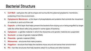
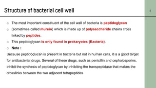
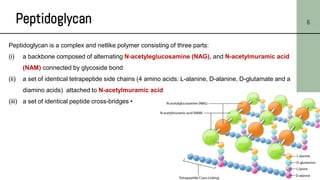
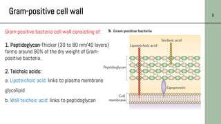
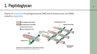
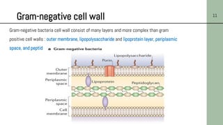
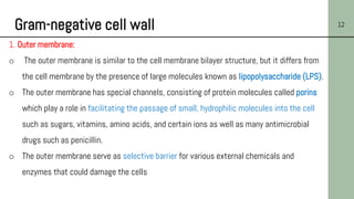
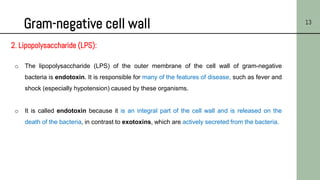
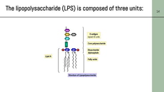
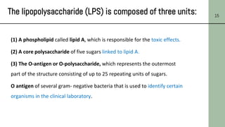
1. Ministry of Education
King Abdul Aziz University
Faculty of Sciences
Department of Biological Sciences
التعليم وزارة
عبدالعزيز الملك جامعة
العلوم كلية
االحياء علوم قسم
3. 3
1. Cell Wall - wall gives the cell its shape and surrounds the plasma/cytoplasmic membrane,
protecting it from the environment.
2. Cytoplasmic Membrane - a thin layer of phospholipids and proteins that controls the movement
of nutrients in and out of the cell
3. Capsule - a third layer that helps prevent the bacteria from drying out or being engulfed by larger
cells like white blood cells (only present in some types of bacteria)
4. Cytoplasm - a gel-like material in which the ribosomes and genetic material are suspended
5. Nucleoid - a mass of genetic material (DNA)
6. Plasmids - genetic material (DNA)
7. Ribosomes - structures that make proteins
8. Flagellum - structure that helps the bacteria move around and sense their environment
9. Pili - hair-like structures that help bacteria attach to surfaces and other bacteria
Bacterial Structure
4. 1. Cell Wall 4
Cell wall function:
o The cell wall of bacterial cell is a complex , rigid structure responsible for shape of
bacteria.
o Cell walls of bacteria protect them from mechanical damage and osmotic lysis.
o Bacterial cell walls are usually essential for the growth and division of bacteria.
5. Structure of bacterial cell wall 5
o The most important constituent of the cell wall of bacteria is peptidoglycan
o (sometimes called murein) which is made up of polysaccharide chains cross
linked by peptides.
o This peptidoglycan is only found in prokaryotes (Bacteria).
o Note :
Because peptidoglycan is present in bacteria but not in human cells, it is a good target
for antibacterial drugs. Several of these drugs, such as penicillin and cephalosporins,
inhibit the synthesis of peptidoglycan by inhibiting the transpeptidase that makes the
crosslinks between the two adjacent tetrapeptides
6. Peptidoglycan 6
Peptidoglycan is a complex and netlike polymer consisting of three parts:
(i) a backbone composed of alternating N-acetyleglucosamine (NAG), and N-acetylmuramic acid
(NAM) connected by glycoside bond
(ii) a set of identical tetrapeptide side chains (4 amino acids: L-alanine, D-alanine, D-glutamate and a
diamino acids) attached to N-acetylmuramic acid
(iii) a set of identical peptide cross-bridges •
7. Walls of Gram-positive and Gram-negative bacteria 7
• The cell wall of Gram-positive bacteria:
The Peptidoglycan may consist of as many as
40 layers (30–80 nm thick).
• The cell wall of Gram-negative bacteria:
Here the Peptidoglycan is only about 2-8 nm
thick .
8. Gram-positive cell wall 8
Gram-positive bacteria cell wall consisting of:
1. Peptidoglycan-Thicker (30 to 80 nm/40 layers)
forms around 90% of the dry weight of Gram-
positive bacteria.
2. Teichoic acids:
a. Lipoteichoic acid: links to plasma membrane
glycolipid
b. Wall teichoic acid: links to peptidoglycan
9. 1. Peptidoglycan 9
Polymer of disaccharide N-acetylglucosamine (NAG) and N-acetylmuramic acid (NAM)
Linked by polypeptides
10. 2. Teichoic Acid 10
Teichoic acids are negatively charged polysaccharides; they are polymers of
ribitol phosphate and cross-link to peptidoglycan.
1. Teichoic acid is believed to play several important
roles for the cell, such as generation of the negative
charge of the cell.
2. Teichoic acid contributes to the overall rigidity of the
cell wall, which is important for the maintenance of the
cell shape, particularly in rod-shaped organisms.
3. Teichoic acids appear to play a role in resistance to
harsh conditions such as high temperatures and high
salt concentrations, as well as to β-lactam antibiotics.
11. Gram-negative cell wall 11
Gram-negative bacteria cell wall consist of many layers and more complex than gram
positive cell walls : outer membrane, lipopolysaccharide and lipoprotein layer, periplasmic
space, and peptidoglycan layer
12. Gram-negative cell wall 12
1. Outer membrane:
o The outer membrane is similar to the cell membrane bilayer structure, but it differs from
the cell membrane by the presence of large molecules known as lipopolysaccharide (LPS).
o The outer membrane has special channels, consisting of protein molecules called porins
which play a role in facilitating the passage of small, hydrophilic molecules into the cell
such as sugars, vitamins, amino acids, and certain ions as well as many antimicrobial
drugs such as penicillin.
o The outer membrane serve as selective barrier for various external chemicals and
enzymes that could damage the cells
13. Gram-negative cell wall 13
2. Lipopolysaccharide (LPS):
o The lipopolysaccharide (LPS) of the outer membrane of the cell wall of gram-negative
bacteria is endotoxin. It is responsible for many of the features of disease, such as fever and
shock (especially hypotension) caused by these organisms.
o It is called endotoxin because it is an integral part of the cell wall and is released on the
death of the bacteria, in contrast to exotoxins, which are actively secreted from the bacteria.
15. 15
The lipopolysaccharide (LPS) is composed of three units:
(1) A phospholipid called lipid A, which is responsible for the toxic effects.
(2) A core polysaccharide of five sugars linked to lipid A.
(3) The O-antigen or O-polysaccharide, which represents the outermost
part of the structure consisting of up to 25 repeating units of sugars.
O antigen of several gram- negative bacteria that is used to identify certain
organisms in the clinical laboratory.
16. Gram-negative cell wall 16
Function of LPS:
• Helping to stabilize the outer membrane
• Assist in the attachment of bacteria to other surfaces
• Formation of biofilm
• Restrict the entry of antibiotics and other toxic substances
• Protection from host immunological barriers like such as phagocytosis
• LPS plays a role in the host's response to pathogenic gram-negative
bacteria
• Lipid A portion is toxic causing endotoxic shock
17. Gram-negative cell wall- contd. 17
3. Lipoprotein layer: known as Braun’s lipoprotein
connects the peptidoglycan to outer membrane.
4. Periplasmic space: the space located between
the outer surface of the cell membrane and the inner
surface of the outer membrane, and it contains the
gram-negative peptidoglycan.
5. Peptidoglycan thin of few layers. The dry weight
only 10% of Gram-negative strains
18. Comparison of Cell Walls of
Gram-Positive and Gram-Negative Bacteria 18
Component Gram-Positive Cell Gram- Negative Cell
Peptidoglycan Thicker; multilayer Thinner; single layer
Teichoic acids Yes No
Outer membrane or
Lipopolysaccharide
(endotoxin)
No Yes
Periplasmic space No Yes
19. Cell Walls of Acid-Fast Bacteria 19
Acid fast bacteria like (Mycobacterium and Nocardia)
have an unusual cell wall, contain large amounts of lipid
substances within their cell walls called mycolic acids.
These acids resist staining by ordinary methods such as a
Gram stain.
The stains used are the red colored Carbol fuchsin that
stains the bacteria, these bacteria resist decolorization with
acid-alcohol after being stained with Carbol fuchsin.