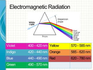
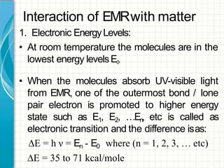
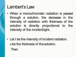
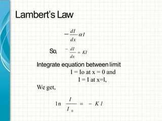
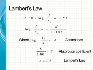
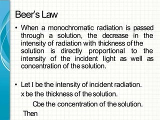
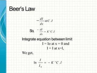
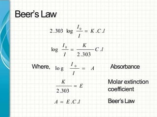
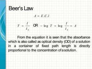
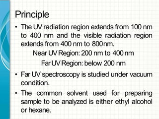
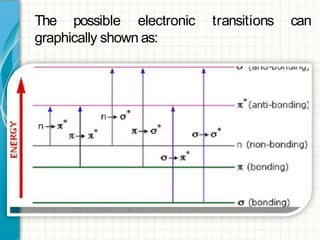
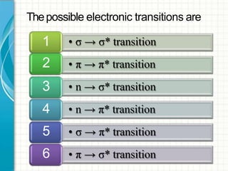
UV / VISIBLE SPECTROSCOPY
Dr. Nilesh Thakare
Spectroscopy
• It is the branch of science that deals with the
study of interaction of matter with light.
OR
• It is the branch of science that deals with the
study of interaction of electromagnetic
radiation with matter.
Electromagnetic
Radiation
Electromagnetic Radiation
• Electromagnetic radiation consist of discrete
packages of energy which are called as
photons.
• Aphoton consists of an oscillating electric field
(E)& an oscillating magnetic field (M) which
are perpendicular to eachother.
Electromagnetic Radiation
• Frequency(ν):
– It is defined as the number of times electrical field
radiation oscillates in onesecond.
– Theunit for frequency is Hertz(Hz).
1 Hz=1 cycle per second
• Wavelength (λ):
– It is the distance between two nearest parts of the
wave in the same phase i.e. distance between two
nearest crest or troughs.
Electromagnetic Radiation
• The relationship between wavelength &
frequency canbe written as:
c=ν λ
• Asphoton is subjected to energy,so
E=h ν =h c/ λ
Electromagnetic Radiation
Violet 400 - 420 nm Yellow 570 - 585 nm
Indigo 420 - 440 nm Orange 585 - 620 nm
Blue 440 - 490 nm Red 620 - 780 nm
Green 490 - 570 nm
Principles of
Spectroscopy
Principles of Spectroscopy
• The principle is based on the measurement of
spectrum of a sample containing atoms /
molecules.
• Spectrum is a graph of intensity of absorbed or
emitted radiation by sample verses frequency
(ν) or wavelength(λ).
• Spectrometer is an instrument design to
measure the spectrum of acompound.
Principles of Spectroscopy
1. Absorption Spectroscopy:
• An analytical technique which concerns with
the measurement of absorption of
electromagnetic radiation.
• e.g. UV (185 - 400 nm) / Visible (400 - 800 nm)
Spectroscopy, IRSpectroscopy (0.76 - 15μm)
Interaction of
EMR with
Matter
Interaction of EMRwith matter
1. Electronic EnergyLevels:
• At room temperature the molecules are in the
lowest energy levelsE0.
• When the molecules absorb UV-visible light
from EMR, one of the outermost bond / lone
pair electron is promoted to higher energy
state such as E1, E2, …En, etc is called as
electronic transition and the difference isas:
∆E = h ν = En - E0 where (n = 1, 2, 3, … etc)
∆E = 35 to 71 kcal/mole
Lambert’
s Law
Lambert’s Law
• When a monochromatic radiation is passed
through a solution, the decrease in the
intensity of radiation with thickness of the
solution is directly proportional to the
intensity of the incidentlight.
• Let I be the intensity of incident radiation.
xbe the thickness of thesolution.
Then
Lambert’s Law
 dI
I
dx
So,  KI
 dI
dx
Integrate equation betweenlimit
I = Io at x = 0 and
I = I at x=l,
We get,
I
I 0
l n   K l
Lambert’s Law
I
  K l
I 0
2 . 3 0 3 lo g
l
KI
2 . 3 0 3
lo g
I 0
 
Absorbance
I
 A
I 0
Where, l o g
K
 E
2.303
A  E .l
Absorption coefficient
Lambert’s Law
Beer’
s Law
Beer’sLaw
• When a monochromatic radiation is passed
through a solution, the decrease in the
intensity of radiation with thickness ofthe
solution
intensity
is directly proportional
of the incident light as
to the
well as
concentration of thesolution.
• Let I be the intensity of incident radiation.
xbe the thickness of thesolution.
Cbe the concentration of thesolution.
Then
Beer’sLaw
 dI
 C .I
dx
So,  K 'C .I
 dI
dx
Integrate equation betweenlimit
I = Io at x = 0 and
I = I at x=l,
We get,
I
I 0
ln   K ' C .l
Beer’sLaw
I
2 .303 log
I0
 K .C .l
K
I
C .l
2 .303
log
I 0

Where, Absorbance
I
 A
I 0
lo g
K
 E
2.303
A  E .C .l
Molar extinction
coefficient
Beer’sLaw
Beer’sLaw
A  E .C .l
I
T  OR
I
 A log T  log
I 0 I 0
From the equation it is seen that the absorbance
which is also called as optical density (OD) of a solution
in a container of fixed path length is directly
proportional to the concentration ofasolution.
PRINCIPLES OF
UV - VISIBLE
SPECTROSCOPY
Principle
• The UVradiation region extends from 100 nm
to 400 nm and the visible radiation region
extends from 400 nm to 800nm.
Near UVRegion:200 nm to 400nm
FarUVRegion:below 200 nm
• Far UV spectroscopy is studied under vacuum
condition.
• The common solvent used for preparing
sample to be analyzed is either ethyl alcohol
or hexane.
Electronic
Transitions
transitions canThe possible electronic
graphically shown as:
• σ → σ* transition
• π → π* transition
• n → σ* transition
• n → π* transition
• σ → π* transition
• π → σ* transition
1
2
3
4
5
6
Thepossible electronic transitions are
this
• σ electron from orbital is excited to
corresponding anti-bonding orbital σ*.
• The energy required is large for
transition.
• e.g. Methane (CH4) has C-H bond only and
can undergo σ → σ* transition and shows
absorbance maxima at 125nm.
• σ → σ* transition1
• π electron in a bonding orbital is excited to
corresponding anti-bonding orbital π*.
• Compounds containing multiple bonds like
alkenes, alkynes, carbonyl, nitriles, aromatic
compounds, etc undergo π→π* transitions.
• e.g. Alkenes generally absorb in the region
170 to 205nm.
• π → π* transition2
• Saturated compounds containing atoms with
lone pair of electrons like O, N, S and
halogens are capable of n →σ* transition.
• These transitions usually requires less energy
than σ→σ* transitions.
• The number of organic functional groups
with n →σ* peaksin UVregion is small (150
– 250 nm).
• n → σ* transition3
• An electron from non-bonding orbital is
promoted to anti-bonding π*orbital.
• Compounds containing double bond
involving hetero atoms (C=O, C≡N, N=O)
undergo suchtransitions.
• n → π* transitions require minimum energy
and show absorption at longer wavelength
around 300 nm.
• n → π* transition4
•These electronic transitions are
transitions & are only theoreticallypossible.
•Thus,n →π* & π→π* electronic transitions
show absorption in region
which is accessible
above 200 nm
to UV-visible
spectrophotometer.
•The UV spectrum is of only a few broad of
absorption.
• σ → π* transition
• π → σ* transition 6
forbidden
5
&
Termsused
in
UV/ Visible
Spectroscopy
Chromophore
Thepart of amolecule responsible for imparting
color, are called aschromospheres.
OR
Thefunctional groups containing multiple bonds
capable of absorbing radiations above 200 nm
due to n →π* & π→π* transitions.
e.g. NO2,N=O,C=O,C=N,C≡N, C=C, C=S, etc
Chromophore
Tointerpretate UV– visible spectrum following
points should benoted:
1. Non-conjugated alkenes show an intense
absorption below 200 nm & are therefore
inaccessible to UVspectrophotometer.
2. Non-conjugated carbonyl group compound
give a weak absorption band in the 200 - 300
nm region.
Chromophore
e.g.
C
H3C CH3
and that cyclohexane hasλmax =291nm.
When double bonds are conjugated in
a compound λmax is shifted to
longer wavelength.
O
e.g. Acetone which has λmax =279 nmO
H2C
1,5 - hexadiene hasλmax =178 nm
2,4 - hexadiene hasλmax =227nm
CH2
H3C
CH3
Chromophore
3. Conjugation of C=Cand carbonyl group shifts
the λmax of both groups to longerwavelength.
e.g. Ethylene hasλmax =171 nm
Acetone hasλmax =279nm
C
H3C CH3
O
H 2C CH2
C
CH3
Crotonaldehyde hasλmax =290 nm
O
H2C
Auxochrome
The functional groups attached to a
chromophore which modifies the ability of the
chromophore to absorb light , altering the
wavelength or intensity ofabsorption.
OR
Thefunctional group with non-bondingelectrons
that does not absorb radiation in near UVregion
but when attached to achromophore alters the
wavelength & intensity ofabsorption.
Auxochrome
e.g. Benzeneλmax =255nm
Phenol λmax =270nm
Aniline λmax =280nm
OH
NH2
Absorption
& Intensity
Shifts
• When absorption maxima (λmax) of a
compound shifts to longer wavelength, it is
known asbathochromic shift or redshift.
• The effect is due to presence of an auxochrome
or by the changeofsolvent.
• e.g. An auxochrome group like –OH, -OCH3
causes absorption of compound at longer
wavelength.
• Bathochromic Shift (Red Shift)1
• In alkaline medium, p-nitrophenol shows red
shift. Because negatively charged oxygen
delocalizes more effectively than the unshared
pair of electron.
p-nitrophenol
λmax =255nm λmax =265nm
• Bathochromic Shift (Red Shift)1
O H
N
+
O O
-
O H
Alk a lin e
m e d iu m -
O
N
+
- -
O O
• When absorption maxima (λmax) of a
compound shifts to shorter wavelength, it is
known ashypsochromic shift or blueshift.
• The effect is due to presence of an group
causes removal of conjugation or by the
changeof solvent.
• Hypsochromic Shift (Blue Shift)2
• Aniline shows blue shift in acidic medium, it
loses conjugation.
Aniline
λmax =280nm λmax =265nm
• Hypsochromic Shift (Blue Shift)2
N H 2 +
H
A cid ic
m e d iu m
+
N H 3 Cl
-
• When absorption intensity (ε) of a compound is
increased, it is known ashyperchromicshift.
• If auxochrome introduces to the compound,
the intensity of absorption increases.
Pyridine
λmax =257nm
2-methyl pyridine
λmax =260nm
• Hyperchromic Effect3
N N CH3
• When absorption intensity (ε) of acompoundis
decreased, it is known ashypochromicshift.
Naphthalene
ε =19000
CH3
2-methyl naphthalene
ε =10250
• Hypochromic Effect4
Absorbance(A)
Shifts and Effects
Hyperchromic shift
Red
shift
Blu
e
shift
Hypochromic shift
λmax
Wavelength ( λ)
APPLICATIONS OF
UV / VISIBLE
SPECTROSCOPY
Applications
• Qualitative & QuantitativeAnalysis:
– It is used for characterizing aromatic compounds
and conjugated olefins.
– It can be used to find out molar concentration of the
solute understudy.
• Detection of impurities:
– It is one of the important method to detect
impurities in organicsolvents.
• Detection of isomers arepossible.
• Determination of molecular weight using Beer’s
law.
REFERENCES
ReferenceBooks
• Introduction to Spectroscopy
– DonaldA. Pavia
• Elementary OrganicSpectroscopy
– Y.R.Sharma
• PhysicalChemistry
– Puri, Sharma& Pathaniya
Resources
• http://www2.chemistry.msu.edu/faculty/reu
sch/VirtTxtJml/Spectrpy/UV-
Vis/spectrum.htm
• http://en.wikipedia.org/wiki/Ultraviolet%E2
%80%93visible_spectroscopy
• http://teaching.shu.ac.uk/hwb/chemistry/tut
orials/molspec/uvvisab1.htm

Uv visible spectroscopy

  • 1.
    UV / VISIBLESPECTROSCOPY Dr. Nilesh Thakare
  • 2.
    Spectroscopy • It isthe branch of science that deals with the study of interaction of matter with light. OR • It is the branch of science that deals with the study of interaction of electromagnetic radiation with matter.
  • 3.
  • 4.
    Electromagnetic Radiation • Electromagneticradiation consist of discrete packages of energy which are called as photons. • Aphoton consists of an oscillating electric field (E)& an oscillating magnetic field (M) which are perpendicular to eachother.
  • 6.
    Electromagnetic Radiation • Frequency(ν): –It is defined as the number of times electrical field radiation oscillates in onesecond. – Theunit for frequency is Hertz(Hz). 1 Hz=1 cycle per second • Wavelength (λ): – It is the distance between two nearest parts of the wave in the same phase i.e. distance between two nearest crest or troughs.
  • 7.
    Electromagnetic Radiation • Therelationship between wavelength & frequency canbe written as: c=ν λ • Asphoton is subjected to energy,so E=h ν =h c/ λ
  • 8.
  • 9.
    Electromagnetic Radiation Violet 400- 420 nm Yellow 570 - 585 nm Indigo 420 - 440 nm Orange 585 - 620 nm Blue 440 - 490 nm Red 620 - 780 nm Green 490 - 570 nm
  • 10.
  • 11.
    Principles of Spectroscopy •The principle is based on the measurement of spectrum of a sample containing atoms / molecules. • Spectrum is a graph of intensity of absorbed or emitted radiation by sample verses frequency (ν) or wavelength(λ). • Spectrometer is an instrument design to measure the spectrum of acompound.
  • 12.
    Principles of Spectroscopy 1.Absorption Spectroscopy: • An analytical technique which concerns with the measurement of absorption of electromagnetic radiation. • e.g. UV (185 - 400 nm) / Visible (400 - 800 nm) Spectroscopy, IRSpectroscopy (0.76 - 15μm)
  • 13.
  • 14.
    Interaction of EMRwithmatter 1. Electronic EnergyLevels: • At room temperature the molecules are in the lowest energy levelsE0. • When the molecules absorb UV-visible light from EMR, one of the outermost bond / lone pair electron is promoted to higher energy state such as E1, E2, …En, etc is called as electronic transition and the difference isas: ∆E = h ν = En - E0 where (n = 1, 2, 3, … etc) ∆E = 35 to 71 kcal/mole
  • 15.
  • 16.
    Lambert’s Law • Whena monochromatic radiation is passed through a solution, the decrease in the intensity of radiation with thickness of the solution is directly proportional to the intensity of the incidentlight. • Let I be the intensity of incident radiation. xbe the thickness of thesolution. Then
  • 17.
    Lambert’s Law  dI I dx So, KI  dI dx Integrate equation betweenlimit I = Io at x = 0 and I = I at x=l, We get, I I 0 l n   K l
  • 18.
    Lambert’s Law I  K l I 0 2 . 3 0 3 lo g l KI 2 . 3 0 3 lo g I 0   Absorbance I  A I 0 Where, l o g K  E 2.303 A  E .l Absorption coefficient Lambert’s Law
  • 19.
  • 20.
    Beer’sLaw • When amonochromatic radiation is passed through a solution, the decrease in the intensity of radiation with thickness ofthe solution intensity is directly proportional of the incident light as to the well as concentration of thesolution. • Let I be the intensity of incident radiation. xbe the thickness of thesolution. Cbe the concentration of thesolution. Then
  • 21.
    Beer’sLaw  dI  C.I dx So,  K 'C .I  dI dx Integrate equation betweenlimit I = Io at x = 0 and I = I at x=l, We get, I I 0 ln   K ' C .l
  • 22.
    Beer’sLaw I 2 .303 log I0 K .C .l K I C .l 2 .303 log I 0  Where, Absorbance I  A I 0 lo g K  E 2.303 A  E .C .l Molar extinction coefficient Beer’sLaw
  • 23.
    Beer’sLaw A  E.C .l I T  OR I  A log T  log I 0 I 0 From the equation it is seen that the absorbance which is also called as optical density (OD) of a solution in a container of fixed path length is directly proportional to the concentration ofasolution.
  • 24.
    PRINCIPLES OF UV -VISIBLE SPECTROSCOPY
  • 25.
    Principle • The UVradiationregion extends from 100 nm to 400 nm and the visible radiation region extends from 400 nm to 800nm. Near UVRegion:200 nm to 400nm FarUVRegion:below 200 nm • Far UV spectroscopy is studied under vacuum condition. • The common solvent used for preparing sample to be analyzed is either ethyl alcohol or hexane.
  • 26.
  • 27.
    transitions canThe possibleelectronic graphically shown as:
  • 28.
    • σ →σ* transition • π → π* transition • n → σ* transition • n → π* transition • σ → π* transition • π → σ* transition 1 2 3 4 5 6 Thepossible electronic transitions are
  • 29.
    this • σ electronfrom orbital is excited to corresponding anti-bonding orbital σ*. • The energy required is large for transition. • e.g. Methane (CH4) has C-H bond only and can undergo σ → σ* transition and shows absorbance maxima at 125nm. • σ → σ* transition1
  • 30.
    • π electronin a bonding orbital is excited to corresponding anti-bonding orbital π*. • Compounds containing multiple bonds like alkenes, alkynes, carbonyl, nitriles, aromatic compounds, etc undergo π→π* transitions. • e.g. Alkenes generally absorb in the region 170 to 205nm. • π → π* transition2
  • 31.
    • Saturated compoundscontaining atoms with lone pair of electrons like O, N, S and halogens are capable of n →σ* transition. • These transitions usually requires less energy than σ→σ* transitions. • The number of organic functional groups with n →σ* peaksin UVregion is small (150 – 250 nm). • n → σ* transition3
  • 32.
    • An electronfrom non-bonding orbital is promoted to anti-bonding π*orbital. • Compounds containing double bond involving hetero atoms (C=O, C≡N, N=O) undergo suchtransitions. • n → π* transitions require minimum energy and show absorption at longer wavelength around 300 nm. • n → π* transition4
  • 33.
    •These electronic transitionsare transitions & are only theoreticallypossible. •Thus,n →π* & π→π* electronic transitions show absorption in region which is accessible above 200 nm to UV-visible spectrophotometer. •The UV spectrum is of only a few broad of absorption. • σ → π* transition • π → σ* transition 6 forbidden 5 &
  • 34.
  • 35.
    Chromophore Thepart of amoleculeresponsible for imparting color, are called aschromospheres. OR Thefunctional groups containing multiple bonds capable of absorbing radiations above 200 nm due to n →π* & π→π* transitions. e.g. NO2,N=O,C=O,C=N,C≡N, C=C, C=S, etc
  • 36.
    Chromophore Tointerpretate UV– visiblespectrum following points should benoted: 1. Non-conjugated alkenes show an intense absorption below 200 nm & are therefore inaccessible to UVspectrophotometer. 2. Non-conjugated carbonyl group compound give a weak absorption band in the 200 - 300 nm region.
  • 37.
    Chromophore e.g. C H3C CH3 and thatcyclohexane hasλmax =291nm. When double bonds are conjugated in a compound λmax is shifted to longer wavelength. O e.g. Acetone which has λmax =279 nmO H2C 1,5 - hexadiene hasλmax =178 nm 2,4 - hexadiene hasλmax =227nm CH2 H3C CH3
  • 38.
    Chromophore 3. Conjugation ofC=Cand carbonyl group shifts the λmax of both groups to longerwavelength. e.g. Ethylene hasλmax =171 nm Acetone hasλmax =279nm C H3C CH3 O H 2C CH2 C CH3 Crotonaldehyde hasλmax =290 nm O H2C
  • 39.
    Auxochrome The functional groupsattached to a chromophore which modifies the ability of the chromophore to absorb light , altering the wavelength or intensity ofabsorption. OR Thefunctional group with non-bondingelectrons that does not absorb radiation in near UVregion but when attached to achromophore alters the wavelength & intensity ofabsorption.
  • 40.
    Auxochrome e.g. Benzeneλmax =255nm Phenolλmax =270nm Aniline λmax =280nm OH NH2
  • 41.
  • 43.
    • When absorptionmaxima (λmax) of a compound shifts to longer wavelength, it is known asbathochromic shift or redshift. • The effect is due to presence of an auxochrome or by the changeofsolvent. • e.g. An auxochrome group like –OH, -OCH3 causes absorption of compound at longer wavelength. • Bathochromic Shift (Red Shift)1
  • 44.
    • In alkalinemedium, p-nitrophenol shows red shift. Because negatively charged oxygen delocalizes more effectively than the unshared pair of electron. p-nitrophenol λmax =255nm λmax =265nm • Bathochromic Shift (Red Shift)1 O H N + O O - O H Alk a lin e m e d iu m - O N + - - O O
  • 45.
    • When absorptionmaxima (λmax) of a compound shifts to shorter wavelength, it is known ashypsochromic shift or blueshift. • The effect is due to presence of an group causes removal of conjugation or by the changeof solvent. • Hypsochromic Shift (Blue Shift)2
  • 46.
    • Aniline showsblue shift in acidic medium, it loses conjugation. Aniline λmax =280nm λmax =265nm • Hypsochromic Shift (Blue Shift)2 N H 2 + H A cid ic m e d iu m + N H 3 Cl -
  • 47.
    • When absorptionintensity (ε) of a compound is increased, it is known ashyperchromicshift. • If auxochrome introduces to the compound, the intensity of absorption increases. Pyridine λmax =257nm 2-methyl pyridine λmax =260nm • Hyperchromic Effect3 N N CH3
  • 48.
    • When absorptionintensity (ε) of acompoundis decreased, it is known ashypochromicshift. Naphthalene ε =19000 CH3 2-methyl naphthalene ε =10250 • Hypochromic Effect4
  • 49.
    Absorbance(A) Shifts and Effects Hyperchromicshift Red shift Blu e shift Hypochromic shift λmax Wavelength ( λ)
  • 50.
    APPLICATIONS OF UV /VISIBLE SPECTROSCOPY
  • 51.
    Applications • Qualitative &QuantitativeAnalysis: – It is used for characterizing aromatic compounds and conjugated olefins. – It can be used to find out molar concentration of the solute understudy. • Detection of impurities: – It is one of the important method to detect impurities in organicsolvents. • Detection of isomers arepossible. • Determination of molecular weight using Beer’s law.
  • 52.
  • 53.
    ReferenceBooks • Introduction toSpectroscopy – DonaldA. Pavia • Elementary OrganicSpectroscopy – Y.R.Sharma • PhysicalChemistry – Puri, Sharma& Pathaniya
  • 54.