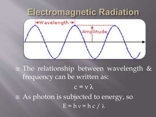
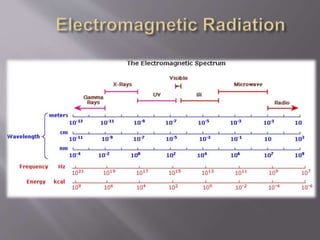
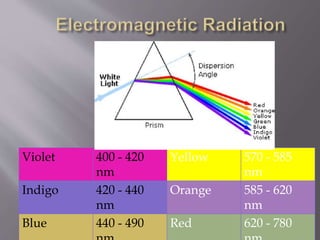
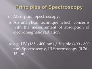
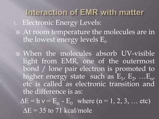
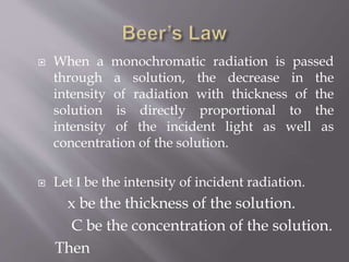
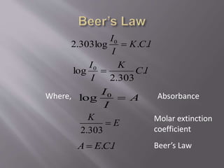
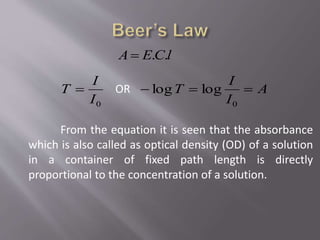
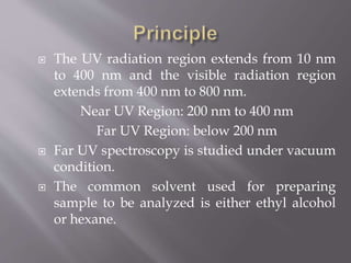
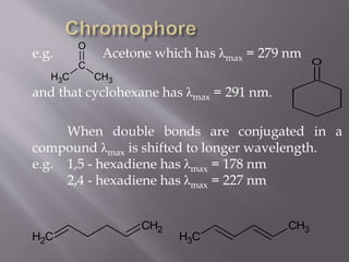
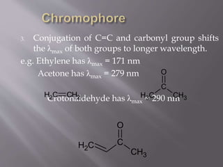
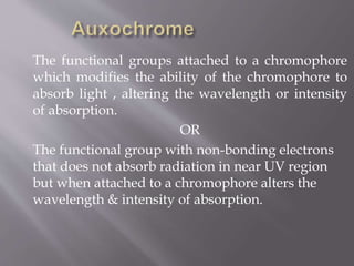
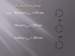
Presentation
ON
UV Spectroscopy
By Rokon
 It is the branch of science that deals with the
study of interaction of matter with light.
OR
 It is the branch of science that deals with the
study of interaction of electromagnetic
radiation with matter.
 Electromagnetic radiation consist of
discrete packages of energy which are
called as photons.
 A photon consists of an oscillating electric
field (E) & an oscillating magnetic field
(M) which are perpendicular to each
other.
 Frequency (ν):
 It is defined as the number of times electrical
field radiation oscillates in one second.
 The unit for frequency is Hertz (Hz).
1 Hz = 1 cycle per second
 Wavelength (λ):
 It is the distance between two nearest parts of
the wave in the same phase i.e. distance
between two nearest crest or troughs.
 The relationship between wavelength &
frequency can be written as:
c = ν λ
 As photon is subjected to energy, so
E = h ν = h c / λ
Violet 400 - 420
nm
Yellow 570 - 585
nm
Indigo 420 - 440
nm
Orange 585 - 620
nm
Blue 440 - 490 Red 620 - 780
 The principle is based on the
measurement of spectrum of a sample
containing atoms / molecules.
 Spectrum is a graph of intensity of
absorbed or emitted radiation by sample
verses frequency (ν) or wavelength (λ).
 Spectrometer is an instrument design to
measure the spectrum of a compound.
1. Absorption Spectroscopy:
 An analytical technique which concerns
with the measurement of absorption of
electromagnetic radiation.
 e.g. UV (185 - 400 nm) / Visible (400 - 800
nm) Spectroscopy, IR Spectroscopy (0.76 -
15 μm)
2. Emission Spectroscopy:
 An analytical technique in which
emission (of a particle or radiation) is
dispersed according to some property of
the emission & the amount of dispersion
is measured.
 e.g. Mass Spectroscopy
1. Electronic Energy Levels:
 At room temperature the molecules are in
the lowest energy levels E0.
 When the molecules absorb UV-visible
light from EMR, one of the outermost
bond / lone pair electron is promoted to
higher energy state such as E1, E2, …En,
etc is called as electronic transition and
the difference is as:
∆E = h ν = En - E0 where (n = 1, 2, 3, … etc)
∆E = 35 to 71 kcal/mole
2. Vibrational Energy Levels:
 These are less energy level than electronic
energy levels.
 The spacing between energy levels are
relatively small i.e. 0.01 to 10 kcal/mole.
 e.g. when IR radiation is absorbed,
molecules are excited from one
vibrational level to another or it vibrates
with higher amplitude.
3. Rotational Energy Levels:
 These energy levels are quantized &
discrete.
 The spacing between energy levels are
even smaller than vibrational energy
levels.
∆Erotational < ∆Evibrational < ∆Eelectronic
 When a monochromatic radiation is passed
through a solution, the decrease in the
intensity of radiation with thickness of the
solution is directly proportional to the
intensity of the incident light.
 Let I be the intensity of incident radiation.
x be the thickness of the solution.
Then
I
dx
dI

So, KI
dx
dI

Integrate equation between limit
I = Io at x = 0 and
I = I at x=l,
We get,
Kl
I
I

0
ln
Kl
I
I

0
log303.2
l
K
I
I
303.2
log
0

Where, AbsorbanceA
I
I
0
log
E
K

303.2
lEA .
Absorption coefficient
Lambert’s Law
 When a monochromatic radiation is passed
through a solution, the decrease in the
intensity of radiation with thickness of the
solution is directly proportional to the
intensity of the incident light as well as
concentration of the solution.
 Let I be the intensity of incident radiation.
x be the thickness of the solution.
C be the concentration of the solution.
Then
IC
dx
dI
.
So, ICK
dx
dI
.'
Integrate equation between limit
I = Io at x = 0 and
I = I at x=l,
We get,
lCK
I
I
.'ln
0

lCK
I
I
..log303.2 0

lC
K
I
I
.
303.2
log 0

Where, AbsorbanceA
I
I
0
log
E
K

303.2
lCEA ..
Molar extinction
coefficient
Beer’s Law
lCEA ..
0I
I
T  OR A
I
I
T 
0
loglog
From the equation it is seen that the absorbance
which is also called as optical density (OD) of a solution
in a container of fixed path length is directly
proportional to the concentration of a solution.
 The UV radiation region extends from 10 nm
to 400 nm and the visible radiation region
extends from 400 nm to 800 nm.
Near UV Region: 200 nm to 400 nm
Far UV Region: below 200 nm
 Far UV spectroscopy is studied under vacuum
condition.
 The common solvent used for preparing
sample to be analyzed is either ethyl alcohol
or hexane.
• σ → σ* transition1
• π → π* transition2
• n → σ* transition3
• n → π* transition4
• σ → π* transition5
• π → σ* transition6
• σ electron from orbital is excited to
corresponding anti-bonding orbital σ*.
• The energy required is large for this
transition.
• e.g. Methane (CH4) has C-H bond only
and can undergo σ → σ* transition and
shows absorbance maxima at 125 nm.
• σ → σ* transition1
• π electron in a bonding orbital is excited
to corresponding anti-bonding orbital π*.
• Compounds containing multiple bonds
like alkenes, alkynes, carbonyl, nitriles,
aromatic compounds, etc undergo π → π*
transitions.
• e.g. Alkenes generally absorb in the
• π → π* transition2
• Saturated compounds containing atoms
with lone pair of electrons like O, N, S
and halogens are capable of n → σ*
transition.
• These transitions usually requires less
energy than σ → σ* transitions.
• The number of organic functional groups
with n → σ* peaks in UV region is small
• n → σ* transition3
• An electron from non-bonding orbital is
promoted to anti-bonding π* orbital.
• Compounds containing double bond
involving hetero atoms (C=O, C≡N, N=O)
undergo such transitions.
• n → π* transitions require minimum
energy and show absorption at longer
wavelength around 300 nm.
• n → π* transition4
•These electronic transitions are forbidden
transitions & are only theoretically
possible.
•Thus, n → π* & π → π* electronic
transitions show absorption in region
above 200 nm which is accessible to UV-
visible spectrophotometer.
•The UV spectrum is of only a few broad of
• σ → π* transition5
• π → σ* transition 6&
The part of a molecule responsible for imparting
color, are called as chromospheres.
OR
The functional groups containing multiple bonds
capable of absorbing radiations above 200 nm due
to n → π* & π → π* transitions.
e.g. NO2, N=O, C=O, C=N, C≡N, C=C, C=S, etc
To interpretate UV – visible spectrum following
points should be noted:
1. Non-conjugated alkenes show an intense
absorption below 200 nm & are therefore
inaccessible to UV spectrophotometer.
2. Non-conjugated carbonyl group compound
give a weak absorption band in the 200 - 300 nm
region.
e.g. Acetone which has λmax = 279 nm
and that cyclohexane has λmax = 291 nm.
When double bonds are conjugated in a
compound λmax is shifted to longer wavelength.
e.g. 1,5 - hexadiene has λmax = 178 nm
2,4 - hexadiene has λmax = 227 nm
CH3
C
CH3
O
O
CH2
CH2
CH3
CH3
3. Conjugation of C=C and carbonyl group shifts
the λmax of both groups to longer wavelength.
e.g. Ethylene has λmax = 171 nm
Acetone has λmax = 279 nm
Crotonaldehyde has λmax = 290 nmCH3
C
CH3
O
CH2 CH2
C
CH3
O
CH2
The functional groups attached to a chromophore
which modifies the ability of the chromophore to
absorb light , altering the wavelength or intensity
of absorption.
OR
The functional group with non-bonding electrons
that does not absorb radiation in near UV region
but when attached to a chromophore alters the
wavelength & intensity of absorption.
e.g. Benzene λmax = 255 nm
Phenol λmax = 270 nm
Aniline λmax = 280 nm
OH
NH2
• When absorption maxima (λmax) of a
compound shifts to longer wavelength, it is
known as bathochromic shift or red shift.
• The effect is due to presence of an
auxochrome or by the change of solvent.
• e.g. An auxochrome group like –OH, -
OCH3 causes absorption of compound at
longer wavelength.
• Bathochromic Shift (Red Shift)1
• In alkaline medium, p-nitrophenol shows
red shift. Because negatively charged
oxygen delocalizes more effectively than
the unshared pair of electron.
p-nitrophenol
λmax = 255 nm λmax = 265 nm
• Bathochromic Shift (Red Shift)1
OH
N
+ O
-
O
OH
-
Alkaline
medium
O
-
N
+ O
-
O
• When absorption maxima (λmax) of a
compound shifts to shorter wavelength, it
is known as hypsochromic shift or blue
shift.
• The effect is due to presence of an group
causes removal of conjugation or by the
change of solvent.
• Hypsochromic Shift (Blue Shift)2
• Aniline shows blue shift in acidic medium,
it loses conjugation.
Aniline
λmax = 280 nm λmax = 265 nm
• Hypsochromic Shift (Blue Shift)2
NH2
H
+
Acidic
medium
NH3
+
Cl
-
• When absorption intensity (ε) of a
compound is increased, it is known as
hyperchromic shift.
• If auxochrome introduces to the
compound, the intensity of absorption
increases.
Pyridine 2-methyl
• Hyperchromic Effect3
N N CH3
• When absorption intensity (ε) of a
compound is decreased, it is known as
hypochromic shift.
Naphthalene 2-methyl
naphthalene
ε = 19000 ε = 10250
CH3
• Hypochromic Effect4
Wavelength ( λ )
Absorbance(A)
Hyperchromic shift
Hypochromic shift
Red
shift
Blue
shift
λmax
 Qualitative & Quantitative Analysis:
 It is used for characterizing aromatic compounds
and conjugated olefins.
 It can be used to find out molar concentration of
the solute under study.
 Detection of impurities:
 It is one of the important method to detect
impurities in organic solvents.
 Detection of isomers are possible.
 Determination of molecular weight using
Beer’s law.
 Introduction to Spectroscopy
 Donald A. Pavia
 Elementary Organic Spectroscopy
 Y. R. Sharma
 Physical Chemistry
 Puri, Sharma & Pathaniya
 http://www2.chemistry.msu.edu/faculty/re
usch/VirtTxtJml/Spectrpy/UV-
Vis/spectrum.htm
 http://en.wikipedia.org/wiki/Ultraviolet%E
2%80%93visible_spectroscopy
 http://teaching.shu.ac.uk/hwb/chemistry/tu
torials/molspec/uvvisab1.htm
Md Rokunuzzman
Batch : 12th
Roll :328

Uv spectroscopy

  • 1.
  • 2.
     It isthe branch of science that deals with the study of interaction of matter with light. OR  It is the branch of science that deals with the study of interaction of electromagnetic radiation with matter.
  • 3.
     Electromagnetic radiationconsist of discrete packages of energy which are called as photons.  A photon consists of an oscillating electric field (E) & an oscillating magnetic field (M) which are perpendicular to each other.
  • 5.
     Frequency (ν): It is defined as the number of times electrical field radiation oscillates in one second.  The unit for frequency is Hertz (Hz). 1 Hz = 1 cycle per second  Wavelength (λ):  It is the distance between two nearest parts of the wave in the same phase i.e. distance between two nearest crest or troughs.
  • 6.
     The relationshipbetween wavelength & frequency can be written as: c = ν λ  As photon is subjected to energy, so E = h ν = h c / λ
  • 8.
    Violet 400 -420 nm Yellow 570 - 585 nm Indigo 420 - 440 nm Orange 585 - 620 nm Blue 440 - 490 Red 620 - 780
  • 9.
     The principleis based on the measurement of spectrum of a sample containing atoms / molecules.  Spectrum is a graph of intensity of absorbed or emitted radiation by sample verses frequency (ν) or wavelength (λ).  Spectrometer is an instrument design to measure the spectrum of a compound.
  • 10.
    1. Absorption Spectroscopy: An analytical technique which concerns with the measurement of absorption of electromagnetic radiation.  e.g. UV (185 - 400 nm) / Visible (400 - 800 nm) Spectroscopy, IR Spectroscopy (0.76 - 15 μm)
  • 11.
    2. Emission Spectroscopy: An analytical technique in which emission (of a particle or radiation) is dispersed according to some property of the emission & the amount of dispersion is measured.  e.g. Mass Spectroscopy
  • 12.
    1. Electronic EnergyLevels:  At room temperature the molecules are in the lowest energy levels E0.  When the molecules absorb UV-visible light from EMR, one of the outermost bond / lone pair electron is promoted to higher energy state such as E1, E2, …En, etc is called as electronic transition and the difference is as: ∆E = h ν = En - E0 where (n = 1, 2, 3, … etc) ∆E = 35 to 71 kcal/mole
  • 13.
    2. Vibrational EnergyLevels:  These are less energy level than electronic energy levels.  The spacing between energy levels are relatively small i.e. 0.01 to 10 kcal/mole.  e.g. when IR radiation is absorbed, molecules are excited from one vibrational level to another or it vibrates with higher amplitude.
  • 14.
    3. Rotational EnergyLevels:  These energy levels are quantized & discrete.  The spacing between energy levels are even smaller than vibrational energy levels. ∆Erotational < ∆Evibrational < ∆Eelectronic
  • 15.
     When amonochromatic radiation is passed through a solution, the decrease in the intensity of radiation with thickness of the solution is directly proportional to the intensity of the incident light.  Let I be the intensity of incident radiation. x be the thickness of the solution. Then
  • 16.
    I dx dI  So, KI dx dI  Integrate equationbetween limit I = Io at x = 0 and I = I at x=l, We get, Kl I I  0 ln
  • 17.
  • 18.
     When amonochromatic radiation is passed through a solution, the decrease in the intensity of radiation with thickness of the solution is directly proportional to the intensity of the incident light as well as concentration of the solution.  Let I be the intensity of incident radiation. x be the thickness of the solution. C be the concentration of the solution. Then
  • 19.
    IC dx dI . So, ICK dx dI .' Integrate equationbetween limit I = Io at x = 0 and I = I at x=l, We get, lCK I I .'ln 0 
  • 20.
    lCK I I ..log303.2 0  lC K I I . 303.2 log 0  Where,AbsorbanceA I I 0 log E K  303.2 lCEA .. Molar extinction coefficient Beer’s Law
  • 21.
    lCEA .. 0I I T OR A I I T  0 loglog From the equation it is seen that the absorbance which is also called as optical density (OD) of a solution in a container of fixed path length is directly proportional to the concentration of a solution.
  • 22.
     The UVradiation region extends from 10 nm to 400 nm and the visible radiation region extends from 400 nm to 800 nm. Near UV Region: 200 nm to 400 nm Far UV Region: below 200 nm  Far UV spectroscopy is studied under vacuum condition.  The common solvent used for preparing sample to be analyzed is either ethyl alcohol or hexane.
  • 24.
    • σ →σ* transition1 • π → π* transition2 • n → σ* transition3 • n → π* transition4 • σ → π* transition5 • π → σ* transition6
  • 25.
    • σ electronfrom orbital is excited to corresponding anti-bonding orbital σ*. • The energy required is large for this transition. • e.g. Methane (CH4) has C-H bond only and can undergo σ → σ* transition and shows absorbance maxima at 125 nm. • σ → σ* transition1
  • 26.
    • π electronin a bonding orbital is excited to corresponding anti-bonding orbital π*. • Compounds containing multiple bonds like alkenes, alkynes, carbonyl, nitriles, aromatic compounds, etc undergo π → π* transitions. • e.g. Alkenes generally absorb in the • π → π* transition2
  • 27.
    • Saturated compoundscontaining atoms with lone pair of electrons like O, N, S and halogens are capable of n → σ* transition. • These transitions usually requires less energy than σ → σ* transitions. • The number of organic functional groups with n → σ* peaks in UV region is small • n → σ* transition3
  • 28.
    • An electronfrom non-bonding orbital is promoted to anti-bonding π* orbital. • Compounds containing double bond involving hetero atoms (C=O, C≡N, N=O) undergo such transitions. • n → π* transitions require minimum energy and show absorption at longer wavelength around 300 nm. • n → π* transition4
  • 29.
    •These electronic transitionsare forbidden transitions & are only theoretically possible. •Thus, n → π* & π → π* electronic transitions show absorption in region above 200 nm which is accessible to UV- visible spectrophotometer. •The UV spectrum is of only a few broad of • σ → π* transition5 • π → σ* transition 6&
  • 30.
    The part ofa molecule responsible for imparting color, are called as chromospheres. OR The functional groups containing multiple bonds capable of absorbing radiations above 200 nm due to n → π* & π → π* transitions. e.g. NO2, N=O, C=O, C=N, C≡N, C=C, C=S, etc
  • 31.
    To interpretate UV– visible spectrum following points should be noted: 1. Non-conjugated alkenes show an intense absorption below 200 nm & are therefore inaccessible to UV spectrophotometer. 2. Non-conjugated carbonyl group compound give a weak absorption band in the 200 - 300 nm region.
  • 32.
    e.g. Acetone whichhas λmax = 279 nm and that cyclohexane has λmax = 291 nm. When double bonds are conjugated in a compound λmax is shifted to longer wavelength. e.g. 1,5 - hexadiene has λmax = 178 nm 2,4 - hexadiene has λmax = 227 nm CH3 C CH3 O O CH2 CH2 CH3 CH3
  • 33.
    3. Conjugation ofC=C and carbonyl group shifts the λmax of both groups to longer wavelength. e.g. Ethylene has λmax = 171 nm Acetone has λmax = 279 nm Crotonaldehyde has λmax = 290 nmCH3 C CH3 O CH2 CH2 C CH3 O CH2
  • 34.
    The functional groupsattached to a chromophore which modifies the ability of the chromophore to absorb light , altering the wavelength or intensity of absorption. OR The functional group with non-bonding electrons that does not absorb radiation in near UV region but when attached to a chromophore alters the wavelength & intensity of absorption.
  • 35.
    e.g. Benzene λmax= 255 nm Phenol λmax = 270 nm Aniline λmax = 280 nm OH NH2
  • 37.
    • When absorptionmaxima (λmax) of a compound shifts to longer wavelength, it is known as bathochromic shift or red shift. • The effect is due to presence of an auxochrome or by the change of solvent. • e.g. An auxochrome group like –OH, - OCH3 causes absorption of compound at longer wavelength. • Bathochromic Shift (Red Shift)1
  • 38.
    • In alkalinemedium, p-nitrophenol shows red shift. Because negatively charged oxygen delocalizes more effectively than the unshared pair of electron. p-nitrophenol λmax = 255 nm λmax = 265 nm • Bathochromic Shift (Red Shift)1 OH N + O - O OH - Alkaline medium O - N + O - O
  • 39.
    • When absorptionmaxima (λmax) of a compound shifts to shorter wavelength, it is known as hypsochromic shift or blue shift. • The effect is due to presence of an group causes removal of conjugation or by the change of solvent. • Hypsochromic Shift (Blue Shift)2
  • 40.
    • Aniline showsblue shift in acidic medium, it loses conjugation. Aniline λmax = 280 nm λmax = 265 nm • Hypsochromic Shift (Blue Shift)2 NH2 H + Acidic medium NH3 + Cl -
  • 41.
    • When absorptionintensity (ε) of a compound is increased, it is known as hyperchromic shift. • If auxochrome introduces to the compound, the intensity of absorption increases. Pyridine 2-methyl • Hyperchromic Effect3 N N CH3
  • 42.
    • When absorptionintensity (ε) of a compound is decreased, it is known as hypochromic shift. Naphthalene 2-methyl naphthalene ε = 19000 ε = 10250 CH3 • Hypochromic Effect4
  • 43.
    Wavelength ( λ) Absorbance(A) Hyperchromic shift Hypochromic shift Red shift Blue shift λmax
  • 44.
     Qualitative &Quantitative Analysis:  It is used for characterizing aromatic compounds and conjugated olefins.  It can be used to find out molar concentration of the solute under study.  Detection of impurities:  It is one of the important method to detect impurities in organic solvents.  Detection of isomers are possible.  Determination of molecular weight using Beer’s law.
  • 45.
     Introduction toSpectroscopy  Donald A. Pavia  Elementary Organic Spectroscopy  Y. R. Sharma  Physical Chemistry  Puri, Sharma & Pathaniya
  • 46.
  • 47.
    Md Rokunuzzman Batch :12th Roll :328