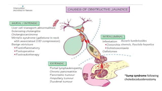
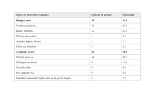
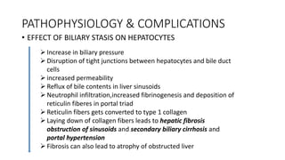
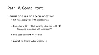
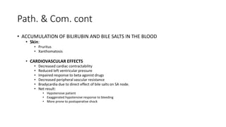
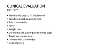
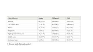
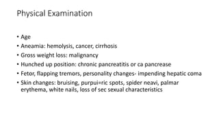
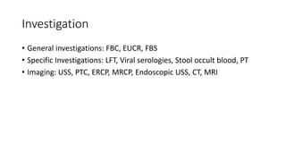
SURGICAL JAUNDICE
A SYNOPSIS
by
Dr. C. O. Alumona
OUTLINE
• DEFINITION OF TERMS
• EPIDEMIOLOGY
• THE HEPATO-BILIARY SYSTEM (Normal Anatomy & Physiology)
• CAUSES
• PATHOPHYSIOLOGY & COMPLICATIONS
• CLINICAL EVALUATION
• CLASSIFICATION & MANAGEMENT
DEFINITION OF TERMS
• Bile: exocrine secretion of the liver produced by hepatocytes (chol. Bili,
salts)
• Bilirubin: 0.2-1.2mg/dl (3.4-6.8umol/L)conj. Vs unconj., delta-Bilirubin
• JAUNDICE: The clinical syndrome associated with hyper-bilirubinaemia.
Yellowish pigmentation of elastin containing tissues {sclera & mucous
membrane (@3mg/dl), skin (@6mg/dl)}
• Surgical jaundice:. Failure of normal amount of bile to reach intestine due
to mechanical obstruction of the extra hepatic biliary tree or within the
porta hepatis. Any jaundice that is amenable to surgical treatment.
OBSTRUCTIVE JAUNDICE VS SURGICAL JAUNDICE
• Some forms of obstructive jaundice are not amenable to surgery
• Intrahepatic cholestasis occuring at levels of hepatocytes or canalicular membrane eg
hepatocellular ds (hepatitis- viral or drug induced), drug induced cholestasis, biliary
cirrhosis and alcoholic liver ds
• Conjugated hyperbilirubineamia is not synonymous with surgical jaundice
• In hepatocellular ds, interference in the three levels of bilirubin metabolism ie uptake,
conjugation and excretion occurs
• Certain causes of Jaundice are not due to cholestasis but are amenable to
surgery eg HS
OUR FOCUS, THEREFORE, IS ON EXTRA HEPATIC CAUSES OF CHOLESTASIS
EPIDEMIOLOGY
• Incidence: ~ 5 cases per 1000 people
• Race:
• Gall stones: Native Americans, Hispanics and Europeans > Africans and Asians
• Malignant causes: Africa, East Asia, Northern India, South America
• Sex
• Gall Stones: females > males
ANATOMY & PHYSIOLOGY OF THE BILIARY SYSTEM
• ANATOMY PIC: X? BROWSE
• PHYSIO PATHWAY: X? BROWSE
• Normal bile secretion: 500-1000ml/day
• Normal secretory pressure of bile is 15-25 cm of water
• CCK
50%
50%
Anat & Physio therapy
Biliverdin reductase
Glucoronyl transferase
4mg/day
CAUSES AND CLASSIFICATIONS
*Sump syndrome following
choledocoduodenostomy
PATHOPHYSIOLOGY & COMPLICATIONS
• EFFECT OF BILIARY STASIS ON HEPATOCYTES
Increase in biliary pressure
Disruption of tight junctions between hepatocytes and bile duct
cells
increased permeability
Reflux of bile contents in liver sinusoids
Neutrophil infiltration,increased fibrinogenesis and deposition of
reticulin fiberes in portal triad
Reticulin fibers gets converted to type 1 collagen
Laying down of collagen fibers leads to hepatic fibrosis
obstruction of sinusoids and secondary biliary cirrhosis and
portal hypertension
Fibrosis can also lead to atrophy of obstructed liver
Path. & Comp. cont
• FAILURE OF BILE TO REACH INTESTINE
• Fat malabsorption with steatorrhea
• Poor absorption of fat soluble vitamins (A,D,E,K)
• Disordered hemostasis with prolonged PT
• Pale Stool: absent stercobilin
• Absent or decreased urobilinogen
Path. & Com. cont
• ACCUMULATION OF BILIRUBIN AND BILE SALTS IN THE BLOOD
• Skin:
• Pruritus
• Xanthomatosis
• CARDIOVASCULAR EFFECTS
• Decreased cardiac contractability
• Reduced left ventricular pressure
• Impaired response to beta agonist drugs
• Decreased peripheral vascular resistance
• Bradycardia due to direct effect of bile salts on SA node.
• Net result:
• Hypotensive patient
• Exaggerated hypotensive response to bleeding
• More prone to postoperative shock
Path. & Com. cont
• HEPATO-RENAL SYNDROME (HRS): worsening of serum creatinine to levels above 1.5 mg/dL
within 2 weeks for type 1 HRS and over several months for type 2 HRS
• 10 % incidence with 70 % mortality.
• Factors responsible include;
• Decresed cardiac function
• Increased levels of ANP resulting in hypovolemia
• Obstructive role of bilirubin mainly in distal tubule and pro inflammatory effects of bile salts
• Resulting in;
• Renal vasoconstriction
• Shunting of blood from cortex
• Activation of complement system with peritubular and glomerular fibrin deposition leading to
tubular and cortical necrosis
• Defects in cellular immunity
• Impaired T cell proliferation
• Decreased neutophil chemotaxis
• Defective bacterial phagocytosis
• Depressed function of RE system ie Kupffer cells
• WOUND HEALING
• Delayed wound healing
• High incidence of wound dehiscence
• Decresed activity of enzyme Propyl hydroxylase in the skin that helps in
incorporation of proline in collagen
• Defective synthesis of collagen
• COAGULATION FACTOR DEFECTS
• Prolongation of Prothrombin time
• Loss of calcium
• Endotoxin induced damage to factor XI ,XII ,platelets
• Low grade DIC with increased fibrin degradation products
• Thrombocytopenia from hyperspleenism
• Decreased absroption of fat solube vitamins A,D,E,K
WORK UP AND MANAGEMENT OF
POSTHEPATIC JAUNDICE
CLINICAL EVALUATION
HISTORY
• Previous dyspepsia, fat intolerance
• Jaundice: onset, course, itching
• Pain: characterize
• Fever
• Weight loss
• Dark urine and clay or pale coloured stool
• Travel to endemic areas
• Contact with jaundiced pt
• Drug intake eg
• Charcot triad, Raynaud pentad
Physical Examination
• Age
• Aneamia: hemolysis, cancer, cirrhosis
• Gross weight loss: malignancy
• Hunched up position: chronic pancreatitis or ca pancrease
• Fetor, flapping tremors, personality changes- impending hepatic coma
• Skin changes: bruising, purpui=ric spots, spider neavi, palmar
erythema, white nails, loss of sec sexual characteristics
Investigation
• General investigations: FBC, EUCR, FBS
• Specific Investigations: LFT, Viral serologies, Stool occult blood, PT
• Imaging: USS, PTC, ERCP, MRCP, Endoscopic USS, CT, MRI
LFT in Jaundice
TEST SOURCE PATTERN IN GALL
STONE
OBSTRUCTION
PATTERN IN
MALIGNANT
OBSTRUCTION
PATTERN IN ACUTE
HEPATITIS
Total Bilirubin Heamolysis,
Hepatocytes
processing
Elevated but
typicaly <10mg/dl
Elevated commonly
>10mg/dl
Elevated
Direct bilirubin Hepatocyte
conjugation
Elevated Markedly Elevated Mildly Elevated
Indirect bilirubin RBC turnover,
hepatocytes
processing
Minimally Elevated Elevated Elevated
ALP Biliary epithelium,
bone
Elevated Markedly Elevated Minimally Elevated
ALT,AST Hepatocytes Minimally Elevated Minimally Elevated Markedly Elevated
GGT Biliary epithelium Elevated Markedly Elevated Minimally Elevated
LFT PATTERN IN SURGICAL VS MEDICAL JAUNDICE
ALP
• Elevated in nearly 100% of cases of extra hepatic obstruction except
in cases of intermittent obstruction
• Values usually greater than 3x the upper limit, may exceed 5x
• Values less than 3x the upper limit is evidence against complete extra
hepatic obstruction
Transaminases: AST and ALT
• They are serum enzymes that provides evidence of hepato cellular
damage
• ALT primarily found in the liver while AST is also found in the heart,
kidney and skeletal muscle
• AST is not usualy raised in extra hepatic obstruction
GGT and 5’Nucleotidase
• Correlates with ALP levels
• Most sensitive indicator of biliary tract disease
• Levels are independent of age hence it a better indicator of
obstruction in children
• Helpful in diagnosis of acute biliary obstruction as ALP lags behind the
onset of obstruction
• 5’Nucleotidase: The principal value is to confirm the hepatic origin of
an elevated ALP
IMAGING
• USS: >98% specific, > 95% sensitive.
• Detects calculi, features of cholecystitis,
• Assess biliary tract: intrahepatic dilatations (>4mm), extrahepatic dilatations
(>10mm)
• Used intra op to asses intrahepatic lesions, assess resectability and determine
involvement of vascular structures
Patterns of Bile duct Instruction
• Complete our Progressive: seen in CA head of Pancreas
,cholangiocarcinoma, etc, impacted stone in CBD, cbd ligation.
• Intermittent Obstruction : produces fluctuating jaundice ; gall stones
,periampulary tumours, intrabiliary parasites
• Chronic incomplete obstruction : strictures of CBG; congenital,
iatrogenic , sclerosing cholangitis, post radiotherapy
• Segmental obstruction : one or more isolated segment of the intra
hepatic biliary tree ate obstructed. Iatrogenic , hepathodochlithiasis ,
Sclerosing cholangitis , cholangiocarcinoma
DUCTAL OBSTRUCTION
SUSPECTED CHOLANGITIS SUSPECTED CHOLEDOCHOLITHIASIS
WITHOUT CHOLANGITIS
OTHER LESIONS OTHER THAN
CHOLEDOCHOLITHIASIS
• Charcot triad, Renaud
pentad
• Adequate resuscitation
• Appropriate antibiotics
• ERCP for diagnosis and
trx
• Transhepatic drainage
or surgery if ERCP is
not available
• Mainstay of trx:
antibiotics plus
establishment of
adequate biliary
drainage
• Strongly suspected in: episodic
jaundice, pain, USS finding of gall
or CBD stone
• Cholecystectomy with
preoperative ERCP or intra op
cholangiography
• No gallstones are seen
• Less acute clinical
presentations eg
constant abd or back
pain
• Associated conditional
sym. Eg weight loss,
fatigue, long standing
anorexia
• Possible causes:
classified based on level
of obstruction: Upper,
middle & distal third
OTHER LESIONS OTHER THAN CHOLEDOCHOLITHIASIS
• Palliation: Left
Hepaticojejunostomy
• Resection for cure
• Palliation: Roux-en-Y
choledochojejunostomy
• Resection for cure
• Palliation:
Hepaticojejunostomy
(distal to hepatic duct
convergence)
• Resection for cure
POST OPERATIVE COMPLICATIONS
REFRRENCES
• Phillipo L Chalya etal, Emmanuel S Kanumba, Mabula Mchembe: Etiological spectrum and treatment outcome of
Obstructive jaundice at a University teaching Hospital in northwestern Tanzania: A diagnostic and therapeutic challenges.
BioMed Central Ltd. 23 May 2011
• Di Bisceglie AM, Oettle GJ, Hodkinson HJ, Segal I: Obstructive jaundice in the South African black population.
(https://www.ncbi.nlm.nih.gov/pubmed/3782751)
• Thiago Gomes Romano, Jose mauro vieira junior: Do Biliary Salts Have Role on Acute Kidney Injury Development? Journal
of clinical Medicine Research 2015 Sep; 7(9): 667–671. (https://www.ncbi.nlm.nih.gov/pmc/articles/PMC4522982/#)
THANK YOU
QUESTIONS & CLOSING REMARKS

Surgical Jaundice: A synopsis

  • 1.
  • 2.
    OUTLINE • DEFINITION OFTERMS • EPIDEMIOLOGY • THE HEPATO-BILIARY SYSTEM (Normal Anatomy & Physiology) • CAUSES • PATHOPHYSIOLOGY & COMPLICATIONS • CLINICAL EVALUATION • CLASSIFICATION & MANAGEMENT
  • 3.
    DEFINITION OF TERMS •Bile: exocrine secretion of the liver produced by hepatocytes (chol. Bili, salts) • Bilirubin: 0.2-1.2mg/dl (3.4-6.8umol/L)conj. Vs unconj., delta-Bilirubin • JAUNDICE: The clinical syndrome associated with hyper-bilirubinaemia. Yellowish pigmentation of elastin containing tissues {sclera & mucous membrane (@3mg/dl), skin (@6mg/dl)} • Surgical jaundice:. Failure of normal amount of bile to reach intestine due to mechanical obstruction of the extra hepatic biliary tree or within the porta hepatis. Any jaundice that is amenable to surgical treatment.
  • 4.
    OBSTRUCTIVE JAUNDICE VSSURGICAL JAUNDICE • Some forms of obstructive jaundice are not amenable to surgery • Intrahepatic cholestasis occuring at levels of hepatocytes or canalicular membrane eg hepatocellular ds (hepatitis- viral or drug induced), drug induced cholestasis, biliary cirrhosis and alcoholic liver ds • Conjugated hyperbilirubineamia is not synonymous with surgical jaundice • In hepatocellular ds, interference in the three levels of bilirubin metabolism ie uptake, conjugation and excretion occurs • Certain causes of Jaundice are not due to cholestasis but are amenable to surgery eg HS OUR FOCUS, THEREFORE, IS ON EXTRA HEPATIC CAUSES OF CHOLESTASIS
  • 5.
    EPIDEMIOLOGY • Incidence: ~5 cases per 1000 people • Race: • Gall stones: Native Americans, Hispanics and Europeans > Africans and Asians • Malignant causes: Africa, East Asia, Northern India, South America • Sex • Gall Stones: females > males
  • 6.
    ANATOMY & PHYSIOLOGYOF THE BILIARY SYSTEM • ANATOMY PIC: X? BROWSE • PHYSIO PATHWAY: X? BROWSE • Normal bile secretion: 500-1000ml/day • Normal secretory pressure of bile is 15-25 cm of water • CCK 50% 50%
  • 7.
  • 8.
  • 9.
    CAUSES AND CLASSIFICATIONS *Sumpsyndrome following choledocoduodenostomy
  • 11.
    PATHOPHYSIOLOGY & COMPLICATIONS •EFFECT OF BILIARY STASIS ON HEPATOCYTES Increase in biliary pressure Disruption of tight junctions between hepatocytes and bile duct cells increased permeability Reflux of bile contents in liver sinusoids Neutrophil infiltration,increased fibrinogenesis and deposition of reticulin fiberes in portal triad Reticulin fibers gets converted to type 1 collagen Laying down of collagen fibers leads to hepatic fibrosis obstruction of sinusoids and secondary biliary cirrhosis and portal hypertension Fibrosis can also lead to atrophy of obstructed liver
  • 12.
    Path. & Comp.cont • FAILURE OF BILE TO REACH INTESTINE • Fat malabsorption with steatorrhea • Poor absorption of fat soluble vitamins (A,D,E,K) • Disordered hemostasis with prolonged PT • Pale Stool: absent stercobilin • Absent or decreased urobilinogen
  • 13.
    Path. & Com.cont • ACCUMULATION OF BILIRUBIN AND BILE SALTS IN THE BLOOD • Skin: • Pruritus • Xanthomatosis • CARDIOVASCULAR EFFECTS • Decreased cardiac contractability • Reduced left ventricular pressure • Impaired response to beta agonist drugs • Decreased peripheral vascular resistance • Bradycardia due to direct effect of bile salts on SA node. • Net result: • Hypotensive patient • Exaggerated hypotensive response to bleeding • More prone to postoperative shock
  • 14.
    Path. & Com.cont • HEPATO-RENAL SYNDROME (HRS): worsening of serum creatinine to levels above 1.5 mg/dL within 2 weeks for type 1 HRS and over several months for type 2 HRS • 10 % incidence with 70 % mortality. • Factors responsible include; • Decresed cardiac function • Increased levels of ANP resulting in hypovolemia • Obstructive role of bilirubin mainly in distal tubule and pro inflammatory effects of bile salts • Resulting in; • Renal vasoconstriction • Shunting of blood from cortex • Activation of complement system with peritubular and glomerular fibrin deposition leading to tubular and cortical necrosis
  • 15.
    • Defects incellular immunity • Impaired T cell proliferation • Decreased neutophil chemotaxis • Defective bacterial phagocytosis • Depressed function of RE system ie Kupffer cells • WOUND HEALING • Delayed wound healing • High incidence of wound dehiscence • Decresed activity of enzyme Propyl hydroxylase in the skin that helps in incorporation of proline in collagen • Defective synthesis of collagen
  • 16.
    • COAGULATION FACTORDEFECTS • Prolongation of Prothrombin time • Loss of calcium • Endotoxin induced damage to factor XI ,XII ,platelets • Low grade DIC with increased fibrin degradation products • Thrombocytopenia from hyperspleenism • Decreased absroption of fat solube vitamins A,D,E,K
  • 17.
    WORK UP ANDMANAGEMENT OF POSTHEPATIC JAUNDICE
  • 18.
    CLINICAL EVALUATION HISTORY • Previousdyspepsia, fat intolerance • Jaundice: onset, course, itching • Pain: characterize • Fever • Weight loss • Dark urine and clay or pale coloured stool • Travel to endemic areas • Contact with jaundiced pt • Drug intake eg
  • 19.
    • Charcot triad,Raynaud pentad
  • 20.
    Physical Examination • Age •Aneamia: hemolysis, cancer, cirrhosis • Gross weight loss: malignancy • Hunched up position: chronic pancreatitis or ca pancrease • Fetor, flapping tremors, personality changes- impending hepatic coma • Skin changes: bruising, purpui=ric spots, spider neavi, palmar erythema, white nails, loss of sec sexual characteristics
  • 21.
    Investigation • General investigations:FBC, EUCR, FBS • Specific Investigations: LFT, Viral serologies, Stool occult blood, PT • Imaging: USS, PTC, ERCP, MRCP, Endoscopic USS, CT, MRI
  • 22.
    LFT in Jaundice TESTSOURCE PATTERN IN GALL STONE OBSTRUCTION PATTERN IN MALIGNANT OBSTRUCTION PATTERN IN ACUTE HEPATITIS Total Bilirubin Heamolysis, Hepatocytes processing Elevated but typicaly <10mg/dl Elevated commonly >10mg/dl Elevated Direct bilirubin Hepatocyte conjugation Elevated Markedly Elevated Mildly Elevated Indirect bilirubin RBC turnover, hepatocytes processing Minimally Elevated Elevated Elevated ALP Biliary epithelium, bone Elevated Markedly Elevated Minimally Elevated ALT,AST Hepatocytes Minimally Elevated Minimally Elevated Markedly Elevated GGT Biliary epithelium Elevated Markedly Elevated Minimally Elevated
  • 23.
    LFT PATTERN INSURGICAL VS MEDICAL JAUNDICE
  • 24.
    ALP • Elevated innearly 100% of cases of extra hepatic obstruction except in cases of intermittent obstruction • Values usually greater than 3x the upper limit, may exceed 5x • Values less than 3x the upper limit is evidence against complete extra hepatic obstruction
  • 25.
    Transaminases: AST andALT • They are serum enzymes that provides evidence of hepato cellular damage • ALT primarily found in the liver while AST is also found in the heart, kidney and skeletal muscle • AST is not usualy raised in extra hepatic obstruction
  • 26.
    GGT and 5’Nucleotidase •Correlates with ALP levels • Most sensitive indicator of biliary tract disease • Levels are independent of age hence it a better indicator of obstruction in children • Helpful in diagnosis of acute biliary obstruction as ALP lags behind the onset of obstruction • 5’Nucleotidase: The principal value is to confirm the hepatic origin of an elevated ALP
  • 27.
    IMAGING • USS: >98%specific, > 95% sensitive. • Detects calculi, features of cholecystitis, • Assess biliary tract: intrahepatic dilatations (>4mm), extrahepatic dilatations (>10mm) • Used intra op to asses intrahepatic lesions, assess resectability and determine involvement of vascular structures
  • 29.
    Patterns of Bileduct Instruction • Complete our Progressive: seen in CA head of Pancreas ,cholangiocarcinoma, etc, impacted stone in CBD, cbd ligation. • Intermittent Obstruction : produces fluctuating jaundice ; gall stones ,periampulary tumours, intrabiliary parasites • Chronic incomplete obstruction : strictures of CBG; congenital, iatrogenic , sclerosing cholangitis, post radiotherapy • Segmental obstruction : one or more isolated segment of the intra hepatic biliary tree ate obstructed. Iatrogenic , hepathodochlithiasis , Sclerosing cholangitis , cholangiocarcinoma
  • 30.
    DUCTAL OBSTRUCTION SUSPECTED CHOLANGITISSUSPECTED CHOLEDOCHOLITHIASIS WITHOUT CHOLANGITIS OTHER LESIONS OTHER THAN CHOLEDOCHOLITHIASIS • Charcot triad, Renaud pentad • Adequate resuscitation • Appropriate antibiotics • ERCP for diagnosis and trx • Transhepatic drainage or surgery if ERCP is not available • Mainstay of trx: antibiotics plus establishment of adequate biliary drainage • Strongly suspected in: episodic jaundice, pain, USS finding of gall or CBD stone • Cholecystectomy with preoperative ERCP or intra op cholangiography • No gallstones are seen • Less acute clinical presentations eg constant abd or back pain • Associated conditional sym. Eg weight loss, fatigue, long standing anorexia • Possible causes: classified based on level of obstruction: Upper, middle & distal third
  • 31.
    OTHER LESIONS OTHERTHAN CHOLEDOCHOLITHIASIS • Palliation: Left Hepaticojejunostomy • Resection for cure • Palliation: Roux-en-Y choledochojejunostomy • Resection for cure • Palliation: Hepaticojejunostomy (distal to hepatic duct convergence) • Resection for cure
  • 32.
  • 33.
    REFRRENCES • Phillipo LChalya etal, Emmanuel S Kanumba, Mabula Mchembe: Etiological spectrum and treatment outcome of Obstructive jaundice at a University teaching Hospital in northwestern Tanzania: A diagnostic and therapeutic challenges. BioMed Central Ltd. 23 May 2011 • Di Bisceglie AM, Oettle GJ, Hodkinson HJ, Segal I: Obstructive jaundice in the South African black population. (https://www.ncbi.nlm.nih.gov/pubmed/3782751) • Thiago Gomes Romano, Jose mauro vieira junior: Do Biliary Salts Have Role on Acute Kidney Injury Development? Journal of clinical Medicine Research 2015 Sep; 7(9): 667–671. (https://www.ncbi.nlm.nih.gov/pmc/articles/PMC4522982/#)
  • 34.
  • 35.