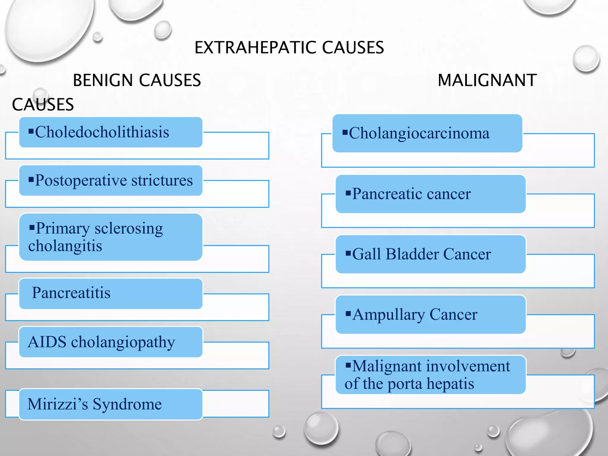
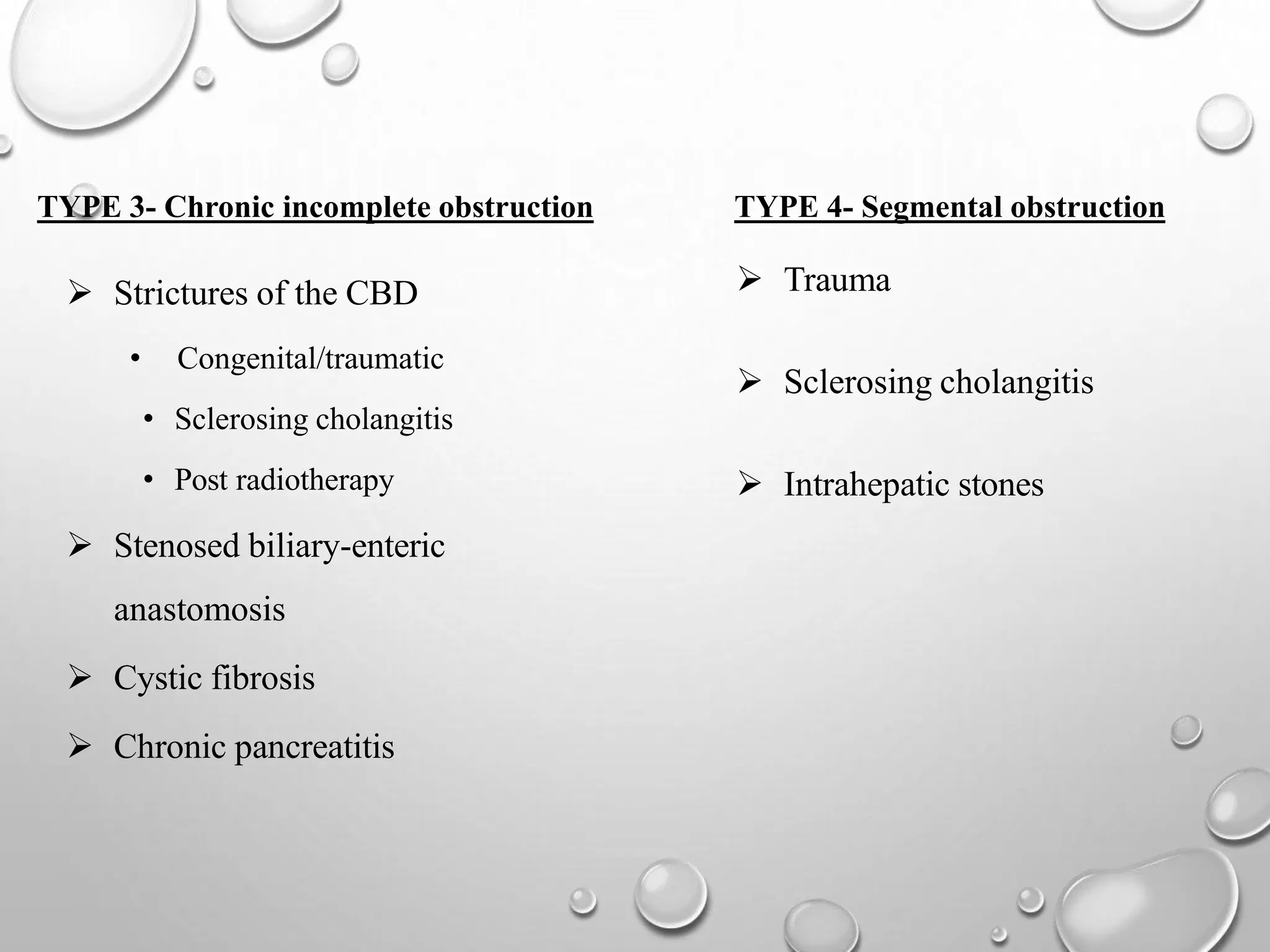
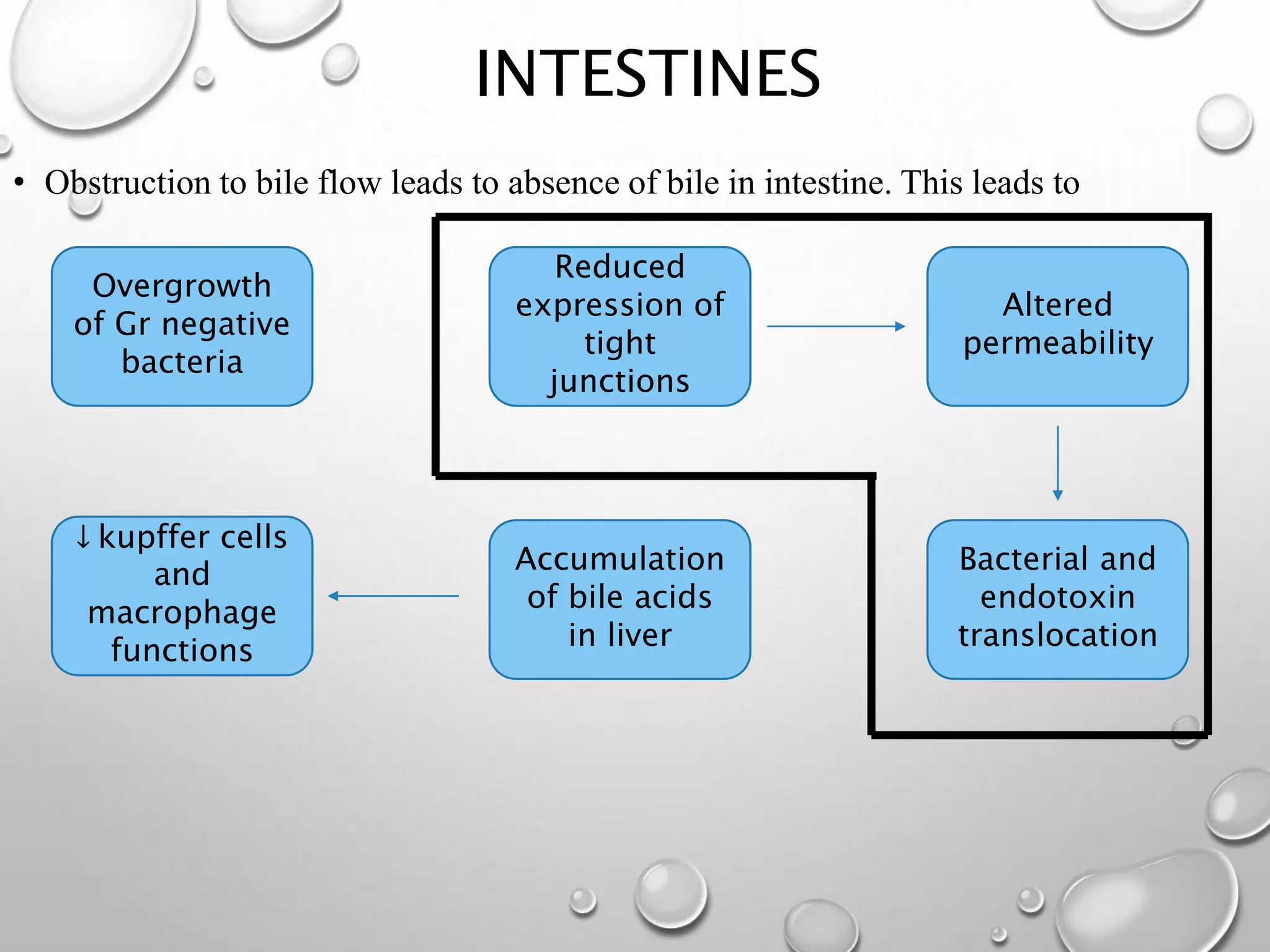
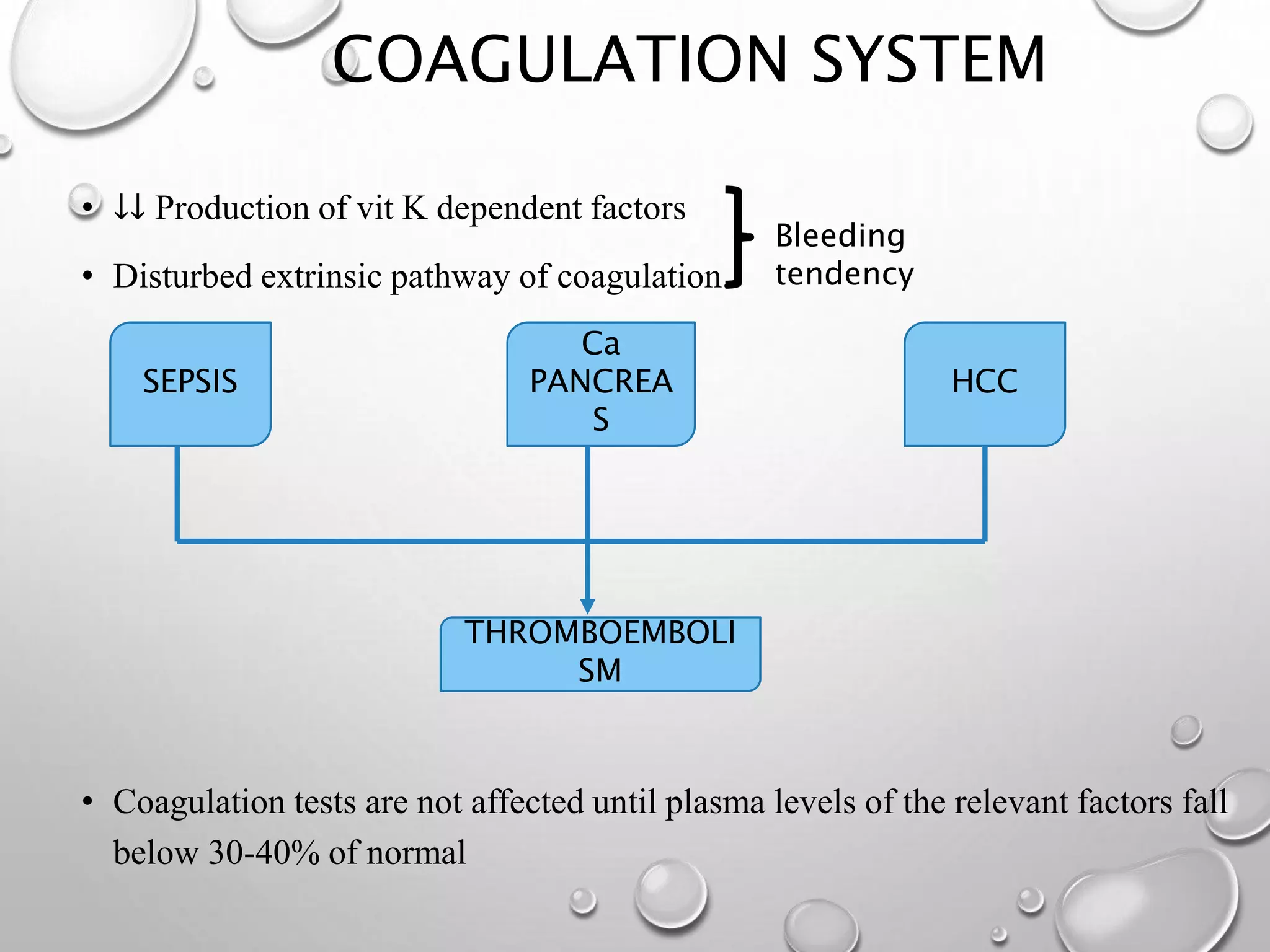
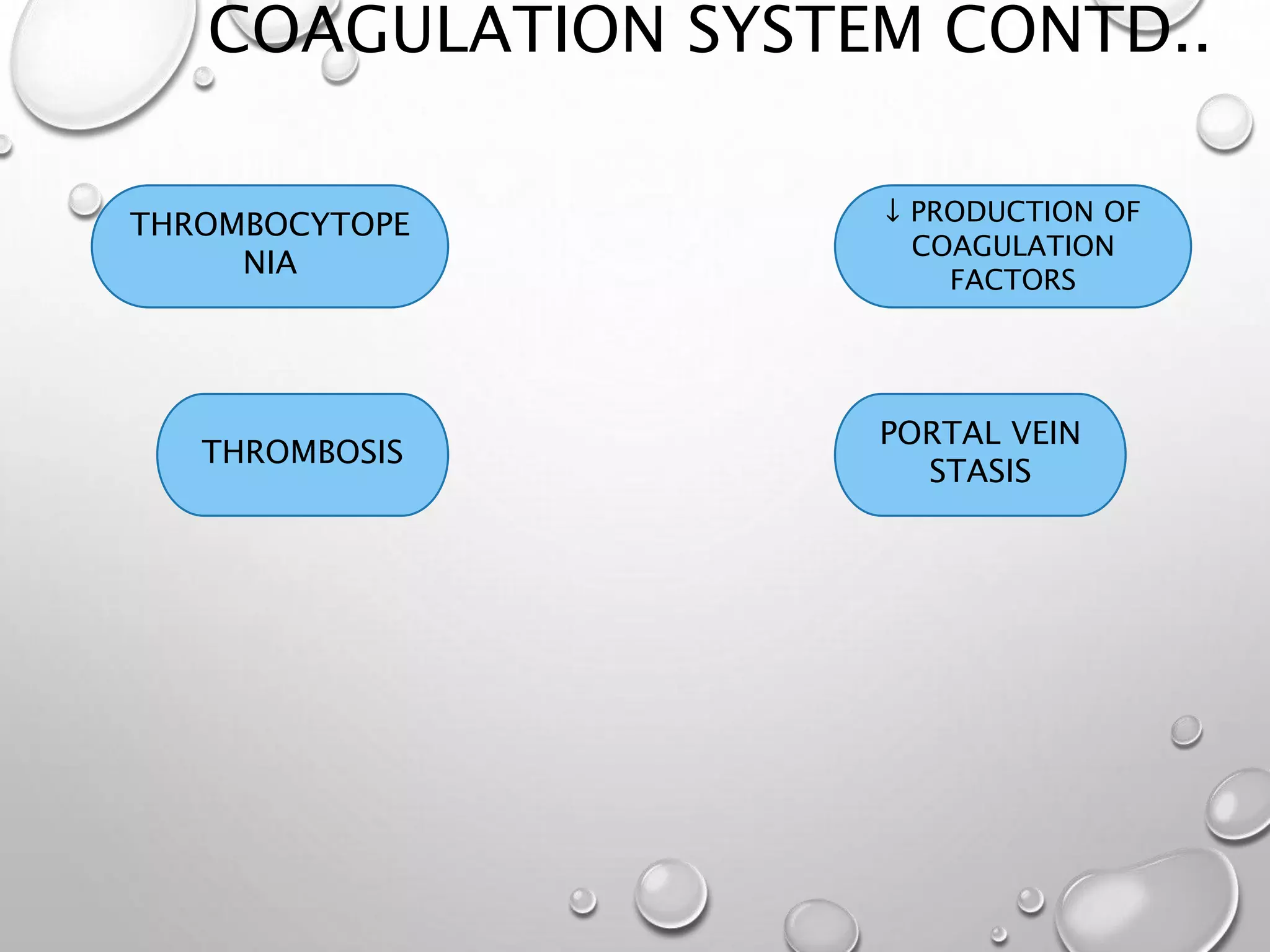
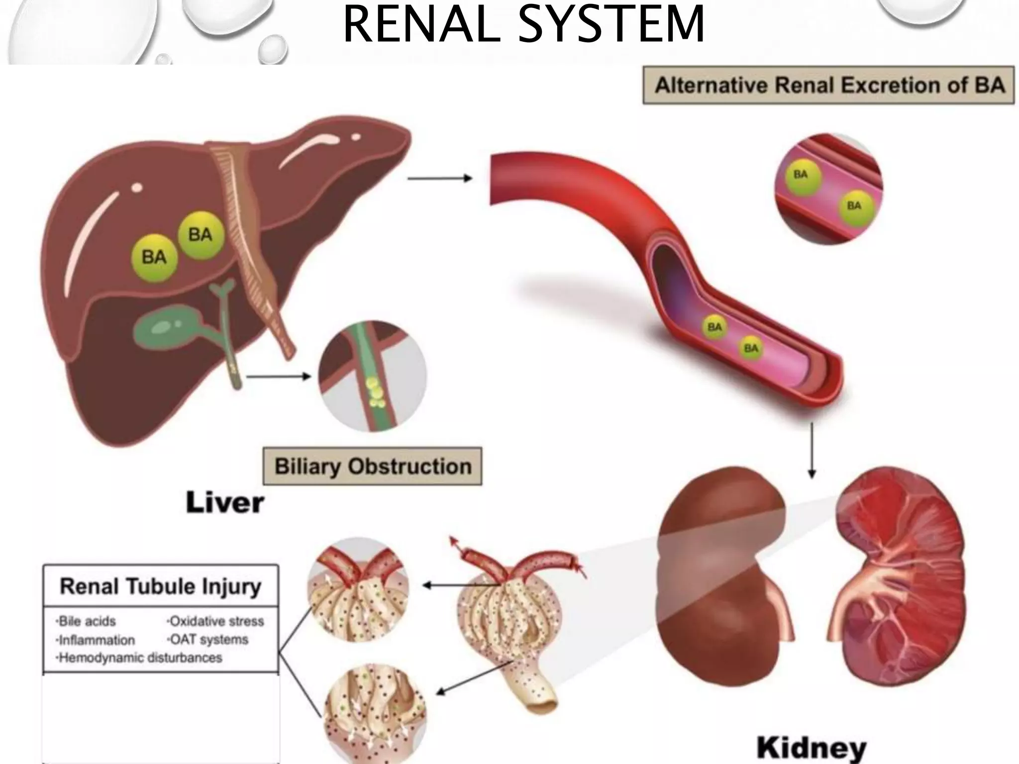
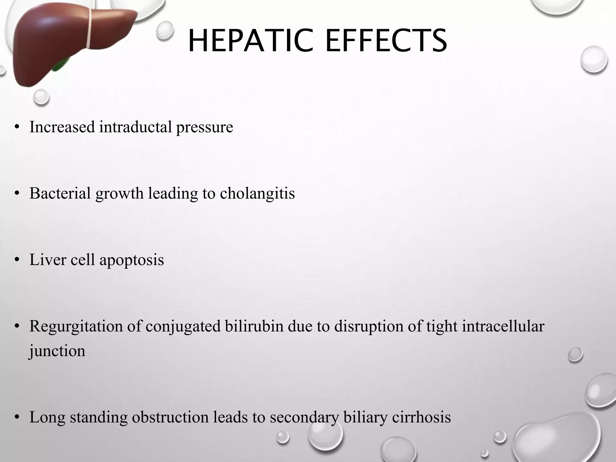
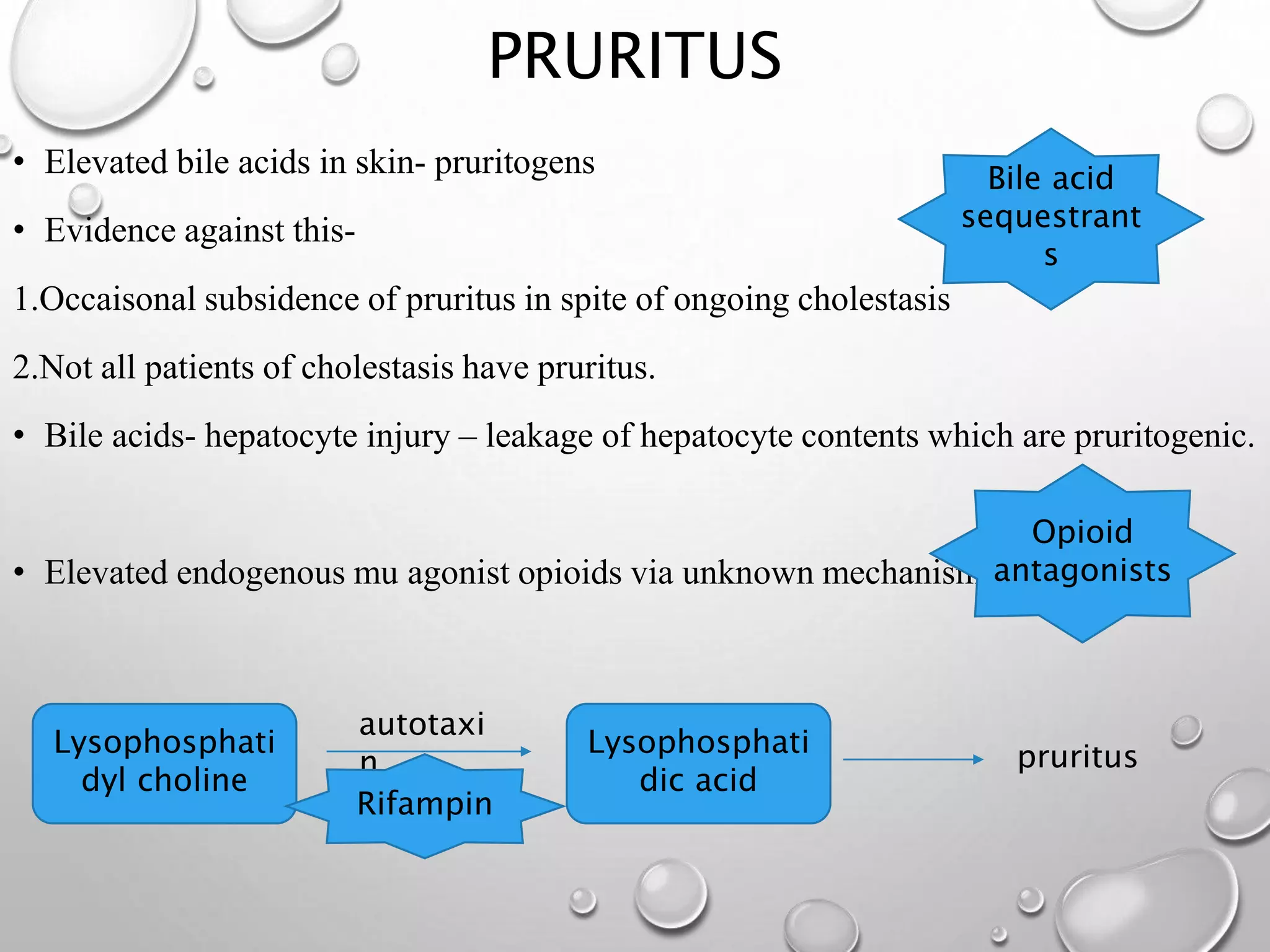
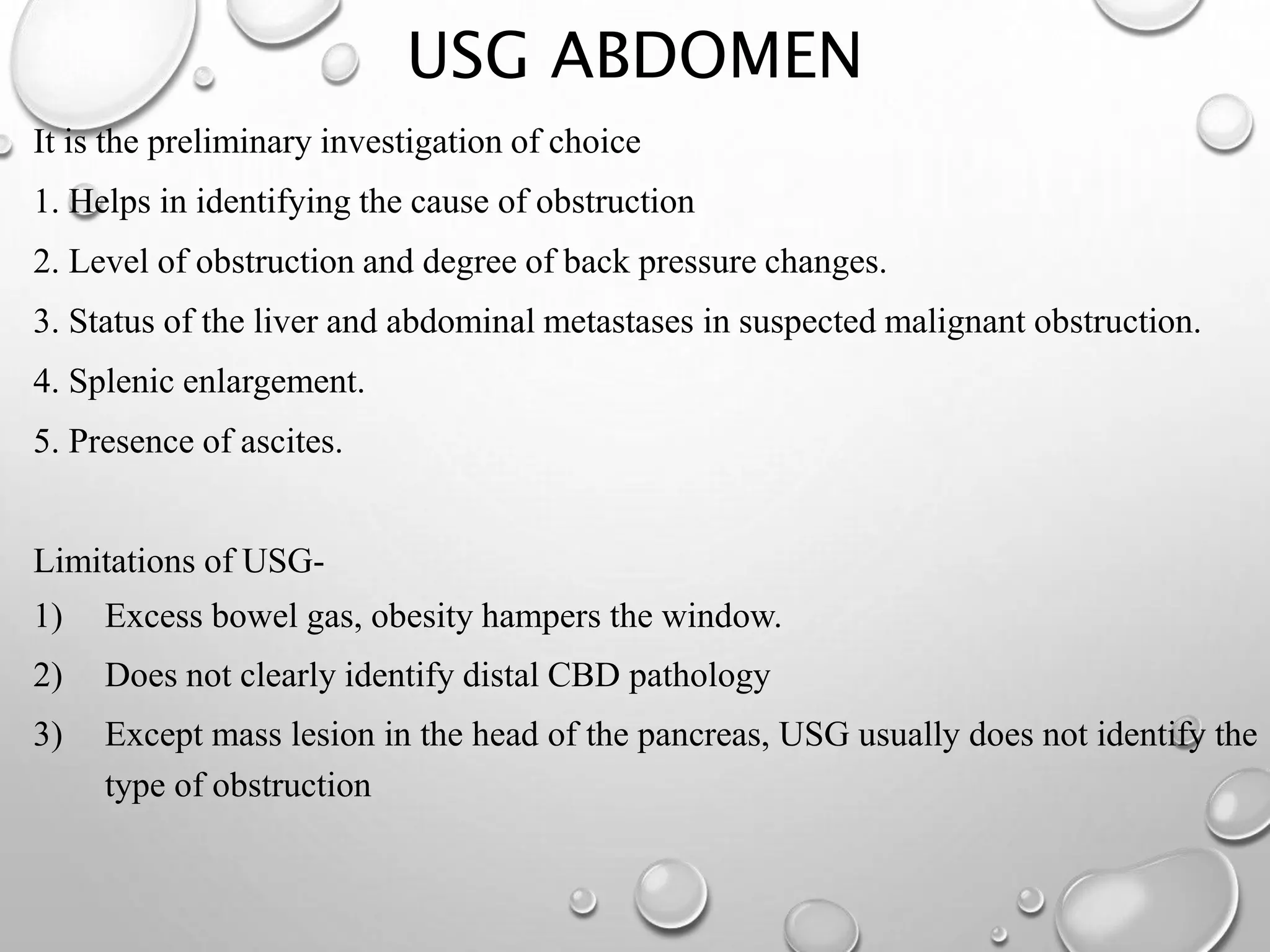
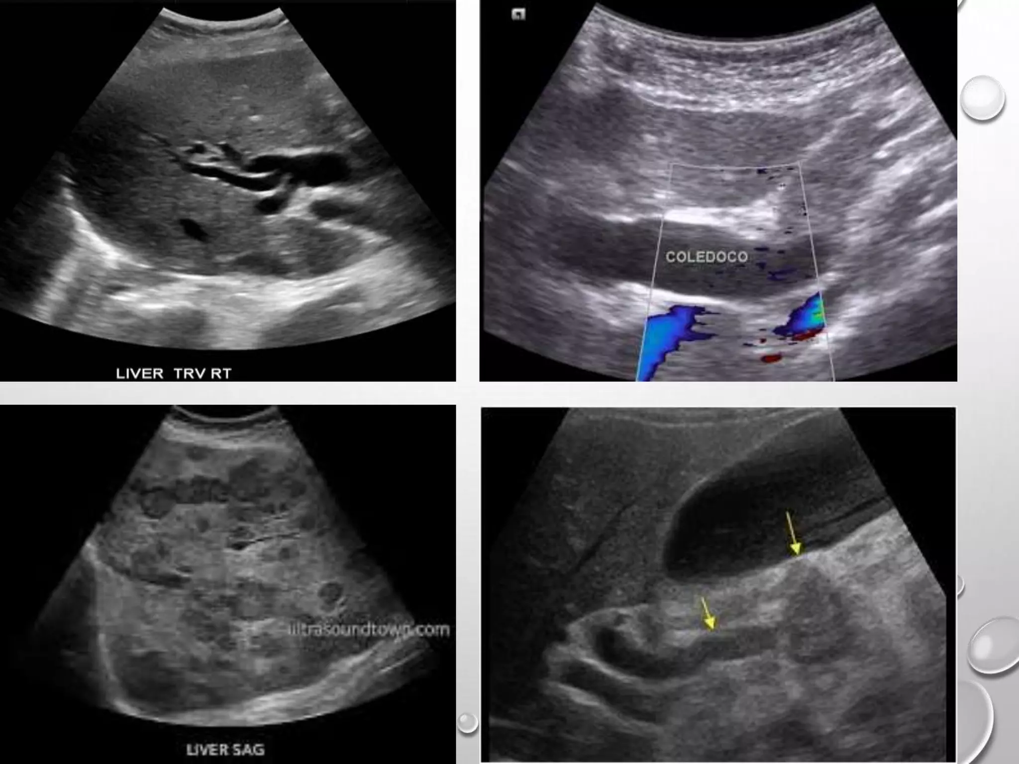
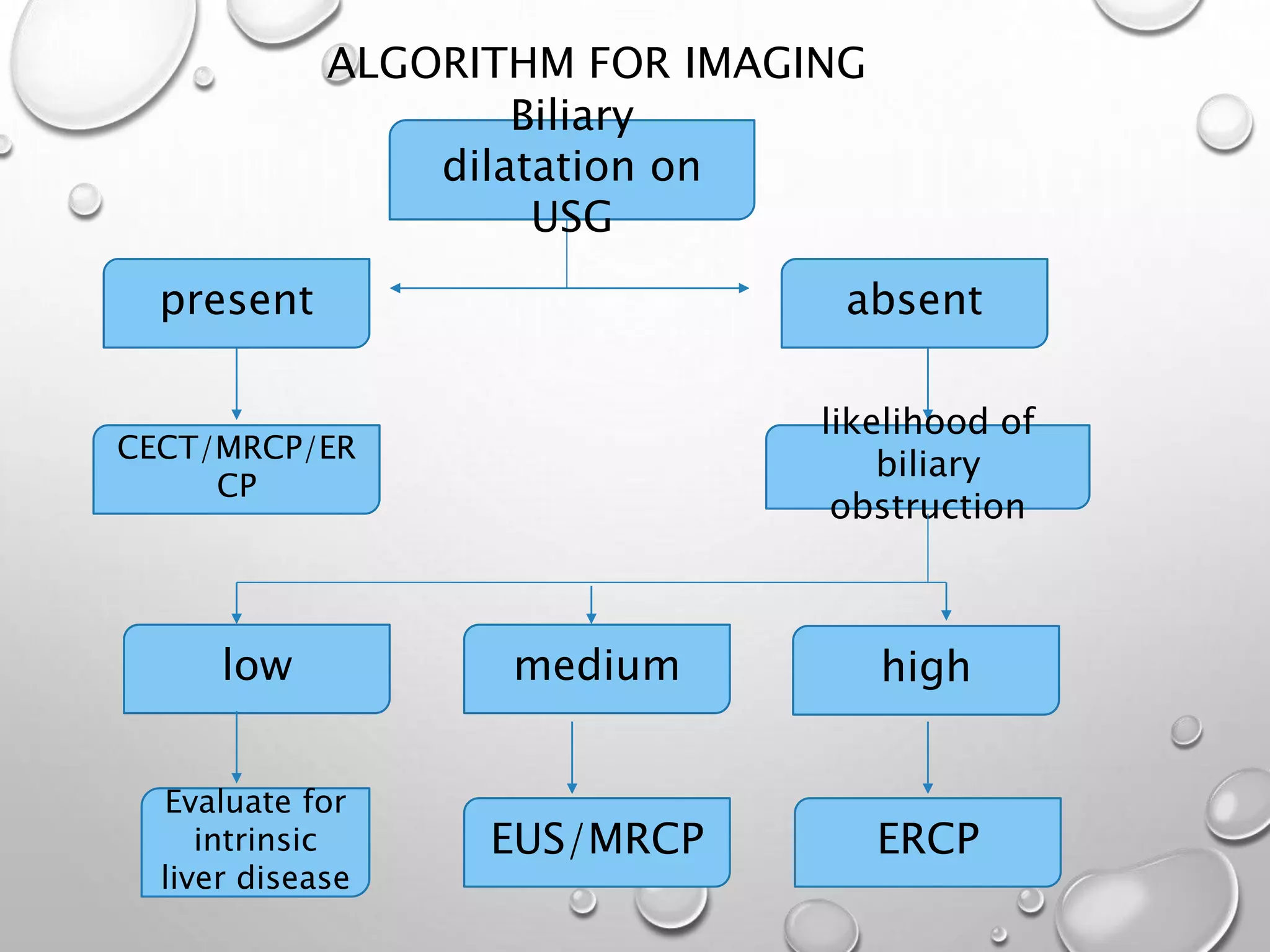
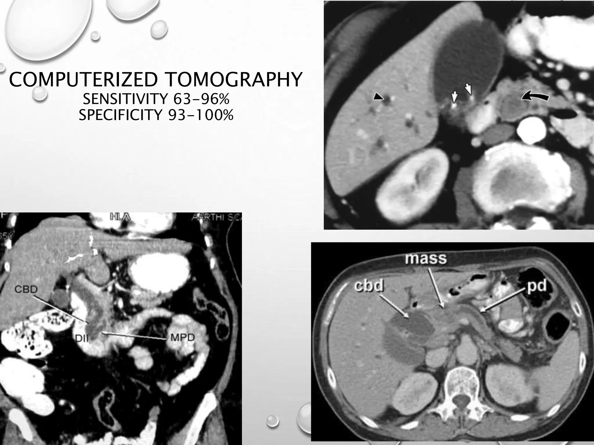
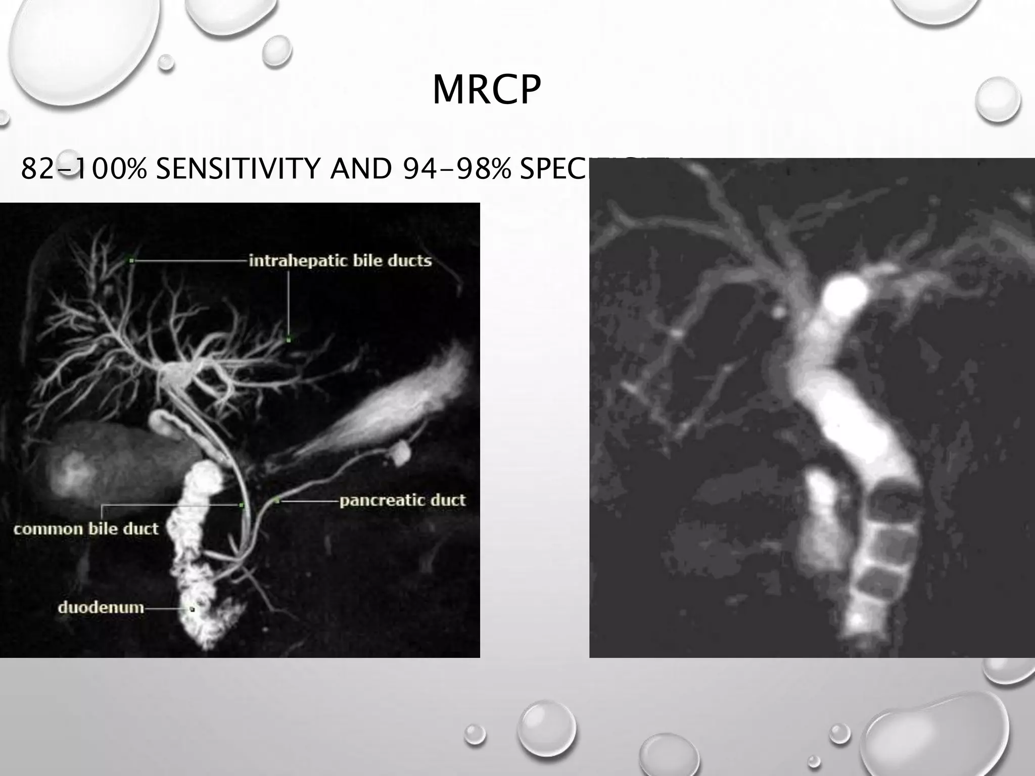
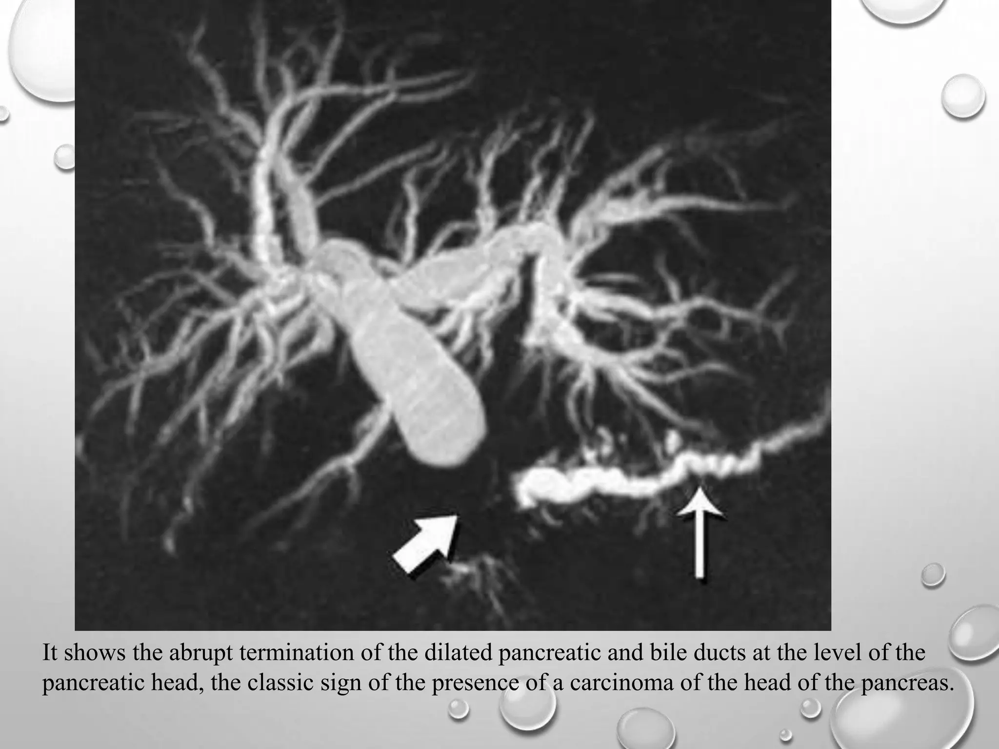
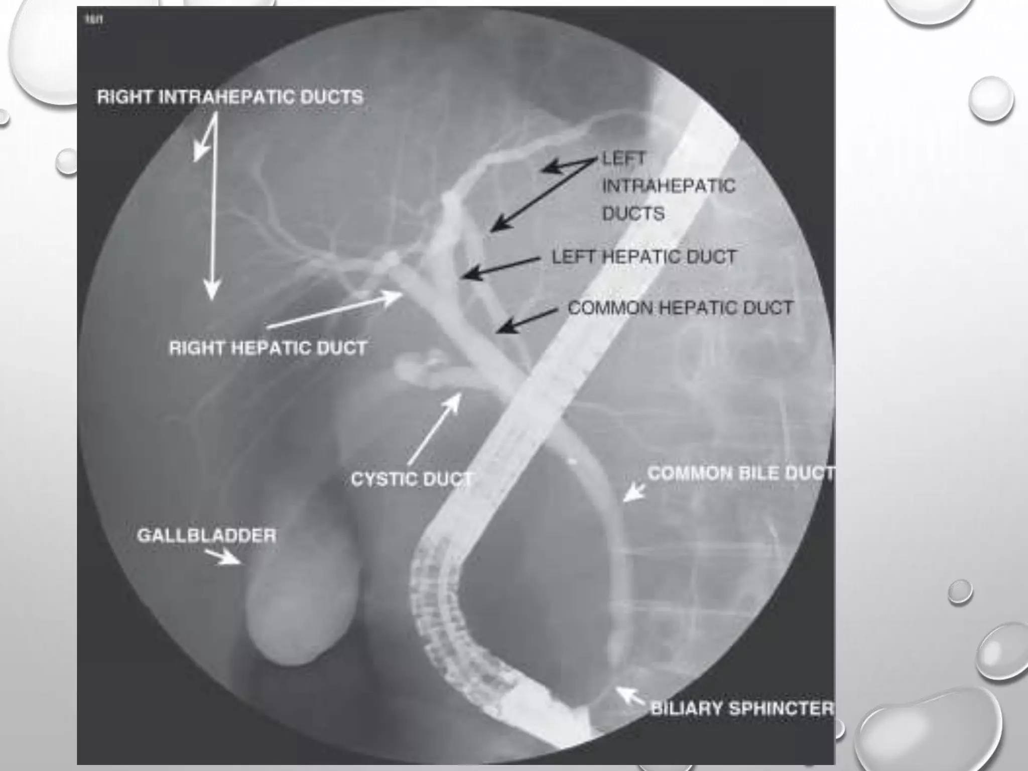
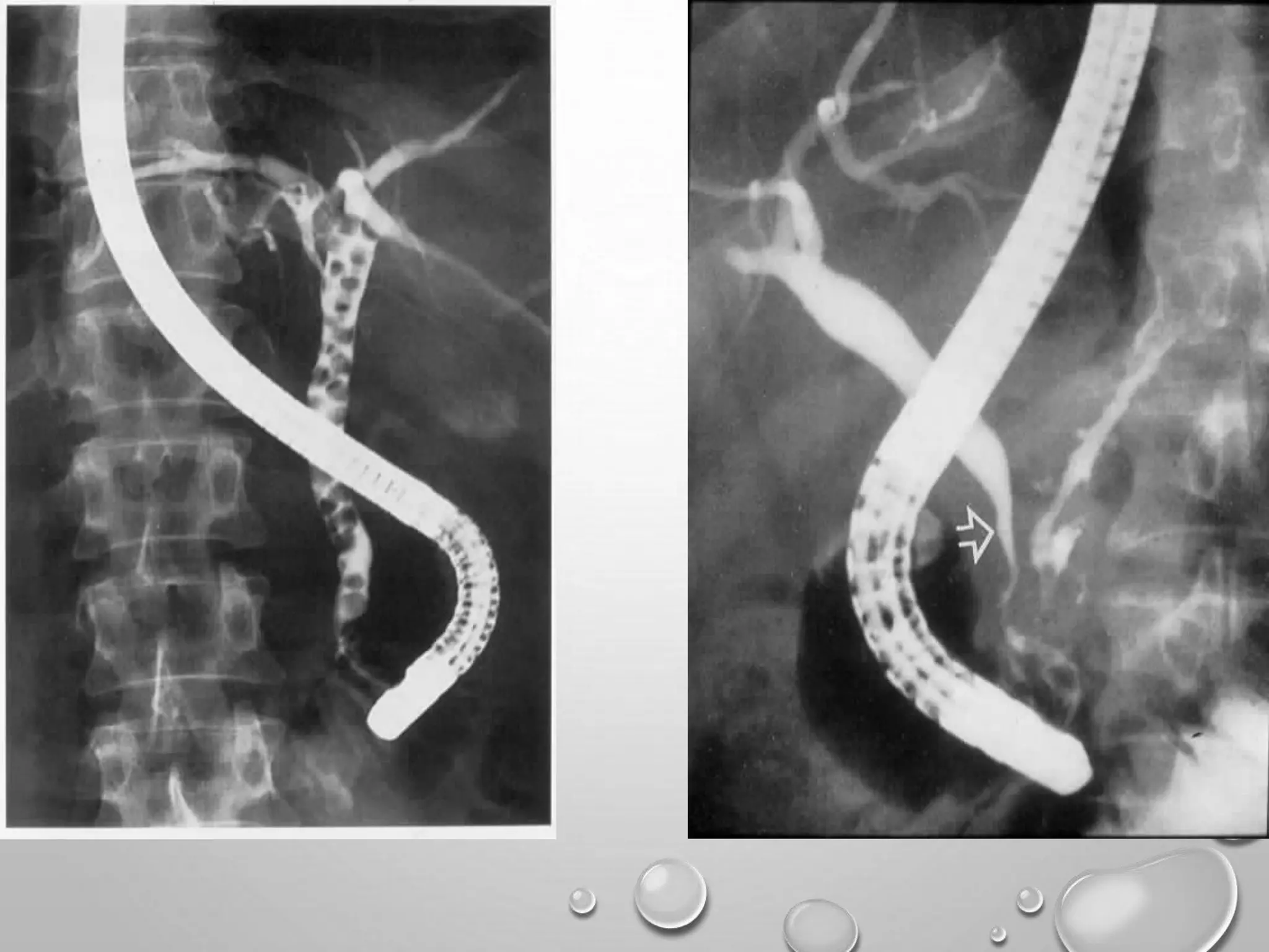
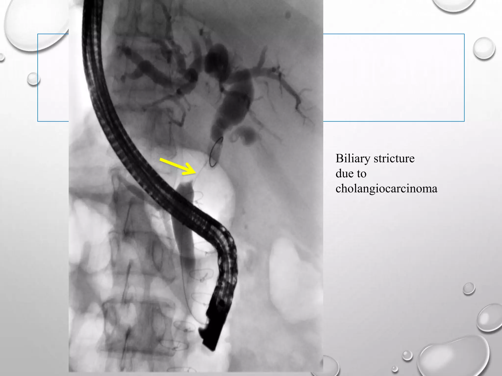
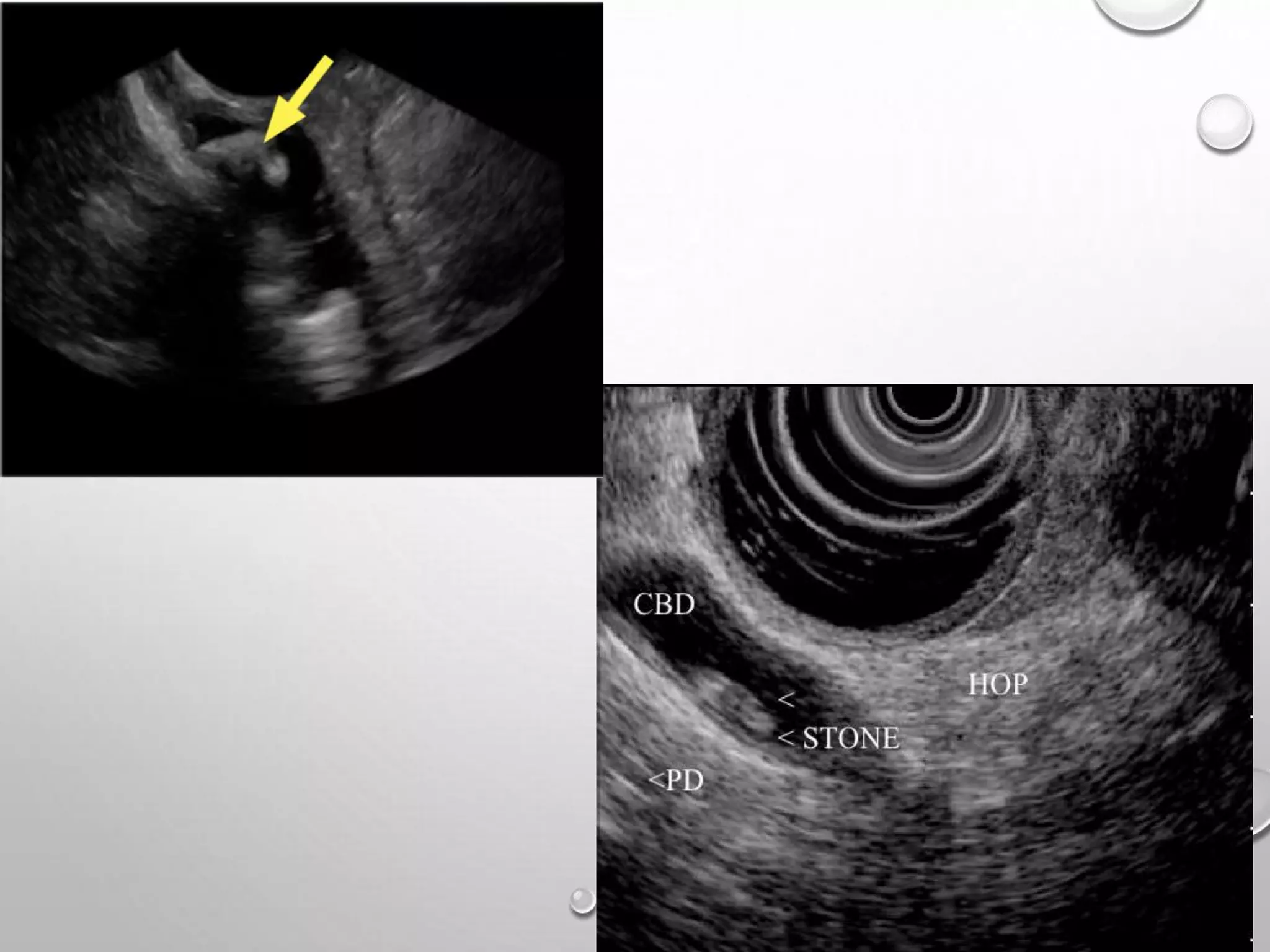
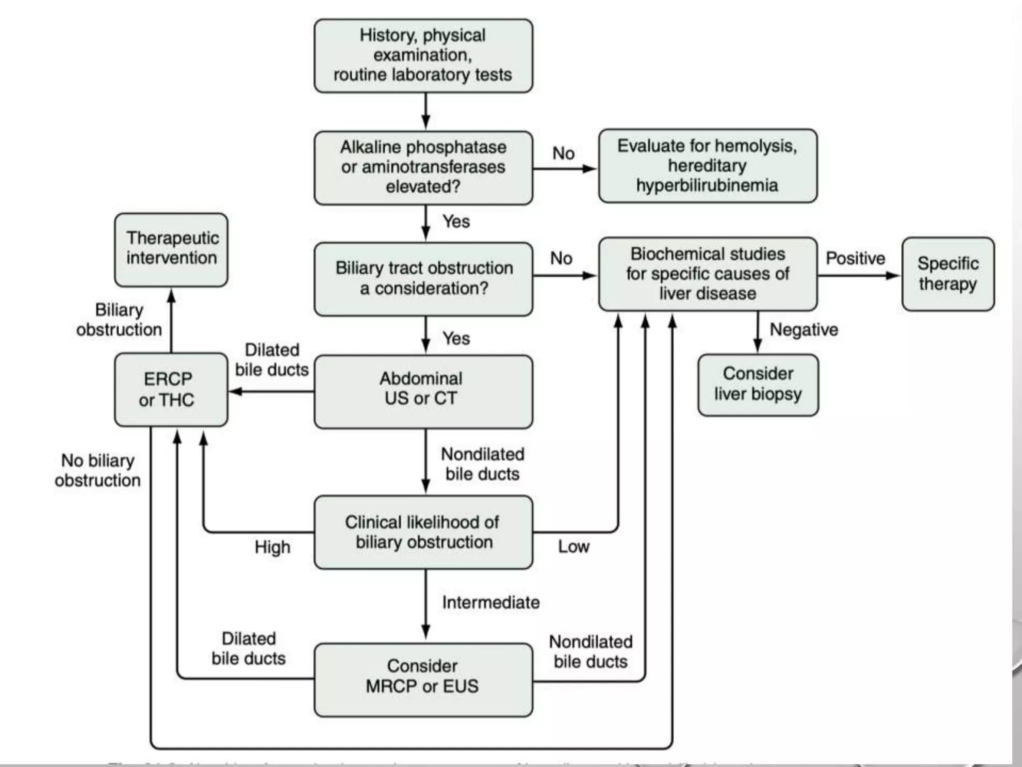
APPROACH TO OBSTRUCTIVE
JAUNDICE
• Jaundice, derived from the word jaune (in 1300 AD), is the yellowish
discoloration of the skin, conjunctivae, and mucous membranes.
• The first description of jaundice is mentioned in EBERS PAPYRUS of Egypt
dating to 3000 BC.
• The word obstructive jaundice was first used in 1935, by A. O. Whipple, an
American surgeon.
Water
Ions
Bile acids
proteins
Bilirubin
Phospholipids
cholesterol
BILE
Bilirubin is the catabolic end product of heme metabolism.
A healthy adult produces an average of 4 mg/kg of bilirubin each day.
Most bilirubin (70% to 80%) results from catabolism of hemoglobin.
The remaining 20% to 30% of bilirubin derived from breakdown of
other heme-containing proteins (e.g., catalase, cytochrome oxidases)
in hepatocytes.
BILIRUBIN METABOLISM
heme
oxygenase
Fe, CO
Biliverdin
reductase
Conversion of erythrocyte-derived hemoglobin to bilirubin
occurs in macrophages in the spleen, bone marrow, and liver
Free hemoglobin, haptoglobin-bound hemoglobin,
are catabolized to bilirubin in hepatocytes.
Has internal Hydrogen bonds
toxic
Insoluble in water
Needs to be eliminated
Bilirubin(Unconjug
ated)
BILIRUBIN IX-
ALPHA-ZZ
ALBUMI
N
TRANSPORTED
TO LIVER
NON COVALENT,
TEMPORARY
BOND
soluble in water
Has no internal Hydrogen
bonds
Is metabolised and excreted
Bilirubin(conjugate
d)
Zakim and boyer’s text book of
hepatology
Rotor
syndrome
Dubin Johnson
syndrome
Gilbert
Criggler Najar
syndrome
CONJUGATE
D BILIRUBIN
COLO
N
UROBILINOGE
N
EXCRETED
BY
KIDNEYS
20%
REABSORBED
De-
conjugati
on
Excreted
in feces
Backflow of
conjugated bilirubin
Water soluble
Large free fraction
Weak bond with
Albumin
Excreted by kidneys
Glomerular
filtration
Unconjugated bilirubin never appears in
the urine
Cholestasis is defined as disturbances in bile flow caused by diseases either in the hepatocytes,
intrahepatic bile ducts or extrahepatic biliary system.
INTRAHEPATIC CHOLESTASIS
GRANULOMATOUS
AMYLOIDOSIS
MALIGNANCIES
CYSTIC FIBROSIS(30%)
GVHD
PRIMARY BILIARY
CHOLANGITIS
BENIGN RECURRENT
CHOLESTASIS
PARANEOPLASTIC
TPN
INFILITRATIVE DISEASES
DISEASES WITH
CHOLANGIOCYTE
INJURY
DISEASES WITH
MINIMAL HISTOLOGIC
CHANGES
• The most common Granulomatous disorders that produce jaundice are tuberculosis
and sarcoidosis.
• In PBC, cholangiocyte is the target of immune mediated inflammation,
predominantly seen in middle aged women.
• Erythromycin, septran, amoxicillin–clavulanic can cause cholangiocyte injury leading
to cholestasis.
JAUNDIC
E PUO
SUSPECT
GRANULOMATO
US DISEASE
JAUNDIC
E
ANOREXI
A,
WEIGHT
LOSS
SUSPECT
NEOPLASTIC
DISEASE
Jaundice may accompany cholestasis in the absence of hepatic infiltration or injury to
hepatocytes or cholangiocytes.
Mutations in the genes that encode transport proteins involved in bile formation and
conditions that interfere with the function or expression of such proteins.
EXTRAHEPATIC CAUSES
BENIGN CAUSES MALIGNANT
CAUSES
Choledocholithiasis
Postoperative strictures
Primary sclerosing
cholangitis
Pancreatitis
AIDS cholangiopathy
Mirizzi’s Syndrome
Cholangiocarcinoma
Pancreatic cancer
Gall Bladder Cancer
Ampullary Cancer
Malignant involvement
of the porta hepatis
BENJAMIN CLASSIFICATION OF
CHOLESTATIC JAUNDICE
 Ca Head of Pancreas
 Cholangiocarcinoma
 Ligated CBD
TYPE 1- Complete obstruction
 Choledocholithiasis
 Choledochal cyst
 Duodenal diverticula
 Periampullary tumor
 Papillomas of the bile duct
 Intra biliary parasites
TYPE 2- Intermittent obstruction
 Strictures of the CBD
• Congenital/traumatic
• Sclerosing cholangitis
• Post radiotherapy
 Stenosed biliary-enteric
anastomosis
 Cystic fibrosis
 Chronic pancreatitis
TYPE 3- Chronic incomplete obstruction
 Trauma
 Sclerosing cholangitis
 Intrahepatic stones
TYPE 4- Segmental obstruction
EFFECTS OF BILIARY
OBSTRUCTION
Intestines Coagulation Renal
Liver Dermatologic
INTESTINES
• Obstruction to bile flow leads to absence of bile in intestine. This leads to
Overgrowth
of Gr negative
bacteria
Reduced
expression of
tight
junctions
Altered
permeability
Bacterial and
endotoxin
translocation
Accumulation
of bile acids
in liver
↓ kupffer cells
and
macrophage
functions
COAGULATION SYSTEM
• ↓↓ Production of vit K dependent factors
• Disturbed extrinsic pathway of coagulation.
• Coagulation tests are not affected until plasma levels of the relevant factors fall
below 30-40% of normal
SEPSIS
Ca
PANCREA
S
HCC
THROMBOEMBOLI
SM
Bleeding
tendency
COAGULATION SYSTEM CONTD..
THROMBOCYTOPE
NIA
↓ PRODUCTION OF
COAGULATION
FACTORS
PORTAL VEIN
STASIS
THROMBOSIS
RENAL SYSTEM
AKI
HEPATIC EFFECTS
• Increased intraductal pressure
• Bacterial growth leading to cholangitis
• Liver cell apoptosis
• Regurgitation of conjugated bilirubin due to disruption of tight intracellular
junction
• Long standing obstruction leads to secondary biliary cirrhosis
DERMATOLOGIC
EFFECT ON WOUND HEALING
• Delayed wound healing and high incidence of wound dehiscence.
• This is due to decreased activity of enzyme propyl hydroxylase in the skin
• Normally, this enzyme helps in incorporation of proline into collagen
• Decreased activity leads defective synthesis of collagen
PRURITUS
• Elevated bile acids in skin- pruritogens
• Evidence against this-
1.Occaisonal subsidence of pruritus in spite of ongoing cholestasis
2.Not all patients of cholestasis have pruritus.
• Bile acids- hepatocyte injury – leakage of hepatocyte contents which are pruritogenic.
• Elevated endogenous mu agonist opioids via unknown mechanism in CLD.
Lysophosphati
dyl choline
Lysophosphati
dic acid
autotaxi
n pruritus
Bile acid
sequestrant
s
Opioid
antagonists
Rifampin
CLINICAL EVALUATION
• THE MAIN AIMS OF CLINICAL EVALUATION ARE
7. POSTULATE AN INVESTIGATION ALGORITHM BASED ON THE CLINICAL
fiNDINGS.
Determining the obstructive
nature of jaundice.
The need for multiorgan system
support.
Impact of jaundice on other organ
systems.
If malignant then whether the
disease is metastatic in nature.
Whether the etiology is benign or
malignant in nature.
Identifying the cause for
obstructive jaundice.
HISTORY
Jaundice with pruritus, clay coloured stools (acholic stools) and high coloured urine is seen in
the obstructive pattern of jaundice.
• Pain abdomen before the onset of jaundice --- choledocholithiasis or cholecystitis causing
mirrizi syndrome
• Fever -- highly suggestive of cholangitis
• The onset of jaundice after any hepatobiliary surgery--- bile duct injury, bile leak.
• Any history of critical illness or shock that can cause cholestasis of sepsis.
• Any recent new medication use, especially antibiotics which can cause cholestasis
• Jaundice with nausea, vomiting, and prodrome phase -- viral hepatitis. History of travel,
sexual contact, blood transfusion, major surgery, intravenous (IV) drug use can support this
diagnosis
• Family or personal history of any autoimmune disease can point toward
PSC or PBC
• Severe pain and associated symptoms suggestive of pancreatitis can also
cause transient obstructive jaundice.
• Painless progressive jaundice, Weight loss, bone pains, and abdominal
distension due to ascites are suggestive of malignant etiology.
• History of abdominal masses is highly suggestive of malignancy.
• Jaundice in IBD should always raise suspicion of primary sclerosing
cholangitis
• Patient giving history of recurrent episodes of malaise and pruritus in
association with jaundice, starting from 2nd decade of life- BRIC OF A
BILIARY STRICTURE. HISTORY OF INflAMMATORY BOWEL
DISEASE PRESENTING AS JAUNDICE IS SUGGESTIVE OF
CLINICAL EVALUATION
• Clinical evaluation has a positive predictive value of only about 75%.
• About 25% of patients with suspected obstruction actually have hepatocellular
disease.
• General examination should be done to look for the presence of stigmata of chronic
liver disease
• White nails
• Muehrcke nails
• Clubbing
• Palmer erythema
• Dupuytren contracture
• Carpel tunnel syndrome
• Loss axillary and pubic hairs
• Spider nevi
• Jaundice
• Glossy lip and tongue
• Gynecomastia
• Testicular atrophy
• Petechial rash
• Dry skin, paper money skin
EXAMINATION
Large, nodular liver suggests possible hepatic metastasis.
Tender hepatomegaly can be seen in viral hepatitis, congestive heart failure, and
alcoholic hepatitis.
In choledocholithiasis and cholecystitis, right upper quadrant (RUQ) tenderness is
appreciated.
Palpable, enlarged, painless gallbladder can suggest malignant etiology.
Splenomegaly can be seen due to massive hemolysis or portal HTN.
Ascites can be observed in cirrhosis due to portal HTN.
• Curvoiser’s law- a palpable non-tender gallbladder in the presence of jaundice is
unlikely to be due to gallstones. It usually indicates a neoplastic stricture obstructing
the distal common bile duct.
• Exceptions-
• 1.Double impaction: stones, simultaneously occluding the cystic duct and the distal
common bile duct (CBD). The stone in the CBD causes obstructive jaundice and a
synchronous stone in the cystic duct leads to mucocele or empyema of gall bladder.
• 2. Pancreatic calculus obstructing the ampulla of vater
• 3. Oriental cholangiohepatitis (ductal stones formed secondary to liver fluke
infestation)
INVESTIGATIONS
Determining the severity of illness
Urgency of intervention
Level of care required
Increasing PT/INR: hepatocellular dysfunction.
PT/INR can also be deranged in cholestasis due to the malabsorption of vit. K.
Coagulopathy in obstructive jaundice is rapidly corrected by parenteral administration
of vit k.
Obstructive pattern Hemolytic pattern
Serum Bilirubin
conjugated
unconjugated
+++
+
+
+++
Urobilinogen ↓ ↑
Urinary Bilirubin + 0
Serum ALP ↑ No change
Serum GGTP ↑ No change
Serum 5- nucleotidase ↑ No change
Transaminases Mildly raised Markedly raised
Complete blood count- to look for anemia(malignancies), Leucocytosis (cholangitis).
Liver function tests
• LIVER FUNCTION TESTS – CHOLESTATIC PATTERN
• ALP- Hydrolyze the phosphate esters in alkaline environment, found in Liver,
Placenta, bone, intestine.
• Liver ALP – Canalicular membrane of hepatocytes and apical membrane of
cholangiocytes.
• Elevation of 5′-nucleotidase levels is used in parallel as specific
marker to confirm a suspected hepatic origin of ALP.
• Elevations of ALP and Bil out of proportion to AST and ALT - Cholestasis
bile duct
obstruction
Duct cell
injury
liver
infiltration
Architectural
disarray, damaged
liver lobule
Raised
ALP
History, clinical examination, basic routine
investigations
Ultrasonography of abdomen
USG ABDOMEN
It is the preliminary investigation of choice
1. Helps in identifying the cause of obstruction
2. Level of obstruction and degree of back pressure changes.
3. Status of the liver and abdominal metastases in suspected malignant obstruction.
4. Splenic enlargement.
5. Presence of ascites.
Limitations of USG-
1) Excess bowel gas, obesity hampers the window.
2) Does not clearly identify distal CBD pathology
3) Except mass lesion in the head of the pancreas, USG usually does not identify the
type of obstruction
ALGORITHM FOR IMAGING
Biliary
dilatation on
USG
absent
present
CECT/MRCP/ER
CP
likelihood of
biliary
obstruction
high
medium
low
Evaluate for
intrinsic
liver disease
EUS/MRCP ERCP
• Jaundiced patients with biochemical evidence of cholestasis in whom imaging
studies do not suggest biliary obstruction should be evaluated for underlying liver
disease.
Depending on the disorder suspected, laboratory studies include
• Viral serologies – HBV HCV HEV,EBV,CMV
• AMA (for PBC)
• ANA,ANCA – PSC
• One protocol suggests that if all the tests are negative and still ALP is ≥ 2 times the
normal for minimum of 6 months, liver biopsy is done.
COMPUTERIZED TOMOGRAPHY
SENSITIVITY 63-96%
SPECIFICITY 93-100%
82-100% SENSITIVITY AND 94-98% SPECIFICITY
MRCP
It shows the abrupt termination of the dilated pancreatic and bile ducts at the level of the
pancreatic head, the classic sign of the presence of a carcinoma of the head of the pancreas.
ERCP
 89%-98% sensitivity & 89%-100% specificity.
 ERCP is the procedure of choice in choledocholithiasis and in suspected
ampullary or duodenal lesions in ca pancreas.
 The diagnostic role of ERCP is to collect tissue for biopsy in periampullary
growths or brush cytology samples. The exact level of the block and the severity
of backpressure changes can also be ascertained.
Biliary stricture
due to
cholangiocarcinoma
EUS
• sensitivity 89%-97% and 67%-98% specificity
• Advantage of permitting biopsy and aspiration of suspected malignant lesions
• Most useful in circumstances in which the patient is thought to be at high risk
for complications of ERCP.
• Imaging of bile ducts via EUS is superior to USG and CT.
• Requires passage of a needle through the skin and subcutaneous tissues into the
liver and advancement into a peripheral bile duct.
• When bile is aspirated, a catheter is introduced through the needle, and radiopaque
dye is injected.
• The sensitivity and specificity of percutaneous THC for the diagnosis of biliary tract
disease are comparable with those for ERCP.
• Interventional procedures like balloon dilation and stent placement can be
performed and cholangioscopy is also feasible by this route.
• Particularly technically advantageous when the level of biliary obstruction is
proximal to the common hepatic duct or altered anatomy precludes ERCP .
Percutaneous Transhepatic Cholangiography
PITT’S PROGNOSTIC SCORE
Parameters (one point per factor)
Type of obstruction (benign or malignant)
Age >60 years
Serum albumin < 3gm/dl
Serum bilirubin > 10 mg%
Serum Alkaline phosphatase > 100 IU
Serum creatinine > 1.3 mg%
TLC > 10000/mm 3
Haematocrit < 30
score mortality
≤ 2 0%
3 4%
4 7%
5 44%
6 67%
8 100%
THERAPEUTIC APPROACH
• The therapy is directed at diagnosed condition.
• In a patient with bile duct obstruction, therapy is directed at relieving the obstruction.
• Interventional endoscopic or radiologic approaches include
• sphincterotomy,
• balloon dilation of focal strictures, and
• placement of drains or stents.
• Surgery should be considered for neoplasms, if feasible.
• Preoperative stenting to relieve jaundice is of great help in preparing the patient for
surgery.
• In inoperable cases, palliative stenting is done.
THANK YOU

Approach to obstructive jaundice

  • 1.
  • 2.
    • Jaundice, derivedfrom the word jaune (in 1300 AD), is the yellowish discoloration of the skin, conjunctivae, and mucous membranes. • The first description of jaundice is mentioned in EBERS PAPYRUS of Egypt dating to 3000 BC. • The word obstructive jaundice was first used in 1935, by A. O. Whipple, an American surgeon.
  • 3.
    Water Ions Bile acids proteins Bilirubin Phospholipids cholesterol BILE Bilirubin isthe catabolic end product of heme metabolism. A healthy adult produces an average of 4 mg/kg of bilirubin each day. Most bilirubin (70% to 80%) results from catabolism of hemoglobin. The remaining 20% to 30% of bilirubin derived from breakdown of other heme-containing proteins (e.g., catalase, cytochrome oxidases) in hepatocytes.
  • 4.
    BILIRUBIN METABOLISM heme oxygenase Fe, CO Biliverdin reductase Conversionof erythrocyte-derived hemoglobin to bilirubin occurs in macrophages in the spleen, bone marrow, and liver Free hemoglobin, haptoglobin-bound hemoglobin, are catabolized to bilirubin in hepatocytes.
  • 5.
    Has internal Hydrogenbonds toxic Insoluble in water Needs to be eliminated Bilirubin(Unconjug ated) BILIRUBIN IX- ALPHA-ZZ ALBUMI N TRANSPORTED TO LIVER NON COVALENT, TEMPORARY BOND soluble in water Has no internal Hydrogen bonds Is metabolised and excreted Bilirubin(conjugate d)
  • 6.
    Zakim and boyer’stext book of hepatology
  • 7.
  • 8.
  • 9.
    Backflow of conjugated bilirubin Watersoluble Large free fraction Weak bond with Albumin Excreted by kidneys Glomerular filtration Unconjugated bilirubin never appears in the urine Cholestasis is defined as disturbances in bile flow caused by diseases either in the hepatocytes, intrahepatic bile ducts or extrahepatic biliary system.
  • 10.
    INTRAHEPATIC CHOLESTASIS GRANULOMATOUS AMYLOIDOSIS MALIGNANCIES CYSTIC FIBROSIS(30%) GVHD PRIMARYBILIARY CHOLANGITIS BENIGN RECURRENT CHOLESTASIS PARANEOPLASTIC TPN INFILITRATIVE DISEASES DISEASES WITH CHOLANGIOCYTE INJURY DISEASES WITH MINIMAL HISTOLOGIC CHANGES
  • 11.
    • The mostcommon Granulomatous disorders that produce jaundice are tuberculosis and sarcoidosis. • In PBC, cholangiocyte is the target of immune mediated inflammation, predominantly seen in middle aged women. • Erythromycin, septran, amoxicillin–clavulanic can cause cholangiocyte injury leading to cholestasis. JAUNDIC E PUO SUSPECT GRANULOMATO US DISEASE JAUNDIC E ANOREXI A, WEIGHT LOSS SUSPECT NEOPLASTIC DISEASE
  • 12.
    Jaundice may accompanycholestasis in the absence of hepatic infiltration or injury to hepatocytes or cholangiocytes. Mutations in the genes that encode transport proteins involved in bile formation and conditions that interfere with the function or expression of such proteins.
  • 13.
    EXTRAHEPATIC CAUSES BENIGN CAUSESMALIGNANT CAUSES Choledocholithiasis Postoperative strictures Primary sclerosing cholangitis Pancreatitis AIDS cholangiopathy Mirizzi’s Syndrome Cholangiocarcinoma Pancreatic cancer Gall Bladder Cancer Ampullary Cancer Malignant involvement of the porta hepatis
  • 14.
  • 15.
     Ca Headof Pancreas  Cholangiocarcinoma  Ligated CBD TYPE 1- Complete obstruction  Choledocholithiasis  Choledochal cyst  Duodenal diverticula  Periampullary tumor  Papillomas of the bile duct  Intra biliary parasites TYPE 2- Intermittent obstruction
  • 16.
     Strictures ofthe CBD • Congenital/traumatic • Sclerosing cholangitis • Post radiotherapy  Stenosed biliary-enteric anastomosis  Cystic fibrosis  Chronic pancreatitis TYPE 3- Chronic incomplete obstruction  Trauma  Sclerosing cholangitis  Intrahepatic stones TYPE 4- Segmental obstruction
  • 17.
    EFFECTS OF BILIARY OBSTRUCTION IntestinesCoagulation Renal Liver Dermatologic
  • 18.
    INTESTINES • Obstruction tobile flow leads to absence of bile in intestine. This leads to Overgrowth of Gr negative bacteria Reduced expression of tight junctions Altered permeability Bacterial and endotoxin translocation Accumulation of bile acids in liver ↓ kupffer cells and macrophage functions
  • 19.
    COAGULATION SYSTEM • ↓↓Production of vit K dependent factors • Disturbed extrinsic pathway of coagulation. • Coagulation tests are not affected until plasma levels of the relevant factors fall below 30-40% of normal SEPSIS Ca PANCREA S HCC THROMBOEMBOLI SM Bleeding tendency
  • 20.
    COAGULATION SYSTEM CONTD.. THROMBOCYTOPE NIA ↓PRODUCTION OF COAGULATION FACTORS PORTAL VEIN STASIS THROMBOSIS
  • 21.
  • 22.
    HEPATIC EFFECTS • Increasedintraductal pressure • Bacterial growth leading to cholangitis • Liver cell apoptosis • Regurgitation of conjugated bilirubin due to disruption of tight intracellular junction • Long standing obstruction leads to secondary biliary cirrhosis
  • 23.
    DERMATOLOGIC EFFECT ON WOUNDHEALING • Delayed wound healing and high incidence of wound dehiscence. • This is due to decreased activity of enzyme propyl hydroxylase in the skin • Normally, this enzyme helps in incorporation of proline into collagen • Decreased activity leads defective synthesis of collagen
  • 24.
    PRURITUS • Elevated bileacids in skin- pruritogens • Evidence against this- 1.Occaisonal subsidence of pruritus in spite of ongoing cholestasis 2.Not all patients of cholestasis have pruritus. • Bile acids- hepatocyte injury – leakage of hepatocyte contents which are pruritogenic. • Elevated endogenous mu agonist opioids via unknown mechanism in CLD. Lysophosphati dyl choline Lysophosphati dic acid autotaxi n pruritus Bile acid sequestrant s Opioid antagonists Rifampin
  • 25.
    CLINICAL EVALUATION • THEMAIN AIMS OF CLINICAL EVALUATION ARE 7. POSTULATE AN INVESTIGATION ALGORITHM BASED ON THE CLINICAL fiNDINGS. Determining the obstructive nature of jaundice. The need for multiorgan system support. Impact of jaundice on other organ systems. If malignant then whether the disease is metastatic in nature. Whether the etiology is benign or malignant in nature. Identifying the cause for obstructive jaundice.
  • 26.
    HISTORY Jaundice with pruritus,clay coloured stools (acholic stools) and high coloured urine is seen in the obstructive pattern of jaundice. • Pain abdomen before the onset of jaundice --- choledocholithiasis or cholecystitis causing mirrizi syndrome • Fever -- highly suggestive of cholangitis • The onset of jaundice after any hepatobiliary surgery--- bile duct injury, bile leak. • Any history of critical illness or shock that can cause cholestasis of sepsis. • Any recent new medication use, especially antibiotics which can cause cholestasis • Jaundice with nausea, vomiting, and prodrome phase -- viral hepatitis. History of travel, sexual contact, blood transfusion, major surgery, intravenous (IV) drug use can support this diagnosis
  • 27.
    • Family orpersonal history of any autoimmune disease can point toward PSC or PBC • Severe pain and associated symptoms suggestive of pancreatitis can also cause transient obstructive jaundice. • Painless progressive jaundice, Weight loss, bone pains, and abdominal distension due to ascites are suggestive of malignant etiology. • History of abdominal masses is highly suggestive of malignancy. • Jaundice in IBD should always raise suspicion of primary sclerosing cholangitis • Patient giving history of recurrent episodes of malaise and pruritus in association with jaundice, starting from 2nd decade of life- BRIC OF A BILIARY STRICTURE. HISTORY OF INflAMMATORY BOWEL DISEASE PRESENTING AS JAUNDICE IS SUGGESTIVE OF
  • 28.
    CLINICAL EVALUATION • Clinicalevaluation has a positive predictive value of only about 75%. • About 25% of patients with suspected obstruction actually have hepatocellular disease. • General examination should be done to look for the presence of stigmata of chronic liver disease • White nails • Muehrcke nails • Clubbing • Palmer erythema • Dupuytren contracture • Carpel tunnel syndrome • Loss axillary and pubic hairs • Spider nevi • Jaundice • Glossy lip and tongue • Gynecomastia • Testicular atrophy • Petechial rash • Dry skin, paper money skin
  • 29.
  • 30.
    Large, nodular liversuggests possible hepatic metastasis. Tender hepatomegaly can be seen in viral hepatitis, congestive heart failure, and alcoholic hepatitis. In choledocholithiasis and cholecystitis, right upper quadrant (RUQ) tenderness is appreciated. Palpable, enlarged, painless gallbladder can suggest malignant etiology. Splenomegaly can be seen due to massive hemolysis or portal HTN. Ascites can be observed in cirrhosis due to portal HTN.
  • 31.
    • Curvoiser’s law-a palpable non-tender gallbladder in the presence of jaundice is unlikely to be due to gallstones. It usually indicates a neoplastic stricture obstructing the distal common bile duct. • Exceptions- • 1.Double impaction: stones, simultaneously occluding the cystic duct and the distal common bile duct (CBD). The stone in the CBD causes obstructive jaundice and a synchronous stone in the cystic duct leads to mucocele or empyema of gall bladder. • 2. Pancreatic calculus obstructing the ampulla of vater • 3. Oriental cholangiohepatitis (ductal stones formed secondary to liver fluke infestation)
  • 32.
    INVESTIGATIONS Determining the severityof illness Urgency of intervention Level of care required
  • 33.
    Increasing PT/INR: hepatocellulardysfunction. PT/INR can also be deranged in cholestasis due to the malabsorption of vit. K. Coagulopathy in obstructive jaundice is rapidly corrected by parenteral administration of vit k. Obstructive pattern Hemolytic pattern Serum Bilirubin conjugated unconjugated +++ + + +++ Urobilinogen ↓ ↑ Urinary Bilirubin + 0 Serum ALP ↑ No change Serum GGTP ↑ No change Serum 5- nucleotidase ↑ No change Transaminases Mildly raised Markedly raised Complete blood count- to look for anemia(malignancies), Leucocytosis (cholangitis). Liver function tests
  • 34.
    • LIVER FUNCTIONTESTS – CHOLESTATIC PATTERN • ALP- Hydrolyze the phosphate esters in alkaline environment, found in Liver, Placenta, bone, intestine. • Liver ALP – Canalicular membrane of hepatocytes and apical membrane of cholangiocytes. • Elevation of 5′-nucleotidase levels is used in parallel as specific marker to confirm a suspected hepatic origin of ALP. • Elevations of ALP and Bil out of proportion to AST and ALT - Cholestasis bile duct obstruction Duct cell injury liver infiltration Architectural disarray, damaged liver lobule Raised ALP
  • 35.
    History, clinical examination,basic routine investigations Ultrasonography of abdomen
  • 36.
    USG ABDOMEN It isthe preliminary investigation of choice 1. Helps in identifying the cause of obstruction 2. Level of obstruction and degree of back pressure changes. 3. Status of the liver and abdominal metastases in suspected malignant obstruction. 4. Splenic enlargement. 5. Presence of ascites. Limitations of USG- 1) Excess bowel gas, obesity hampers the window. 2) Does not clearly identify distal CBD pathology 3) Except mass lesion in the head of the pancreas, USG usually does not identify the type of obstruction
  • 38.
    ALGORITHM FOR IMAGING Biliary dilatationon USG absent present CECT/MRCP/ER CP likelihood of biliary obstruction high medium low Evaluate for intrinsic liver disease EUS/MRCP ERCP
  • 39.
    • Jaundiced patientswith biochemical evidence of cholestasis in whom imaging studies do not suggest biliary obstruction should be evaluated for underlying liver disease. Depending on the disorder suspected, laboratory studies include • Viral serologies – HBV HCV HEV,EBV,CMV • AMA (for PBC) • ANA,ANCA – PSC • One protocol suggests that if all the tests are negative and still ALP is ≥ 2 times the normal for minimum of 6 months, liver biopsy is done.
  • 40.
  • 41.
    82-100% SENSITIVITY AND94-98% SPECIFICITY MRCP
  • 42.
    It shows theabrupt termination of the dilated pancreatic and bile ducts at the level of the pancreatic head, the classic sign of the presence of a carcinoma of the head of the pancreas.
  • 43.
    ERCP  89%-98% sensitivity& 89%-100% specificity.  ERCP is the procedure of choice in choledocholithiasis and in suspected ampullary or duodenal lesions in ca pancreas.  The diagnostic role of ERCP is to collect tissue for biopsy in periampullary growths or brush cytology samples. The exact level of the block and the severity of backpressure changes can also be ascertained.
  • 46.
  • 47.
    EUS • sensitivity 89%-97%and 67%-98% specificity • Advantage of permitting biopsy and aspiration of suspected malignant lesions • Most useful in circumstances in which the patient is thought to be at high risk for complications of ERCP. • Imaging of bile ducts via EUS is superior to USG and CT.
  • 49.
    • Requires passageof a needle through the skin and subcutaneous tissues into the liver and advancement into a peripheral bile duct. • When bile is aspirated, a catheter is introduced through the needle, and radiopaque dye is injected. • The sensitivity and specificity of percutaneous THC for the diagnosis of biliary tract disease are comparable with those for ERCP. • Interventional procedures like balloon dilation and stent placement can be performed and cholangioscopy is also feasible by this route. • Particularly technically advantageous when the level of biliary obstruction is proximal to the common hepatic duct or altered anatomy precludes ERCP . Percutaneous Transhepatic Cholangiography
  • 51.
    PITT’S PROGNOSTIC SCORE Parameters(one point per factor) Type of obstruction (benign or malignant) Age >60 years Serum albumin < 3gm/dl Serum bilirubin > 10 mg% Serum Alkaline phosphatase > 100 IU Serum creatinine > 1.3 mg% TLC > 10000/mm 3 Haematocrit < 30 score mortality ≤ 2 0% 3 4% 4 7% 5 44% 6 67% 8 100%
  • 53.
    THERAPEUTIC APPROACH • Thetherapy is directed at diagnosed condition. • In a patient with bile duct obstruction, therapy is directed at relieving the obstruction. • Interventional endoscopic or radiologic approaches include • sphincterotomy, • balloon dilation of focal strictures, and • placement of drains or stents. • Surgery should be considered for neoplasms, if feasible. • Preoperative stenting to relieve jaundice is of great help in preparing the patient for surgery. • In inoperable cases, palliative stenting is done.
  • 54.

Editor's Notes

  • #6 designation indicates that it is derived from protoporphyrin isomer IX, the isomer found in heme and hemoproteins such as hemoglobin, by cleavage of the porphyrin macrocycle at the α-bridge carbon, and that the stereochemical arrangement at the 5- and 15-bridge carbons is in the Z configuration. 2. Ridge tile configuration - This configuration blocks exposure of the molecule’s polar groups to aqueous solvents.
  • #8 BT- Bilirubin Transporter glutathione-S-transferases (GST), UDP-glucuronosyltransferase (UGT1A1) OATP1B1- Organic Anion Transporter Protein MRP – Multidrug Resistant Protein
  • #13 low phospholipid-associated cholelithiasis (LPAC)
  • #25 Intrahepatic cholestasis of pregnancy: 100 percent ●Primary biliary cholangitis: Up to 80 percent by 10 years ●Primary sclerosing cholangitis: 20 to 40 percent of patients at presentation ●Malignant biliary tract obstruction: 45 percent ●Chronic viral hepatitis: 20 percent ●Non-malignant biliary tract obstruction: 17 percent ●Cirrhosis: 7 percent
  • #29 set of one or more pale transverse bands extending all the way across the nail, parallel to the lunula
  • #34 Alkaline phosphatase: •Male: 45 to 115 units/L •Female: 30 to 100 units/L ●Alanine aminotransferase (ALT): •Male: 29 to 33 units/L •Female: 19 to 25 units/L ●Aspartate aminotransferase (AST):  •Male: 10 to 40 units/L •Female: 9 to 32 units/L ●Bilirubin, total: 0.0 to 1.0 mg/dL (0 to 17 micromol/L) ●Bilirubin, direct: 0.0 to 0.4 mg/dL (0 to 7 micromol/L) ●Gamma-glutamyl transpeptidase (GGT): •Male: 8 to 61 units/L •Female: 5 to 36 units/L ●Prothrombin time (PT): 11.0 to 13.7 seconds
  • #41 Fig 1- Curved arrow – soft tissue growth,white arrows – gallstones,black arrow head- intrahepatic biliary dilatation Abnormal IHBD are present when duct diameter exceeds 40% of diameter of adjacent intrahepatic portal veins and when they appear as parallel tubes coursing together upper limit of normal common bile duct (CBD) on ultra sound (US) is 6-8 mm ( lumen) and that of common hepatic duct (CHD) is 6mm. However, on CT scan it is more common to accept a value of 8-10 mm for CBD one may add 1mm to the upper limit of normal CBD for each decade of life after 60 years.