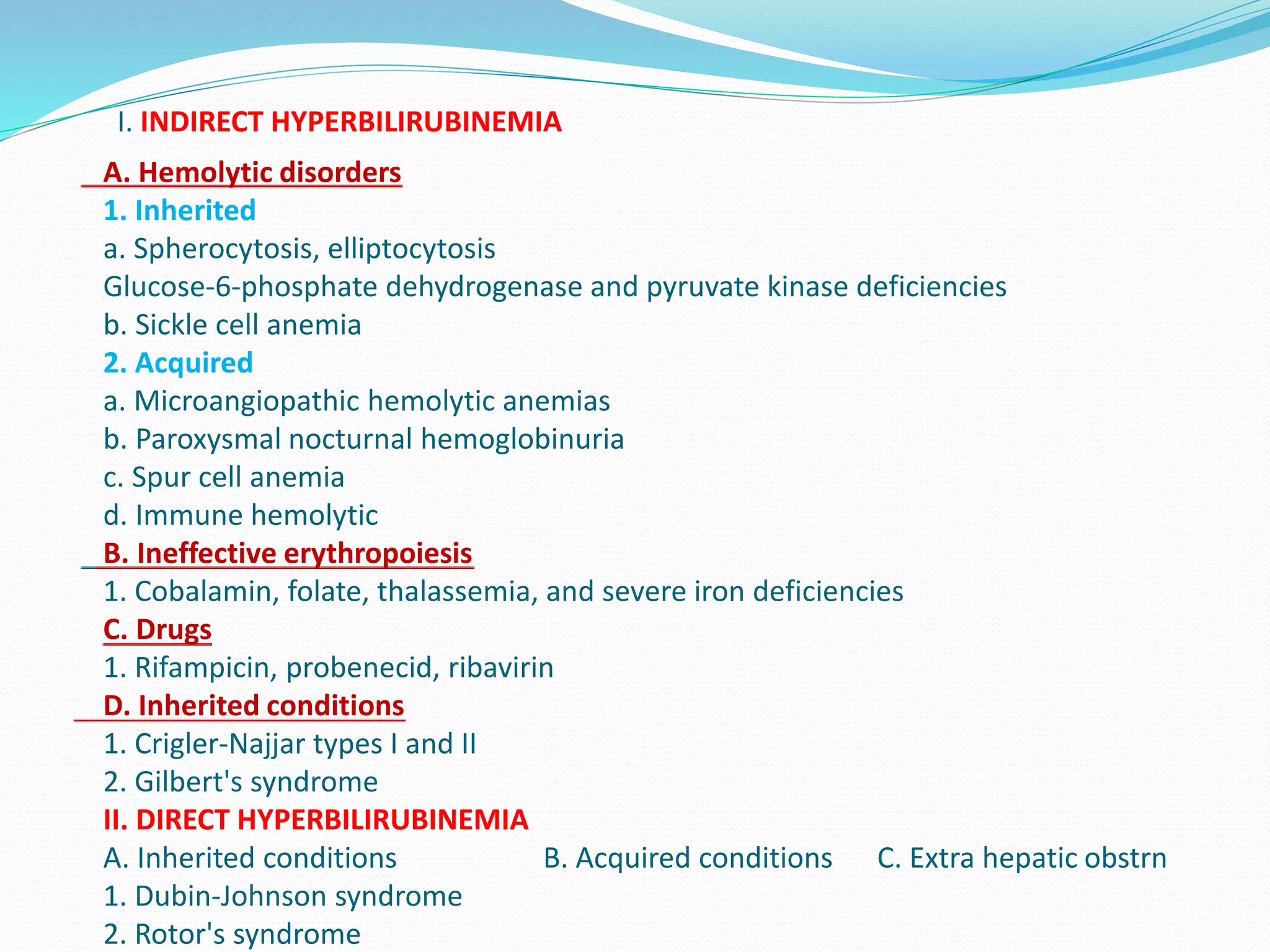
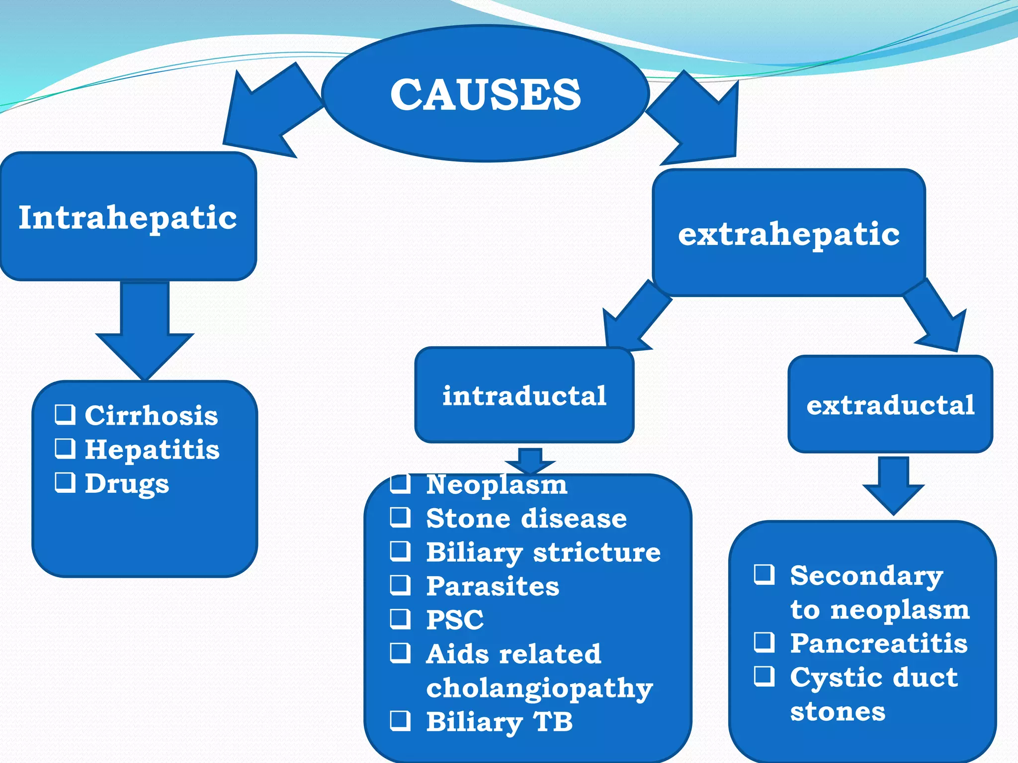
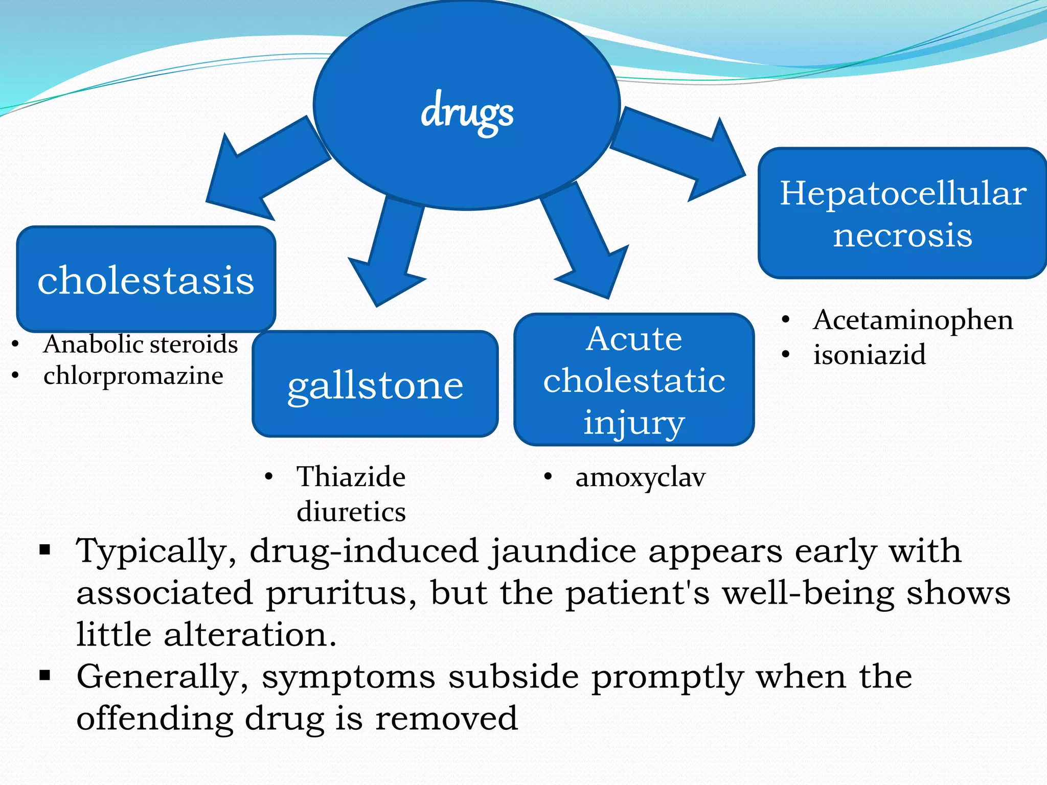
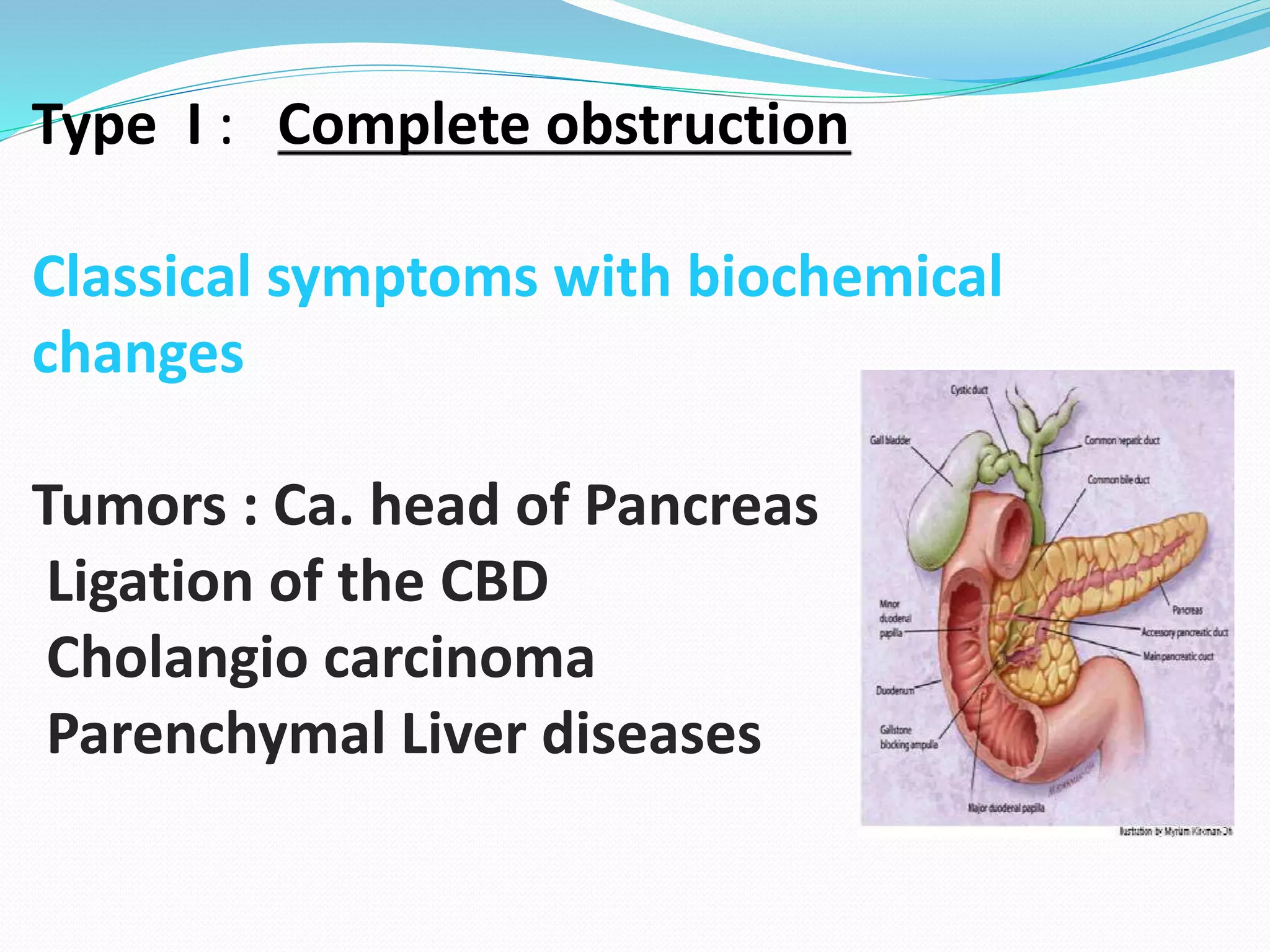
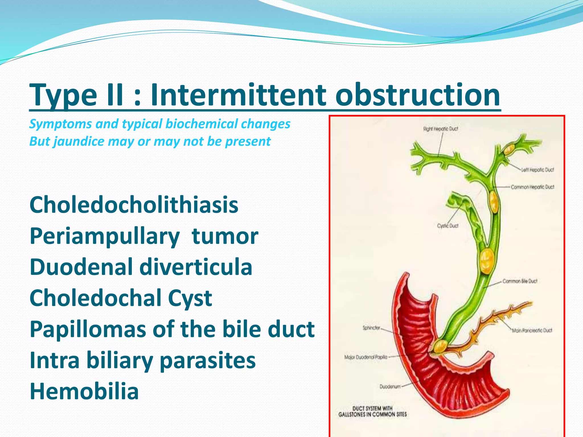
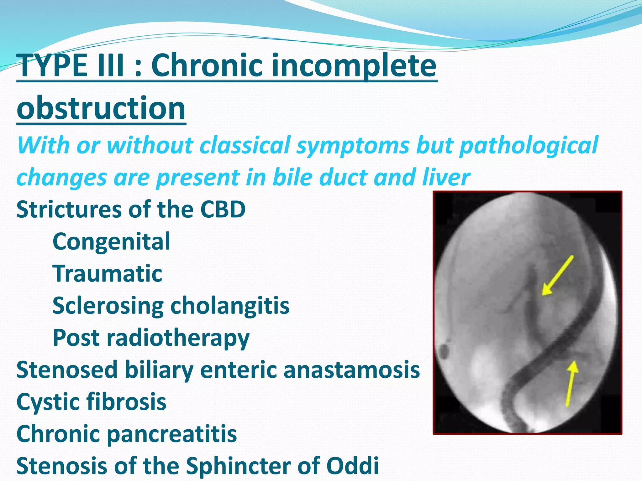
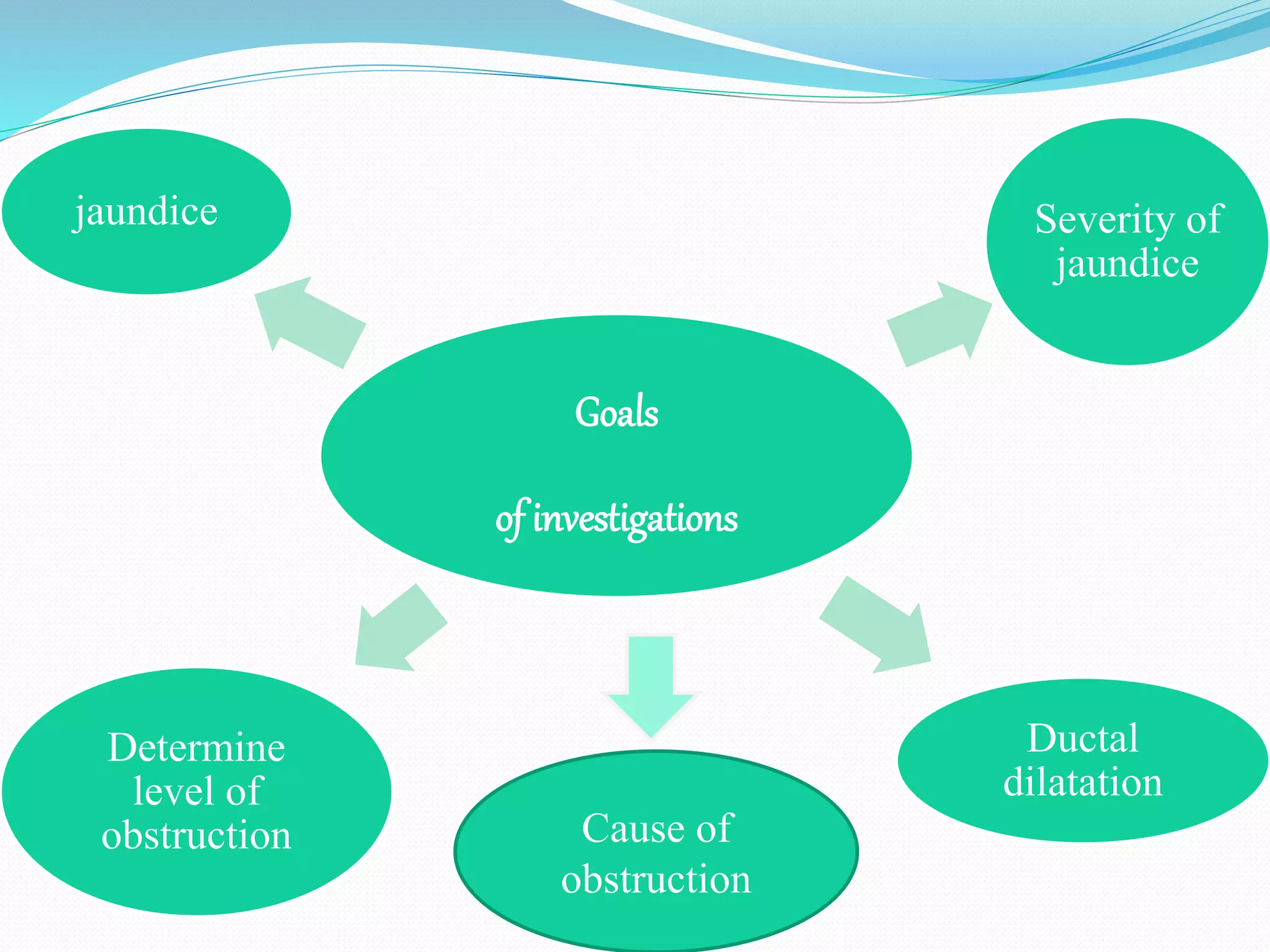
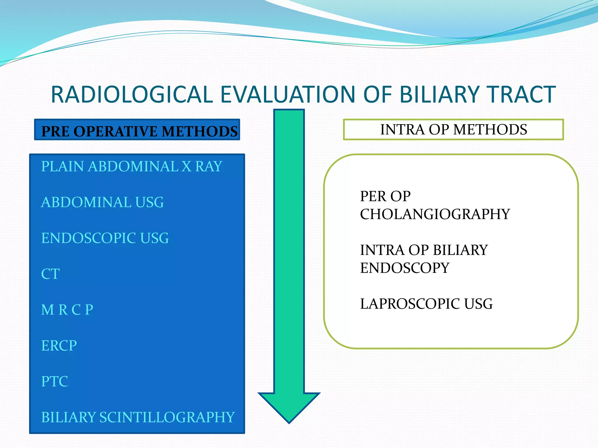
INVESTIGATIONS AND THEIR
RATIONALE
IN OBSTRUCTIVE JAUNDICE
INTRODUCTION
Jaundice, or icterus, is a yellowish discoloration of tissue
resulting from the deposition of bilirubin.
Tissue deposition of bilirubin occurs only in the presence of
serum hyperbilirubinemia and is a sign of either liver disease or,
less often, a hemolytic disorder
I. INDIRECT HYPERBILIRUBINEMIA
A. Hemolytic disorders
1. Inherited
a. Spherocytosis, elliptocytosis
Glucose-6-phosphate dehydrogenase and pyruvate kinase deficiencies
b. Sickle cell anemia
2. Acquired
a. Microangiopathic hemolytic anemias
b. Paroxysmal nocturnal hemoglobinuria
c. Spur cell anemia
d. Immune hemolytic
B. Ineffective erythropoiesis
1. Cobalamin, folate, thalassemia, and severe iron deficiencies
C. Drugs
1. Rifampicin, probenecid, ribavirin
D. Inherited conditions
1. Crigler-Najjar types I and II
2. Gilbert's syndrome
II. DIRECT HYPERBILIRUBINEMIA
A. Inherited conditions B. Acquired conditions C. Extra hepatic obstrn
1. Dubin-Johnson syndrome
2. Rotor's syndrome
CAUSES
Intrahepatic extrahepatic
intraductal extraductal Cirrhosis
 Hepatitis
 Drugs  Neoplasm
 Stone disease
 Biliary stricture
 Parasites
 PSC
 Aids related
cholangiopathy
 Biliary TB
 Secondary
to neoplasm
 Pancreatitis
 Cystic duct
stones
drugs
cholestasis
gallstone
Acute
cholestatic
injury
Hepatocellular
necrosis
• Anabolic steroids
• chlorpromazine
• Thiazide
diuretics
• amoxyclav
• Acetaminophen
• isoniazid
 Typically, drug-induced jaundice appears early with
associated pruritus, but the patient's well-being shows
little alteration.
 Generally, symptoms subside promptly when the
offending drug is removed
Clinical classification Of Obstructive
Jaundice
(Benjamin Classification)
Type I : Complete obstruction
Classical symptoms with biochemical
changes
Tumors : Ca. head of Pancreas
Ligation of the CBD
Cholangio carcinoma
Parenchymal Liver diseases
Type II : Intermittent obstruction
Symptoms and typical biochemical changes
But jaundice may or may not be present
Choledocholithiasis
Periampullary tumor
Duodenal diverticula
Choledochal Cyst
Papillomas of the bile duct
Intra biliary parasites
Hemobilia
TYPE III : Chronic incomplete
obstruction
With or without classical symptoms but pathological
changes are present in bile duct and liver
Strictures of the CBD
Congenital
Traumatic
Sclerosing cholangitis
Post radiotherapy
Stenosed biliary enteric anastamosis
Cystic fibrosis
Chronic pancreatitis
Stenosis of the Sphincter of Oddi
TYPE IV : Segmental Obstruction
one or more segment of intrahepatic biliary tract
is obstructed
Traumatic
Sclerosing cholangitis
Intra hepatic stones
Cholangio carcinoma
INVESTIGATIONS IN OBSTRUCTIVE
JAUNDICE
LABORATORY
INVESTIGATIONS
RADIOLOGICAL
INVESTIGATIONS
Goals
of investigations
Determine
level of
obstruction
Severity of
jaundice
Ductal
dilatation
jaundice
Cause of
obstruction
ROUTINE INVESTIGATIONS
1. HB
2. TLC
3. DLC
4. RFT ( serum urea, serum creatinine, serum sodium, serum
potassium )
5.BLOOD SUGAR
TESTS FOR ASSESSMENT OF LIVER FUNCTION
Tests for liver functioning
Based on
detoxification
& excretory
function
Enzymes
indicating
liver injury
Measure
biosynthetic
function
Damage to
hepatocytes
cholestasis
Serum
bilirubin
Urine
bilirubin
Blood
ammonia Aspartate
aminotransferase
Alanine
aminotransferase
Alkaline
phosphatase
5 nucleotidase
GGT
Serum albumin
Serum globulin
Coagulation
factors
 Bilirubin
Rise by 25-43 micromol/litre/day
Mechanism of hyperbilirubinemia
--- Biliary venous & biliary regurgitation of conjugated bilirubin due to
disruption of tight intracellular junction
--- Trans hepatocytic regurgitation due to reversal of the secretory
polarity of hepatocytes
--- Rupture of dilated canaliculi in to sinusoids due to necrosis of
hepatocytes
BILIRUBIN METABOLISM
SGOT AND SGPT LEVELS
SGOT (AST)/ ASPARTATE TRANSAMINASE
* Marker for hepatocellular toxicity
* Along with ALT is considered biomarker for liver health
* Non specific
* 2 isoenzymes
* Normal Values….
MALES 8-40 IU/L
FEMALES 6-34 IU/L
SGPT ( ALT ) / ALANINE AMINOTRANSFERASE
* Better predictor of hepatic injury than SGOT alone
* Significant elevations in HEPATITIS
INFECTIOUS MONONUCLEOSIS
CHF
* NORMAL VALUES IN
MALES < 50 IU/L
FEMALES < 32 IU/L
ALKALINE PHOSPHATSE
*Most sensitive indicator Of EXTRA HEPATIC BILIARY OBSTRUCTION
* Factor responsible are
Biliary component regurgitation
Increase in hepatic synthesis
* Biliary component is secreted by BILIARY DUCTULAR ENDOTHELIUM
* Normal range 20-140 IU/L
* May remain elevated for a long time even after the obstruction is
relieved
GAMMA GLUTAMYL TRANSFERASE & 5’NUCLEOTIDASE
GGT
* Predominantly used as a marker for liver diseases
* enhanced sensitivity for detection of BILIARY OBSTRUCTION if
correlated with ALKALINE PHOSPHATASE
* NORMAL VALUE 0-51 IU/L
5’ NUCLEOTIDASE
* An enzyme synthesized in liver
* Values if grossly elevated is indicative of biliary obstruction
* NORMAL VALUE 2-17 UNITS/L
Measure biosynthetic function
serum albumin
normal value 3.5 – 5.5 gm /dl
prothrombin time
normal value 12 – 14 sec
URINE ANALYSIS
1 Bile salts
2 Bile pigments
3 Urobilinogen
STOOL EXAMINATION
1 Occult blood
RADIOLOGICAL EVALUATION OF BILIARY TRACT
INTRA OP METHODSPRE OPERATIVE METHODS
PLAIN ABDOMINAL X RAY
ABDOMINAL USG
ENDOSCOPIC USG
CT
M R C P
ERCP
PTC
BILIARY SCINTILLOGRAPHY
PER OP
CHOLANGIOGRAPHY
INTRA OP BILIARY
ENDOSCOPY
LAPROSCOPIC USG
IMAGING GOALS
* To confirm the presence of an extrahepatic obstruction
* To determine the level of the obstruction
* To identify the specific cause of the obstruction
* To provide complementary information relating to the
underlying diagnosis (eg., Staging information in cases of
malignancy).
* What is the best therapeutic approach
PLAIN X RAY
* Cholelithiasis in 10-20 % of patients with radio opaque stones
* Radiolucent gas in a BI and TRI RADIATE FISSURE, in centre of
stone
* May sometimes show rare cases of calcification of GB
(PORCELAIN GB )
* Gas in wall of GB ( EMPHYSEMATOUS CHOLECYSTITIS)
* SPECKLED CALCIFICATION in the head of pancreas suggestive
of CHRONIC PANCREATITIS
* DUCT DILATATION WILL NOT BE REVEALED IN PLAIN FILMS
RADIO OPAQUE STONES IN GALL BALDDER
PORCELAIN GALL BLADDER
GAS IN GALL BLADDER AND ITS WALLS
ABDOMINAL ULTRASONOGRAPHY
* Is the initial imaging modality of choice as
- it is accurate
- readily available
- quick to perform
- inexpensive
OPERATOR DEPENDANT AND MAY GIVE SUBOPTIMAL RESULTS DUE TO
EXCESSIVE BODY FAT AND BOWEL GAS
* Biliary obstruction is characterized by BILIARY DILATATION
THIS DILATATION MAY BE CONSPICUOUSLY ABSENT IN 15 % OF
PATIENTS
* Prospective evaluation of USG suggests that level of obstruction can be
defined in 90 % of the cases
* COLOR FLOW DOPPLER SONOGRAPHY may assist in distinguishing dilated
ducts from Portal venous and Hepatic arterial branches
* Provides useful information about the nature and etiology of BILIARY
OBSTRUCTION
* Mass lesion visualization is possible
THE RELIABILITY WITH WHICH A BENIGN DISEASE MAY BE
DISTINGUISHED FROM A MALIGNANT PROCESS REMAINS UNCLEAR
*Upper limits of normal diameter of
CBD-8mm
CHD-6mm
ENDOSCOPIC ULTRASOUND (EUS)
Combines Endoscopy and US
Higher-frequency ultrasonic waves compared to traditional US (3.5 MHz vs. 20 MHz)
and allows diagnostic tissue sampling via EUS-guided fine-needle aspiration (EUS-
FNA).
EUS has been reported to have up to a 98% diagnostic accuracy in patients with
obstructive jaundice
The sensitivity of EUS for the identification of focal mass lesions in pancreas has
been reported to be superior to that of CT scanning, both traditional and spiral,
particularly for tumors smaller than 3 cm in diameter.
Compared to MRCP for the diagnosis of biliary stricture, EUS has been reported to be
more specific (100% vs. 76%) and to have a much greater positive predictive value
(100% vs. 25%), although the two have equal sensitivity (67%).
The positive yield of eus-fna for cytology in patients with malignant obstruction has
been reported to be as high as 96%.
Endoscopic ultrasonography.
CBD, common bile duct; PD, pancreatic duct.
COMPUTED TOMOGRAPHY
* Unlike USG CT is less affected by body habitus and is less operator
dependant
* It allows visualisation of the liver,
bile ducts, gall bladder and pancreas and is particularly
useful in detecting hepatic and pancreatic lesions and
is the modality of choice in the staging of cancers of the liver,
gall bladder, bile ducts and pancreas.
* It can identify the extent
of the primary tumour and defines its relationship to other
organs and blood vessels
*Improvements in CT technology, such as multidetector scanners,
which allow for three-dimensional reconstruction of the
biliary tree have led to greater diagnostic accuracy and have
increased the accuracy of CT in assessing benign disease.
Computed tomography scan demonstrating a
gallstone
within the gall bladder (arrowed).
Computed tomography scan demonstrating a hilar mass.
Intraductal stones appear as target sign on ct
CT. 75-88% sensitive, 97%specific for Choledocholithiasis
79%sensitive, 100% specific for gallstones
.
MAGNETIC RESONANCE CHOLANGIOPANCREATOGRAPHY (MRCP)
•Noninvasive test to visualize the hepato biliary tree
•No contrast
•Fluid found in the biliary tree is hyper intense on T2-weighted images.
Surrounding structures do not enhance and can be suppressed during image
analysis.
•Sensitive in detecting biliary and pancreatic duct stones, strictures, or dilatations
within the biliary system.
•MRCP combined with conventional MR imaging of the abdomen can provide
information about surrounding structures (eg, pseudocysts, masses).
• ERCP and MRCP similarly effective in detecting malignant hilar and perihilar
obstruction
• MRCP is better able to determine the extent and type of tumor as compared to
ERCP
Absolute contraindications
cardiac pacemaker
cerebral aneurysm clips
ocular or cochlear implants
Fluid stasis in the adjacent duodenum or ascitic fluid
may produce image artifacts on MRCP, making it
difficult to clearly visualize the biliary tree.
MRCP Showing Choledocholithiasis
MRCP is also highly
accurate
 MRCP sensitivity
88-92%, specificity
91-98% in detecting
Choledocholithiasis
Endoscopic retrograde cholangio
pancreatography (ERCP )
 Its an invasive procedure
and has therapeutic
potential.
 Allows biopsy or brush cytology
 Stone extraction or stenting
COMPLICATIONS
 Pancreatitis
 Cholangitis
 Hemorrhage
 Sepsis
CONTRAINDICATIONS
 Unfavorable anatomy
 Pseudo cyst
 Red a/c pancreatitis
ERCP film showing Choledocholithiasis
Endoscopic retrograde cholangiopancreatography: partial
occlusion of the bile duct by a malignant stricture
Percutaneous Transhepatic Cholangiography
(PTC)
 PTC is indicated when
Percutaneous intervention
is needed and ERCP either
is inappropriate or has
failed.
 Can be used to drain biliary
obstructions.
Transhepatic cholangiogram showing a stricture of the
common hepatic duct
Radioisotope scanning
* Technetium-99m (99mTc)-labelled derivatives of iminodiacetic
acid (HIDA, IODIDA) when injected intravenously are selectively
taken up by the retroendothelial cells of the liver and
excreted into the bile.
* This allows for visualisation of the biliary
tree and gall bladder. In 90 per cent of normal individuals the
gall bladder is visualised within 30 minutes following injection
with 100 per cent being seen within 1 hour
* Non-visualisation of the gall bladder is suggestive of acute
cholecystitis. If the patient has a contracted gall bladder as often
seen in chronic cholecystitis, the gall bladder visualisation may
be reduced or delayed.
*Biliary scintigraphy may also be helpful in diagnosing bile
leaks and iatrogenic biliary obstruction.
It can identify and quantitate the leak thus helping the surgeon
determine whether or not an operative or conservative approach
is warranted
Dimethyl iminodiacetic acid (HIDA) scan.
INTRA OPERATIVE TECHNIQUES
A. PER OPERATIVE CHOLANGIOGRAPHY
* During open or laparoscopic cholecystectomy, a catheter can be
placed in the cystic duct and contrast injected directly into the
biliary tree. The technique defines the anatomy and in the main
is used to exclude the presence of stones within the bile ducts
*A single x-ray plate or image
intensifier can be used to obtain and review the images intraoperatively
*In addition, care should be
taken when injecting contrast not to introduce air bubbles into
the system as these may give the appearance of stones and lead
to a false-positive result
Normal common bile duct: gentle The common bile duct is dilated
infusion of contrast with multiple Stones
which passes without hindrance
into the duodenum.
Operative biliary endoscopy (choledochoscopy)
* At operation, a flexible fibre optic endoscope can be passed via
the cystic duct into the common bile duct enabling stone identification
and removal under direct vision
* The technique can
be combined with an x-ray image intensifier to ensure complete
clearance of the biliary tree.
* After exploration of the bile duct,
a tube can be left in the cystic duct remnant or in the common
bile duct (a T-tube) and drainage of the biliary tree established
*After 7–10 days, a track will be established. This track can be
used for the passage of a choledochoscope to remove residual
stones in the awake patient in an endoscopy suite.
LAPROSCOPIC ULTRASONOGRAPHY
* At laparoscopy the use of laparoscopic probe can be
used to image the extra hepatic biliary system
* Useful in BILIARY & PANCREATIC tumor staging and
identify the primary tumors and determine its
relationship to the major vessels such as hepatic artery,
superior mesenteric artery , portal vein and superior
mesenteric vein

Approach to a case of Obstructive jaundice

  • 1.
  • 2.
    INTRODUCTION Jaundice, or icterus,is a yellowish discoloration of tissue resulting from the deposition of bilirubin. Tissue deposition of bilirubin occurs only in the presence of serum hyperbilirubinemia and is a sign of either liver disease or, less often, a hemolytic disorder
  • 3.
    I. INDIRECT HYPERBILIRUBINEMIA A.Hemolytic disorders 1. Inherited a. Spherocytosis, elliptocytosis Glucose-6-phosphate dehydrogenase and pyruvate kinase deficiencies b. Sickle cell anemia 2. Acquired a. Microangiopathic hemolytic anemias b. Paroxysmal nocturnal hemoglobinuria c. Spur cell anemia d. Immune hemolytic B. Ineffective erythropoiesis 1. Cobalamin, folate, thalassemia, and severe iron deficiencies C. Drugs 1. Rifampicin, probenecid, ribavirin D. Inherited conditions 1. Crigler-Najjar types I and II 2. Gilbert's syndrome II. DIRECT HYPERBILIRUBINEMIA A. Inherited conditions B. Acquired conditions C. Extra hepatic obstrn 1. Dubin-Johnson syndrome 2. Rotor's syndrome
  • 4.
    CAUSES Intrahepatic extrahepatic intraductal extraductalCirrhosis  Hepatitis  Drugs  Neoplasm  Stone disease  Biliary stricture  Parasites  PSC  Aids related cholangiopathy  Biliary TB  Secondary to neoplasm  Pancreatitis  Cystic duct stones
  • 5.
    drugs cholestasis gallstone Acute cholestatic injury Hepatocellular necrosis • Anabolic steroids •chlorpromazine • Thiazide diuretics • amoxyclav • Acetaminophen • isoniazid  Typically, drug-induced jaundice appears early with associated pruritus, but the patient's well-being shows little alteration.  Generally, symptoms subside promptly when the offending drug is removed
  • 6.
    Clinical classification OfObstructive Jaundice (Benjamin Classification)
  • 7.
    Type I :Complete obstruction Classical symptoms with biochemical changes Tumors : Ca. head of Pancreas Ligation of the CBD Cholangio carcinoma Parenchymal Liver diseases
  • 8.
    Type II :Intermittent obstruction Symptoms and typical biochemical changes But jaundice may or may not be present Choledocholithiasis Periampullary tumor Duodenal diverticula Choledochal Cyst Papillomas of the bile duct Intra biliary parasites Hemobilia
  • 9.
    TYPE III :Chronic incomplete obstruction With or without classical symptoms but pathological changes are present in bile duct and liver Strictures of the CBD Congenital Traumatic Sclerosing cholangitis Post radiotherapy Stenosed biliary enteric anastamosis Cystic fibrosis Chronic pancreatitis Stenosis of the Sphincter of Oddi
  • 10.
    TYPE IV :Segmental Obstruction one or more segment of intrahepatic biliary tract is obstructed Traumatic Sclerosing cholangitis Intra hepatic stones Cholangio carcinoma
  • 11.
  • 12.
    Goals of investigations Determine level of obstruction Severityof jaundice Ductal dilatation jaundice Cause of obstruction
  • 13.
    ROUTINE INVESTIGATIONS 1. HB 2.TLC 3. DLC 4. RFT ( serum urea, serum creatinine, serum sodium, serum potassium ) 5.BLOOD SUGAR
  • 14.
    TESTS FOR ASSESSMENTOF LIVER FUNCTION
  • 15.
    Tests for liverfunctioning Based on detoxification & excretory function Enzymes indicating liver injury Measure biosynthetic function Damage to hepatocytes cholestasis Serum bilirubin Urine bilirubin Blood ammonia Aspartate aminotransferase Alanine aminotransferase Alkaline phosphatase 5 nucleotidase GGT Serum albumin Serum globulin Coagulation factors
  • 16.
     Bilirubin Rise by25-43 micromol/litre/day Mechanism of hyperbilirubinemia --- Biliary venous & biliary regurgitation of conjugated bilirubin due to disruption of tight intracellular junction --- Trans hepatocytic regurgitation due to reversal of the secretory polarity of hepatocytes --- Rupture of dilated canaliculi in to sinusoids due to necrosis of hepatocytes
  • 17.
  • 18.
    SGOT AND SGPTLEVELS SGOT (AST)/ ASPARTATE TRANSAMINASE * Marker for hepatocellular toxicity * Along with ALT is considered biomarker for liver health * Non specific * 2 isoenzymes * Normal Values…. MALES 8-40 IU/L FEMALES 6-34 IU/L
  • 19.
    SGPT ( ALT) / ALANINE AMINOTRANSFERASE * Better predictor of hepatic injury than SGOT alone * Significant elevations in HEPATITIS INFECTIOUS MONONUCLEOSIS CHF * NORMAL VALUES IN MALES < 50 IU/L FEMALES < 32 IU/L
  • 20.
    ALKALINE PHOSPHATSE *Most sensitiveindicator Of EXTRA HEPATIC BILIARY OBSTRUCTION * Factor responsible are Biliary component regurgitation Increase in hepatic synthesis * Biliary component is secreted by BILIARY DUCTULAR ENDOTHELIUM * Normal range 20-140 IU/L * May remain elevated for a long time even after the obstruction is relieved
  • 21.
    GAMMA GLUTAMYL TRANSFERASE& 5’NUCLEOTIDASE GGT * Predominantly used as a marker for liver diseases * enhanced sensitivity for detection of BILIARY OBSTRUCTION if correlated with ALKALINE PHOSPHATASE * NORMAL VALUE 0-51 IU/L 5’ NUCLEOTIDASE * An enzyme synthesized in liver * Values if grossly elevated is indicative of biliary obstruction * NORMAL VALUE 2-17 UNITS/L
  • 22.
    Measure biosynthetic function serumalbumin normal value 3.5 – 5.5 gm /dl prothrombin time normal value 12 – 14 sec
  • 23.
    URINE ANALYSIS 1 Bilesalts 2 Bile pigments 3 Urobilinogen STOOL EXAMINATION 1 Occult blood
  • 24.
    RADIOLOGICAL EVALUATION OFBILIARY TRACT INTRA OP METHODSPRE OPERATIVE METHODS PLAIN ABDOMINAL X RAY ABDOMINAL USG ENDOSCOPIC USG CT M R C P ERCP PTC BILIARY SCINTILLOGRAPHY PER OP CHOLANGIOGRAPHY INTRA OP BILIARY ENDOSCOPY LAPROSCOPIC USG
  • 25.
    IMAGING GOALS * Toconfirm the presence of an extrahepatic obstruction * To determine the level of the obstruction * To identify the specific cause of the obstruction * To provide complementary information relating to the underlying diagnosis (eg., Staging information in cases of malignancy). * What is the best therapeutic approach
  • 26.
    PLAIN X RAY *Cholelithiasis in 10-20 % of patients with radio opaque stones * Radiolucent gas in a BI and TRI RADIATE FISSURE, in centre of stone * May sometimes show rare cases of calcification of GB (PORCELAIN GB ) * Gas in wall of GB ( EMPHYSEMATOUS CHOLECYSTITIS) * SPECKLED CALCIFICATION in the head of pancreas suggestive of CHRONIC PANCREATITIS * DUCT DILATATION WILL NOT BE REVEALED IN PLAIN FILMS
  • 27.
    RADIO OPAQUE STONESIN GALL BALDDER
  • 28.
  • 29.
    GAS IN GALLBLADDER AND ITS WALLS
  • 30.
    ABDOMINAL ULTRASONOGRAPHY * Isthe initial imaging modality of choice as - it is accurate - readily available - quick to perform - inexpensive OPERATOR DEPENDANT AND MAY GIVE SUBOPTIMAL RESULTS DUE TO EXCESSIVE BODY FAT AND BOWEL GAS * Biliary obstruction is characterized by BILIARY DILATATION THIS DILATATION MAY BE CONSPICUOUSLY ABSENT IN 15 % OF PATIENTS * Prospective evaluation of USG suggests that level of obstruction can be defined in 90 % of the cases
  • 31.
    * COLOR FLOWDOPPLER SONOGRAPHY may assist in distinguishing dilated ducts from Portal venous and Hepatic arterial branches * Provides useful information about the nature and etiology of BILIARY OBSTRUCTION * Mass lesion visualization is possible THE RELIABILITY WITH WHICH A BENIGN DISEASE MAY BE DISTINGUISHED FROM A MALIGNANT PROCESS REMAINS UNCLEAR *Upper limits of normal diameter of CBD-8mm CHD-6mm
  • 33.
    ENDOSCOPIC ULTRASOUND (EUS) CombinesEndoscopy and US Higher-frequency ultrasonic waves compared to traditional US (3.5 MHz vs. 20 MHz) and allows diagnostic tissue sampling via EUS-guided fine-needle aspiration (EUS- FNA). EUS has been reported to have up to a 98% diagnostic accuracy in patients with obstructive jaundice The sensitivity of EUS for the identification of focal mass lesions in pancreas has been reported to be superior to that of CT scanning, both traditional and spiral, particularly for tumors smaller than 3 cm in diameter. Compared to MRCP for the diagnosis of biliary stricture, EUS has been reported to be more specific (100% vs. 76%) and to have a much greater positive predictive value (100% vs. 25%), although the two have equal sensitivity (67%). The positive yield of eus-fna for cytology in patients with malignant obstruction has been reported to be as high as 96%.
  • 34.
    Endoscopic ultrasonography. CBD, commonbile duct; PD, pancreatic duct.
  • 35.
    COMPUTED TOMOGRAPHY * UnlikeUSG CT is less affected by body habitus and is less operator dependant * It allows visualisation of the liver, bile ducts, gall bladder and pancreas and is particularly useful in detecting hepatic and pancreatic lesions and is the modality of choice in the staging of cancers of the liver, gall bladder, bile ducts and pancreas. * It can identify the extent of the primary tumour and defines its relationship to other organs and blood vessels *Improvements in CT technology, such as multidetector scanners, which allow for three-dimensional reconstruction of the biliary tree have led to greater diagnostic accuracy and have increased the accuracy of CT in assessing benign disease.
  • 36.
    Computed tomography scandemonstrating a gallstone within the gall bladder (arrowed).
  • 37.
    Computed tomography scandemonstrating a hilar mass.
  • 38.
    Intraductal stones appearas target sign on ct CT. 75-88% sensitive, 97%specific for Choledocholithiasis 79%sensitive, 100% specific for gallstones
  • 39.
    . MAGNETIC RESONANCE CHOLANGIOPANCREATOGRAPHY(MRCP) •Noninvasive test to visualize the hepato biliary tree •No contrast •Fluid found in the biliary tree is hyper intense on T2-weighted images. Surrounding structures do not enhance and can be suppressed during image analysis. •Sensitive in detecting biliary and pancreatic duct stones, strictures, or dilatations within the biliary system. •MRCP combined with conventional MR imaging of the abdomen can provide information about surrounding structures (eg, pseudocysts, masses). • ERCP and MRCP similarly effective in detecting malignant hilar and perihilar obstruction • MRCP is better able to determine the extent and type of tumor as compared to ERCP
  • 40.
    Absolute contraindications cardiac pacemaker cerebralaneurysm clips ocular or cochlear implants Fluid stasis in the adjacent duodenum or ascitic fluid may produce image artifacts on MRCP, making it difficult to clearly visualize the biliary tree.
  • 41.
  • 42.
    MRCP is alsohighly accurate  MRCP sensitivity 88-92%, specificity 91-98% in detecting Choledocholithiasis
  • 43.
    Endoscopic retrograde cholangio pancreatography(ERCP )  Its an invasive procedure and has therapeutic potential.  Allows biopsy or brush cytology  Stone extraction or stenting COMPLICATIONS  Pancreatitis  Cholangitis  Hemorrhage  Sepsis CONTRAINDICATIONS  Unfavorable anatomy  Pseudo cyst  Red a/c pancreatitis
  • 44.
    ERCP film showingCholedocholithiasis
  • 45.
    Endoscopic retrograde cholangiopancreatography:partial occlusion of the bile duct by a malignant stricture
  • 46.
    Percutaneous Transhepatic Cholangiography (PTC) PTC is indicated when Percutaneous intervention is needed and ERCP either is inappropriate or has failed.  Can be used to drain biliary obstructions.
  • 47.
    Transhepatic cholangiogram showinga stricture of the common hepatic duct
  • 48.
    Radioisotope scanning * Technetium-99m(99mTc)-labelled derivatives of iminodiacetic acid (HIDA, IODIDA) when injected intravenously are selectively taken up by the retroendothelial cells of the liver and excreted into the bile. * This allows for visualisation of the biliary tree and gall bladder. In 90 per cent of normal individuals the gall bladder is visualised within 30 minutes following injection with 100 per cent being seen within 1 hour * Non-visualisation of the gall bladder is suggestive of acute cholecystitis. If the patient has a contracted gall bladder as often seen in chronic cholecystitis, the gall bladder visualisation may be reduced or delayed. *Biliary scintigraphy may also be helpful in diagnosing bile leaks and iatrogenic biliary obstruction.
  • 49.
    It can identifyand quantitate the leak thus helping the surgeon determine whether or not an operative or conservative approach is warranted Dimethyl iminodiacetic acid (HIDA) scan.
  • 50.
    INTRA OPERATIVE TECHNIQUES A.PER OPERATIVE CHOLANGIOGRAPHY * During open or laparoscopic cholecystectomy, a catheter can be placed in the cystic duct and contrast injected directly into the biliary tree. The technique defines the anatomy and in the main is used to exclude the presence of stones within the bile ducts *A single x-ray plate or image intensifier can be used to obtain and review the images intraoperatively *In addition, care should be taken when injecting contrast not to introduce air bubbles into the system as these may give the appearance of stones and lead to a false-positive result
  • 51.
    Normal common bileduct: gentle The common bile duct is dilated infusion of contrast with multiple Stones which passes without hindrance into the duodenum.
  • 52.
    Operative biliary endoscopy(choledochoscopy) * At operation, a flexible fibre optic endoscope can be passed via the cystic duct into the common bile duct enabling stone identification and removal under direct vision * The technique can be combined with an x-ray image intensifier to ensure complete clearance of the biliary tree. * After exploration of the bile duct, a tube can be left in the cystic duct remnant or in the common bile duct (a T-tube) and drainage of the biliary tree established *After 7–10 days, a track will be established. This track can be used for the passage of a choledochoscope to remove residual stones in the awake patient in an endoscopy suite.
  • 53.
    LAPROSCOPIC ULTRASONOGRAPHY * Atlaparoscopy the use of laparoscopic probe can be used to image the extra hepatic biliary system * Useful in BILIARY & PANCREATIC tumor staging and identify the primary tumors and determine its relationship to the major vessels such as hepatic artery, superior mesenteric artery , portal vein and superior mesenteric vein