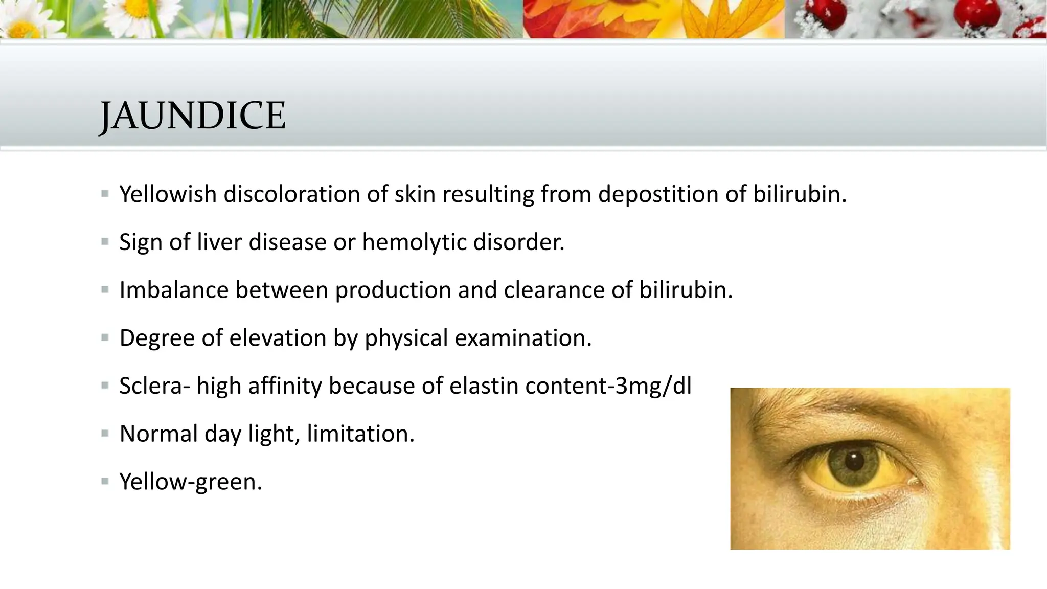
CLINICAL
APPROACH TO
JAUNDICE
DR.R.KARTHIKA-POST GRADUATE
INTERNAL MEDICINE-M1
8/1/14
JAUNDICE
 Yellowish discoloration of skin resulting from depostition of bilirubin.
 Sign of liver disease or hemolytic disorder.
 Imbalance between production and clearance of bilirubin.
 Degree of elevation by physical examination.
 Sclera- high affinity because of elastin content-3mg/dl
 Normal day light, limitation.
 Yellow-green.
 Yellowing of skin
 Carotenoderma-yellow color imparted to the skin because of carotenes.
 Carrots, green leafy vegetables, squash, peaches and oranges.
 Palms, soles, forehead and nasolabial folds.
 Spares the sclera.
 Quinacrine-4-37% yellowish discoloration of skin, also sclera.
PRODUCTION AND METABOLISM OF
BILIRUBIN.
 Tetrapyrrole pigment
 Break down product of heme-ferroprotoporphyrin IX
 70-80% breakdown of hemoglobin from senescent red blood cells.
 Prematurely destroyed erythroid cells in the bone marrow, myoglobin,
cytochromes.
 Reticuluendothelial cells of the spleen and liver.
 Bilirubin is insoluble
 Reversible covalent binding to albumin.
 Hepatocytes – glutathione superfamily.
 Prevents efflux back into serum
 Uridine diphosphate glucuronyl transferase.
 Conjugated bilirubin from the ER to the canalicular membrane of bile duct by
energy dependent mechanism involving MDR protein -2
MEASUREMENT OF SERUM BILIRUBIN
 Van der Bergh reaction.
 Bilirubin is exposed to diazotized sulfanilic acid
 Dipyrrylmethene azopigments which absorbs light maximally at 540nm
 Direct bilirubin.
 Total BR after the addition of alcohol.
 Normal-1mg/dl
 0.3mg-DBR
 DELTA FRACTION-albumin linked bilirubin fraction in pts with cholestasis and
hepatobiliary disease.
 Because of the longer half life of albumin-12-14 days
 Conjugated hyperbilirubinemia donot exhibit bilirubinuria because
 Albumin bound BR not excreted in renal glomeruli
 Serum BR also falls slowly.
MEASUREMENT OF URINE BILIRUBIN
 Unconjuated bilirubin – not found in urine.
 Conjugated bilirubin filtered by the glomerulus and reabsorbed by the prox
tubules.
 Urine dipstick test- ICTOTEST
 False negative in prolonged cholestasis due to conjugated BR bound to
albumin.
APPROACH TO THE PATIENT
 HYPERBILIRUBINEMIA
 Overproduction of BR
 Impaired uptake , conjuagation or excretion of bilirubin.
 Regurgitation of unconjugated or conjugated from damaged hepatocytes or
bileducts.
HISTORY
 Single most important part of the evaluation of the patient with unexplained
jaundice
 Duration of jaundice
 Use or exposure to medication-OTC, physician prescribed
 Complementary or alternative medicine-herbal or vitamin preparations or anabolic
steroids
 Parenteral exposures-transfusions, iv abuse
 Tattoos, sexual activity and alcohol history.
 Loss of weight or appetitite.
 Bleeding diathesis.
HISTORY TAKING
 Recent travel history
 Exposure of patients with jaundice
 Occupational history-contact with rats
 Place of origin-carriage of hepatitis
 Exposure to contaminated foods or water.
 Family history- hemolytic anemias, congenital hyperbilurbinemias and hepatitis
 Travel history
 Dyspepsia , fat intolerance or biliary colic
 Accompanying symptoms- arthralgia, myalgias, rash, anorexia, weight loss,
abdominal pain-choledocholithiasis and ascending cholangitis
 Fever
 Pruritis
 Changes in color of urine and stools
PHYSICAL EXAMINATION
 Assessment of patients nutritional status
 Temporal and proximal muscle wasting- long standing disease like ca or cirrhosis
 Scratch marks, purpura, fetor hepaticus
 Stigmata of chronic liver disease –spider nevi,
 palmar erythema,
 gynecomastia,
 caput medusa,
 dupuytrens contractures,
 parotid enlargement or testicular atrophy.- advanced alcoholic cirrhosis
 Enlarged left supraclavicular lymphnode
 Periumblical nodule-sister mary josephs
 Jvp-right heart failure
 Right sides pleural effusion
 Pedal edema, nails- clubbing and white nails.
 Tremors or flaps, KF ring.
 Abdominal examination-
 Size and consistency of liver and spleen.
 Enlarged left lobe of liver.
 Enlarged nodular liver-malignancy
 Tender hepatomegaly-alcoholic, viral or parasites, amyloid, hepatic
congestion.
 MURPHYS sign-cholecystitis.
 Palpable gall bladder and courvoiser law
 Ascites with jaundice- cirrhosis or malignancy with peritoneal spread
 Abdominal veins.
LABORATORY TESTS
 Total and direct serum bilirubin
 Aminotransferases
 Alkaline phosphatases
 Albumin
 Low-chronic
 Normal acute viral or choledocholithiasis.
 Prothromin time-prolonged jaundice, malabsorption of vit k or significant
hepatocellular dysfunction.
 Failure to correct with parenteral administration of vit k-severe hepatocellular injury.
UNCONJUGATED HYPERBILURUBINEMIA
 HEMOLYTIC DISORDERS
 ACQUIRED OR INHERITED
 Sickle cell anemia, heriditary spherocytosis or thalasemmia etc
 SBR rarely exceed 5mg/dl
 Higher levels when there is coexistent renal or hepatic dysfunction or crisis.
 Pigmented calcium bilirubinate gallstones and choledocholithiasis
 Acquired-HUS, PNH, malaria or babesiosis,
 Ineffective erythropoiesis-cobalamin, folate deficiency or iron def.
 Problem with hepatic uptake and conjugation of BR
 Rifampicin, probenicis, ribavirin
 Genetic- impaired conjugated
 Crigler najjar type I-rare
 Neonated
 SBR>20mg/dl
 Neurological impairment-kernicterus
 Death in infancy or childhood
 Complete absence of UDPGT actitivy
 Type ii
 Common, adulthood, SBR-6-25mg/dl
 Reduced UDPGT activity
 Induced by phenobarbital
 Susceptible to kernicterus under stress or illness
 GILBERT SYNDROME-1/3 UDPGT activity
 Very common-3-12% of general population.
 Mild unconjugated hyper bilirubinemia- less than 6mg/dl
 Serum levels fluctuate and identified during periods of fasting.
 Male predominance of 2-7:1
CONJUGATED HYPERBILIRUBINEMIA
 DUBIN JOHHSON
 ROTOR-hepatic storage of BR
 Asymptomatic jaundice
 Second generation of life
 Multiple drug resistance protein 2
 Altered excretion into bileducts
 Benign.
HEPATOCELLULAR JAUNDICE
 Viral hepatitis-IgM for hep A, Hep B surface antigen, core IgM antibody and
Hepatitis C viral RNA.
 Wilsons disease-ceruloplasmin, urinary copper, serum copper, hepatic copper
 Auto immune hepatitis young and middle aged women of any age
 ANA, SMA, LKM,
 Alcoholic hepatitis- AST:ALT> 2, AST rarely exceed 300IU/L
 Acute viral hepatitis and toxin related hepatitis aminotransferase >500U/L
 Cirrhosis –normal or only slight elevation in aminotranferasses.
DRUG INDUCED HEPATOTOXICITY
 Predictable drug reactions-dose dependent
 and affect all pts who ingest a toxic dose of the drug.
 Eg. Acetaminophen
 Unpredictable or idiosyncratic approach-not dose dependent
 Occurs in minority – eg isoniazid
 Environmental toxins- vinyl chloride, herbal preparations, pyrrolizidine
alkaloids, Jamaica bush tea, mushrooms.
CHOLESTATIC JAUNDICE
 Intra hepatic or extrahepatic cholestasis
 History, physical examination and lab test
 USG-high degree of sensitivity and specificity
 Inexpensive and non invasive
 But not the site or cause of obstruction especially distal CBD due to overlying
bowel gas.
 False negative test- partial obstruction to the common bile ducts
 Cirrhosis
 Primary sclerosing cholangitis-scarring prevent intrahepatic radicals to dilate
 MRCP, ERCP
 CT with MRCP- assessing head of pancreas and choledocholithiasis
 ERCP- gold standard- side viewing endoscopy per orally into the duodenum
 Catheter advanced.
 Injection of dye allows visualization of CBD.
 Removal of CBD stones, placement of stents.
 Fibrosing cholestatic hepatitis- in hepatis B and C
 Alcohol hepatitis
 Drugs- pure cholestasis- anabolic and contraceptive steroids
 Cholestatic hepatitis-chlorpromazine, erythromycin, imipramine, tolbutamide,
sulindac, cimetidine , TMP-SMX, ampicillin, dicloxacillin and clavulinic acid.
 Chronic cholestasis- chlorpromazine and prochlorperazine
 Primary biliary cirrhosis
 Autoimmune- middle aged women
 Progressive destruction of interlobular bile ducts
 Presence of antimitochondrial antibodies- 95%
 Primary sclerosing cholangitis
 Destruction and fibrosis of larger bile ducts
 Intrahepatic and extrahepatic –95%
 Multiple strictures of bile ducts with dilatations proximal to strictures.
 75% inflammatory bowel disease.
 Vanishing bile duct syndrome and adult bile ductopenia-
 Rare
 Decreased number of bile ducts in liver biopsy
 Chronic rejection after liver transplantation and
 Graft versus host reaction after BMT
 Sarcoidosis
 drugs
FAMILIAL
 Progressive intrahepatic cholestasis PFIC1-3 bile salt export pump, multidrug
resistant protein
 Benign recurrent cholestasis
 Autosommal recessive
 Manifests in childhood
 Recurrent episodes of jaundice and pruritis
 Self limited but can be debilitating.
 CHOLESTASIS OF PREGNANCY
 Second or third trimester
 Resolves after delivery
 Cause is unknown
 Inherited and cholestasis can be triggered by estrogen administration.
 TPN
 Non hepatobiliary sepsis
 Bening post operative cholestasis
 Paraneoplastic conditions
 Hodgkins lymphoma, medullary thyroid ca, renal cell ca, sarcoma, prostate
and gi ca
 STAUFFER’S syndrome- intrahepatic cholestasis with renal cell ca
 ICU- sepsis, shock liver , TPN,
 Jaundice with BMT- venoocclusive disease or GVHD
 Plasmodium falcifarum– combination of indirect BR from hemolysis and
cholestatic and hepatocellular jaundice.
 Poor outcomes in jaundice with encephalopathy and renal failure
 Weils disease-severe presentation of leptospirosis-jaundice, with renal failure
ass with headache and myalgias.
EXTRAHEPATIC CHOLESTATSIS
 MALIGNANT- pancreatic, gall bladder , ampullary and cholangiocarcinoma
 Similar to primary sclerosing cholangitis.
 Ampullary ca highest surgical cure rates and present as painless jaundice
 Hilar lymphadenopathy due to metastasis from other ca
 Extra hepatic cholestatis- benign
 Choledocholithiasis
 Mild upper right quadrant pain with only minimal elevation of enzymes to ascending
cholangitis , jaundice, sepsis and shock.
 Strictures.
 Chronic pancreatitis- rarely strictures of distal CBD.
 AIDS cholangiopathy- infection of bile duct epithelium with CMV or crytosporidia-
cholangiographic appreance similar to PSC.
 Elevated Serum alkaline phosphatase – mean 800IU/L
 Mirizzi syndrome
 Parasitic disease like ascariasis
TAKE HOME MESSAGE
 Jaundice is a hallmark of liver disease
 Through clinical examination and history becomes vital in all cases
 Classified as pre hepatic, hepatocellular and cholestatis although overlaps do
occur
 Biochemical and radiological evaluation helps in making a diagnosis.
 This is just a overview
THANK YOU.
SUMMER SECTION
HEADER LAYOUT
Subtitle
AUTUMN SECTION
HEADER LAYOUT
Subtitle
TITLE AND CONTENT LAYOUT WITH LIST
 Add your first bullet point here
 Add your second bullet point here
 Add your third bullet point here
CONTENT WITH
CAPTION LAYOUT
4.3
2.5
3.5
4.5
2.4
4.4
1.8
2.8
2 2
3
5
Category 1 Category 2 Category 3 Category 4
Series 1 Series 2 Series 3
Caption
TWO CONTENT LAYOUT WITH TABLE
 First bullet point here
 Second bullet point here
 Third bullet point here
Group 1 Group 2
Class 1 82 95
Class 2 76 88
Class 3 84 90
CONTENT WITH
CAPTION LAYOUT
WITH SMARTART
Step
3
Title
Step
2
Title
Step
1
Title
Task
description
Task
description
Task
description
Task
description
Task
description
Task
description
Caption
PICTURE WITH
CAPTION LAYOUT
Caption
clinicalapproachtojaundice-140123123013-phpapp01.pptx

clinicalapproachtojaundice-140123123013-phpapp01.pptx

  • 1.
  • 2.
    JAUNDICE  Yellowish discolorationof skin resulting from depostition of bilirubin.  Sign of liver disease or hemolytic disorder.  Imbalance between production and clearance of bilirubin.  Degree of elevation by physical examination.  Sclera- high affinity because of elastin content-3mg/dl  Normal day light, limitation.  Yellow-green.
  • 3.
     Yellowing ofskin  Carotenoderma-yellow color imparted to the skin because of carotenes.  Carrots, green leafy vegetables, squash, peaches and oranges.  Palms, soles, forehead and nasolabial folds.  Spares the sclera.  Quinacrine-4-37% yellowish discoloration of skin, also sclera.
  • 4.
    PRODUCTION AND METABOLISMOF BILIRUBIN.  Tetrapyrrole pigment  Break down product of heme-ferroprotoporphyrin IX  70-80% breakdown of hemoglobin from senescent red blood cells.  Prematurely destroyed erythroid cells in the bone marrow, myoglobin, cytochromes.  Reticuluendothelial cells of the spleen and liver.
  • 6.
     Bilirubin isinsoluble  Reversible covalent binding to albumin.  Hepatocytes – glutathione superfamily.  Prevents efflux back into serum  Uridine diphosphate glucuronyl transferase.  Conjugated bilirubin from the ER to the canalicular membrane of bile duct by energy dependent mechanism involving MDR protein -2
  • 8.
    MEASUREMENT OF SERUMBILIRUBIN  Van der Bergh reaction.  Bilirubin is exposed to diazotized sulfanilic acid  Dipyrrylmethene azopigments which absorbs light maximally at 540nm  Direct bilirubin.  Total BR after the addition of alcohol.  Normal-1mg/dl  0.3mg-DBR  DELTA FRACTION-albumin linked bilirubin fraction in pts with cholestasis and hepatobiliary disease.
  • 9.
     Because ofthe longer half life of albumin-12-14 days  Conjugated hyperbilirubinemia donot exhibit bilirubinuria because  Albumin bound BR not excreted in renal glomeruli  Serum BR also falls slowly.
  • 10.
    MEASUREMENT OF URINEBILIRUBIN  Unconjuated bilirubin – not found in urine.  Conjugated bilirubin filtered by the glomerulus and reabsorbed by the prox tubules.  Urine dipstick test- ICTOTEST  False negative in prolonged cholestasis due to conjugated BR bound to albumin.
  • 11.
    APPROACH TO THEPATIENT  HYPERBILIRUBINEMIA  Overproduction of BR  Impaired uptake , conjuagation or excretion of bilirubin.  Regurgitation of unconjugated or conjugated from damaged hepatocytes or bileducts.
  • 13.
    HISTORY  Single mostimportant part of the evaluation of the patient with unexplained jaundice  Duration of jaundice  Use or exposure to medication-OTC, physician prescribed  Complementary or alternative medicine-herbal or vitamin preparations or anabolic steroids  Parenteral exposures-transfusions, iv abuse  Tattoos, sexual activity and alcohol history.  Loss of weight or appetitite.  Bleeding diathesis.
  • 14.
  • 15.
     Recent travelhistory  Exposure of patients with jaundice  Occupational history-contact with rats  Place of origin-carriage of hepatitis  Exposure to contaminated foods or water.  Family history- hemolytic anemias, congenital hyperbilurbinemias and hepatitis  Travel history  Dyspepsia , fat intolerance or biliary colic  Accompanying symptoms- arthralgia, myalgias, rash, anorexia, weight loss, abdominal pain-choledocholithiasis and ascending cholangitis  Fever  Pruritis  Changes in color of urine and stools
  • 16.
    PHYSICAL EXAMINATION  Assessmentof patients nutritional status  Temporal and proximal muscle wasting- long standing disease like ca or cirrhosis  Scratch marks, purpura, fetor hepaticus  Stigmata of chronic liver disease –spider nevi,  palmar erythema,  gynecomastia,  caput medusa,  dupuytrens contractures,  parotid enlargement or testicular atrophy.- advanced alcoholic cirrhosis
  • 17.
     Enlarged leftsupraclavicular lymphnode  Periumblical nodule-sister mary josephs  Jvp-right heart failure  Right sides pleural effusion  Pedal edema, nails- clubbing and white nails.  Tremors or flaps, KF ring.  Abdominal examination-  Size and consistency of liver and spleen.  Enlarged left lobe of liver.
  • 18.
     Enlarged nodularliver-malignancy  Tender hepatomegaly-alcoholic, viral or parasites, amyloid, hepatic congestion.  MURPHYS sign-cholecystitis.  Palpable gall bladder and courvoiser law  Ascites with jaundice- cirrhosis or malignancy with peritoneal spread  Abdominal veins.
  • 20.
    LABORATORY TESTS  Totaland direct serum bilirubin  Aminotransferases  Alkaline phosphatases  Albumin  Low-chronic  Normal acute viral or choledocholithiasis.  Prothromin time-prolonged jaundice, malabsorption of vit k or significant hepatocellular dysfunction.  Failure to correct with parenteral administration of vit k-severe hepatocellular injury.
  • 21.
    UNCONJUGATED HYPERBILURUBINEMIA  HEMOLYTICDISORDERS  ACQUIRED OR INHERITED  Sickle cell anemia, heriditary spherocytosis or thalasemmia etc  SBR rarely exceed 5mg/dl  Higher levels when there is coexistent renal or hepatic dysfunction or crisis.  Pigmented calcium bilirubinate gallstones and choledocholithiasis  Acquired-HUS, PNH, malaria or babesiosis,  Ineffective erythropoiesis-cobalamin, folate deficiency or iron def.
  • 22.
     Problem withhepatic uptake and conjugation of BR  Rifampicin, probenicis, ribavirin  Genetic- impaired conjugated  Crigler najjar type I-rare  Neonated  SBR>20mg/dl  Neurological impairment-kernicterus  Death in infancy or childhood  Complete absence of UDPGT actitivy
  • 23.
     Type ii Common, adulthood, SBR-6-25mg/dl  Reduced UDPGT activity  Induced by phenobarbital  Susceptible to kernicterus under stress or illness  GILBERT SYNDROME-1/3 UDPGT activity  Very common-3-12% of general population.  Mild unconjugated hyper bilirubinemia- less than 6mg/dl  Serum levels fluctuate and identified during periods of fasting.  Male predominance of 2-7:1
  • 24.
    CONJUGATED HYPERBILIRUBINEMIA  DUBINJOHHSON  ROTOR-hepatic storage of BR  Asymptomatic jaundice  Second generation of life  Multiple drug resistance protein 2  Altered excretion into bileducts  Benign.
  • 25.
    HEPATOCELLULAR JAUNDICE  Viralhepatitis-IgM for hep A, Hep B surface antigen, core IgM antibody and Hepatitis C viral RNA.  Wilsons disease-ceruloplasmin, urinary copper, serum copper, hepatic copper  Auto immune hepatitis young and middle aged women of any age  ANA, SMA, LKM,  Alcoholic hepatitis- AST:ALT> 2, AST rarely exceed 300IU/L  Acute viral hepatitis and toxin related hepatitis aminotransferase >500U/L  Cirrhosis –normal or only slight elevation in aminotranferasses.
  • 26.
    DRUG INDUCED HEPATOTOXICITY Predictable drug reactions-dose dependent  and affect all pts who ingest a toxic dose of the drug.  Eg. Acetaminophen  Unpredictable or idiosyncratic approach-not dose dependent  Occurs in minority – eg isoniazid  Environmental toxins- vinyl chloride, herbal preparations, pyrrolizidine alkaloids, Jamaica bush tea, mushrooms.
  • 27.
    CHOLESTATIC JAUNDICE  Intrahepatic or extrahepatic cholestasis  History, physical examination and lab test  USG-high degree of sensitivity and specificity  Inexpensive and non invasive  But not the site or cause of obstruction especially distal CBD due to overlying bowel gas.  False negative test- partial obstruction to the common bile ducts  Cirrhosis  Primary sclerosing cholangitis-scarring prevent intrahepatic radicals to dilate
  • 28.
     MRCP, ERCP CT with MRCP- assessing head of pancreas and choledocholithiasis  ERCP- gold standard- side viewing endoscopy per orally into the duodenum  Catheter advanced.  Injection of dye allows visualization of CBD.  Removal of CBD stones, placement of stents.
  • 30.
     Fibrosing cholestatichepatitis- in hepatis B and C  Alcohol hepatitis  Drugs- pure cholestasis- anabolic and contraceptive steroids  Cholestatic hepatitis-chlorpromazine, erythromycin, imipramine, tolbutamide, sulindac, cimetidine , TMP-SMX, ampicillin, dicloxacillin and clavulinic acid.  Chronic cholestasis- chlorpromazine and prochlorperazine
  • 31.
     Primary biliarycirrhosis  Autoimmune- middle aged women  Progressive destruction of interlobular bile ducts  Presence of antimitochondrial antibodies- 95%  Primary sclerosing cholangitis  Destruction and fibrosis of larger bile ducts  Intrahepatic and extrahepatic –95%  Multiple strictures of bile ducts with dilatations proximal to strictures.  75% inflammatory bowel disease.
  • 32.
     Vanishing bileduct syndrome and adult bile ductopenia-  Rare  Decreased number of bile ducts in liver biopsy  Chronic rejection after liver transplantation and  Graft versus host reaction after BMT  Sarcoidosis  drugs
  • 33.
    FAMILIAL  Progressive intrahepaticcholestasis PFIC1-3 bile salt export pump, multidrug resistant protein  Benign recurrent cholestasis  Autosommal recessive  Manifests in childhood  Recurrent episodes of jaundice and pruritis  Self limited but can be debilitating.
  • 34.
     CHOLESTASIS OFPREGNANCY  Second or third trimester  Resolves after delivery  Cause is unknown  Inherited and cholestasis can be triggered by estrogen administration.
  • 35.
     TPN  Nonhepatobiliary sepsis  Bening post operative cholestasis  Paraneoplastic conditions  Hodgkins lymphoma, medullary thyroid ca, renal cell ca, sarcoma, prostate and gi ca  STAUFFER’S syndrome- intrahepatic cholestasis with renal cell ca  ICU- sepsis, shock liver , TPN,  Jaundice with BMT- venoocclusive disease or GVHD
  • 36.
     Plasmodium falcifarum–combination of indirect BR from hemolysis and cholestatic and hepatocellular jaundice.  Poor outcomes in jaundice with encephalopathy and renal failure  Weils disease-severe presentation of leptospirosis-jaundice, with renal failure ass with headache and myalgias.
  • 37.
    EXTRAHEPATIC CHOLESTATSIS  MALIGNANT-pancreatic, gall bladder , ampullary and cholangiocarcinoma  Similar to primary sclerosing cholangitis.  Ampullary ca highest surgical cure rates and present as painless jaundice  Hilar lymphadenopathy due to metastasis from other ca
  • 38.
     Extra hepaticcholestatis- benign  Choledocholithiasis  Mild upper right quadrant pain with only minimal elevation of enzymes to ascending cholangitis , jaundice, sepsis and shock.  Strictures.  Chronic pancreatitis- rarely strictures of distal CBD.  AIDS cholangiopathy- infection of bile duct epithelium with CMV or crytosporidia- cholangiographic appreance similar to PSC.  Elevated Serum alkaline phosphatase – mean 800IU/L  Mirizzi syndrome  Parasitic disease like ascariasis
  • 39.
    TAKE HOME MESSAGE Jaundice is a hallmark of liver disease  Through clinical examination and history becomes vital in all cases  Classified as pre hepatic, hepatocellular and cholestatis although overlaps do occur  Biochemical and radiological evaluation helps in making a diagnosis.  This is just a overview
  • 40.
  • 43.
  • 44.
  • 45.
    TITLE AND CONTENTLAYOUT WITH LIST  Add your first bullet point here  Add your second bullet point here  Add your third bullet point here
  • 46.
    CONTENT WITH CAPTION LAYOUT 4.3 2.5 3.5 4.5 2.4 4.4 1.8 2.8 22 3 5 Category 1 Category 2 Category 3 Category 4 Series 1 Series 2 Series 3 Caption
  • 47.
    TWO CONTENT LAYOUTWITH TABLE  First bullet point here  Second bullet point here  Third bullet point here Group 1 Group 2 Class 1 82 95 Class 2 76 88 Class 3 84 90
  • 48.
    CONTENT WITH CAPTION LAYOUT WITHSMARTART Step 3 Title Step 2 Title Step 1 Title Task description Task description Task description Task description Task description Task description Caption
  • 49.