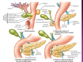
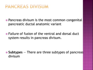
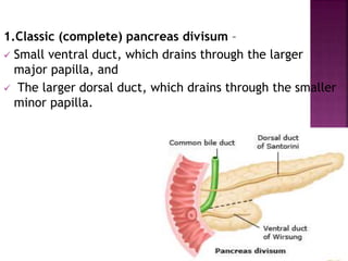
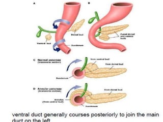
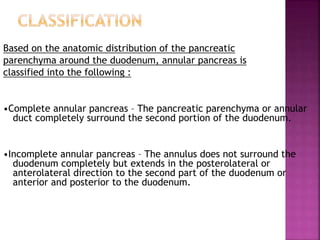
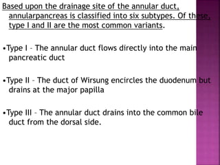
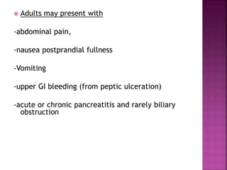
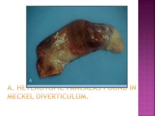
Dr.dipesh k.k
 During the fifth week of gestation, the pancreas
begins forming at the junction of the foregut and
midgut.
 Dorsal bud:-upper part of head ,body and tail-
Accessory pancreatic duct
 Ventral bud:-lower part of head,uncinate process-
main pancreatic duct
 Pancreas divisum is the most common congenital
pancreatic ductal anatomic variant
 Failure of fusion of the ventral and dorsal duct
system results in pancreas divisum.
 Subtypes — There are three subtypes of pancreas
divisum
1.Classic (complete) pancreas divisum –
 Small ventral duct, which drains through the larger
major papilla, and
 The larger dorsal duct, which drains through the smaller
minor papilla.
2.Incomplete pancreas divisum –
 A small branch of the ventral duct
communicates with the dorsal duct.
3.Reverse divisum –
 The accessory duct of Santorini does not
connect with the genu of the main
pancreatic duct.
 Pancreas divisum is associated with
recurrent pancreatitis.
 A recent pediatric study state that among
52 children with relapsing or chronic
pancreatitis, 10 had variants of pancreas
divisum.
EPIDEMIOLOGY
 4–14% of the population autopsy series
 3–8% at ERCP
 9% at MRCP
Lehman GA, Sherman S Gastrointest Endosc Clin N Am 1998
INDIAN DATA
 4.5% male and 6% female cadavers.
Sahni D, et al. TropGastroenterol. 2001 Oct-Dec;22(4):197-201
 Prevalence of 3.7% on ERCP,
 9.2% of patients presented with pancreatitis
Dhar A, et al. Indian J Gastroenterol. 1996 Jan;15(1):7-9
 Higher frequency of SPINK1 gene mutation
compared with healthy controls
Garg PK, et al. J Clin Gastroenterol. 2009 Jul 10
Clinical feature:-
 More than 95 percent asymptomatic
 In some-recurrent acute pancreatitis -
inadequate drainage of pancreatic secretions
via the minor papilla
Gold standard- ERCP
 Short and thin pancreatic ventral duct at the major papilla.
 filling of the dorsal duct at the minor papilla draining pancreas
from the tail to the anterior part of the head.
 NO connection to the ventral duct
Matos C, Metens T, Deviere J, DelhayeM,
Gastrointest Endosc 2001;53:728-33
MRCP
 Dorsal pancreatic duct has a constant caliber
 Crosses the common bile duct anteriorly
 Separated from a smaller ventral duct
 Esp. If secretin stimulated MRCP is used
 Secretin acts as a hydrographic endogenous contrast
agent
Endoscopic ultrasound
 If the ventral duct can be traced from the
major papilla through the body and the tail,
PD usually can be excluded.
SahaiAV. Gastrointest Endosc2002
 Attempt to improve the pancreatic outflow
through the minor papilla.
Options :-
 Endotherapy/ surgical options
Surgical:-
 Transduodenal sphincteroplasty of the minor
papilla with cholecystectomy and major
papilla sphincteroplasty
 Prospective trial (largest surgical trial)
 88 patients
 Sphincteroplasty
 Mean follow-up: 29 months
 74% of patients with acute recurrent pancreatitis
had good response compared to 34% with pain
only
 Restenosis rate : 8%
Warshaw AL, Et al. Evaluation and
treatment of the dominant dorsal duct
Syndrome . Am J Surg1990, 159:59–64
ENDOSCOPIC MANAGEMENT
 Dilation, sphincterotomy, stenting
 Balloon dilation reported a high rate of
pancreatitis and is not recommended
A prospective, randomized trial
 compared long-term dorsal duct stenting to continued
conservative therapy
 19 patients with pancreas divisum with recurrent
pancreatitis
 The stents (3 to 7 cm long with multiple side-holes) were
exchanged every three to four months and were left in
place for one year.
 The stented patients had a much higher rate of
improvement (90 versus 11 percent) due to statistically
significantly reductions in hospitalizations, emergency
department visits, and pancreatitis episodes.
 These benefits generally persisted over a mean 24-
month follow-up period after stent removal.
Ertan A, Gastrointest Endosc2000 Jul;52(1):9-14
 prolonged stent therapy remains largely
experimental and is not generally recommended.
 Associated with stent occlusion or migration,
 Pancreatitis
 pancreatic duct perforation, and
 pseudocyst formation
 Induction of ductal and parenchymal changes
indicating or simulating chronic pancreatitis
Gastrointest Endosc 1996
Sep;44(3):276-82.
ANNULAR PANCREAS:
 2nd most common anamoly
 In which a band of pancreatic tissue surrounds the
descending duodenum, either completely or incompletely,
and is in continuity with the head of the pancreas.
 Annular pancreas results from failure of the ventral bud
to rotate with the duodenum, causing envelopment of
duodenum.
 Three major theories have been proposed to explain the
development of annular pancreas:
1) Adherence of the ventral bud to the duodenal wall prior
to rotation, resulting in its persistence and encirclement
of the duodenum (Lecco's theory).
2) Persistence and enlargement of the left
ventral bud (Baldwin's theory) .
3)Hypertrophy and fusion of the ventral and
dorsal buds before rotation of the gut,
resulting in complete encirclement of the
duodenum.
Based on the anatomic distribution of the pancreatic
parenchyma around the duodenum, annular pancreas is
classified into the following :
•Complete annular pancreas – The pancreatic parenchyma or annular
duct completely surround the second portion of the duodenum.
•Incomplete annular pancreas – The annulus does not surround the
duodenum completely but extends in the posterolateral or
anterolateral direction to the second part of the duodenum or
anterior and posterior to the duodenum.
Based upon the drainage site of the annular duct,
annularpancreas is classified into six subtypes. Of these,
type I and II are the most common variants.
•Type I – The annular duct flows directly into the main
pancreatic duct
•Type II – The duct of Wirsung encircles the duodenum but
drains at the major papilla
•Type III – The annular duct drains into the common bile
duct from the dorsal side.
•Type IV – The annular duct drains into the
common bile duct without the duct of
Wirsung
•Type V – The annular duct drains into the duct
of Santorini from the ventral site
•Type VI – The annular duct drains into the
duct of Santorini with malfusion
 Two thirds of patients remain asymptomatic for life
 One half of patients become symptomatic at birth or
during the first year of life with signs of duodenal
obstruction.
 Adults may present with
-abdominal pain,
-nausea postprandial fullness
-Vomiting
-upper GI bleeding (from peptic ulceration)
-acute or chronic pancreatitis and rarely biliary
obstruction
 series have suggested that patients who
present with obstructive jaundice have an
underlying periampullary malignancy
DIAGNOSIS
 Infants: x-ray abdomen shows double bubble
sign
 In adults: CT Abdomen
TREATMENT:-
preferred surgical approach
 bypass surgery of the annulus,
duodenoduodenostomy, gastrojejunostomy, or a
duodenojejunostomy.
 Resection of the annulus should be avoided
it is associated with complications such as
pancreatitis,pancreatic fistula formation, and
incomplete relief of obstruction.
 In patients presenting with obstructive
jaundice, a thorough investigation must be
undertaken to evaluate for associated
periampullary malignancy.
 PROGNOSIS
40% mortality in infants because of associated
congenital anomalies
ECTOPIC PANCREAS
 Also known as heterotopic pancreatic tissue.
 Most are completely asymptomatic.
Common sites:
 Submucosa of Gastric antrum (70%)
 Proximal Duodenum /jejunum
 Ileum,colon,appendix,mesentry,GB, Meckel’s
 Diverticulum.
THANK YOU

Congenital anamolies of pancrease

  • 1.
  • 2.
     During thefifth week of gestation, the pancreas begins forming at the junction of the foregut and midgut.
  • 4.
     Dorsal bud:-upperpart of head ,body and tail- Accessory pancreatic duct  Ventral bud:-lower part of head,uncinate process- main pancreatic duct
  • 5.
     Pancreas divisumis the most common congenital pancreatic ductal anatomic variant  Failure of fusion of the ventral and dorsal duct system results in pancreas divisum.  Subtypes — There are three subtypes of pancreas divisum
  • 6.
    1.Classic (complete) pancreasdivisum –  Small ventral duct, which drains through the larger major papilla, and  The larger dorsal duct, which drains through the smaller minor papilla.
  • 7.
    2.Incomplete pancreas divisum–  A small branch of the ventral duct communicates with the dorsal duct.
  • 8.
    3.Reverse divisum – The accessory duct of Santorini does not connect with the genu of the main pancreatic duct.
  • 9.
     Pancreas divisumis associated with recurrent pancreatitis.  A recent pediatric study state that among 52 children with relapsing or chronic pancreatitis, 10 had variants of pancreas divisum.
  • 10.
    EPIDEMIOLOGY  4–14% ofthe population autopsy series  3–8% at ERCP  9% at MRCP Lehman GA, Sherman S Gastrointest Endosc Clin N Am 1998
  • 11.
    INDIAN DATA  4.5%male and 6% female cadavers. Sahni D, et al. TropGastroenterol. 2001 Oct-Dec;22(4):197-201  Prevalence of 3.7% on ERCP,  9.2% of patients presented with pancreatitis Dhar A, et al. Indian J Gastroenterol. 1996 Jan;15(1):7-9  Higher frequency of SPINK1 gene mutation compared with healthy controls Garg PK, et al. J Clin Gastroenterol. 2009 Jul 10
  • 12.
    Clinical feature:-  Morethan 95 percent asymptomatic  In some-recurrent acute pancreatitis - inadequate drainage of pancreatic secretions via the minor papilla
  • 13.
    Gold standard- ERCP Short and thin pancreatic ventral duct at the major papilla.  filling of the dorsal duct at the minor papilla draining pancreas from the tail to the anterior part of the head.  NO connection to the ventral duct Matos C, Metens T, Deviere J, DelhayeM, Gastrointest Endosc 2001;53:728-33
  • 15.
    MRCP  Dorsal pancreaticduct has a constant caliber  Crosses the common bile duct anteriorly  Separated from a smaller ventral duct  Esp. If secretin stimulated MRCP is used  Secretin acts as a hydrographic endogenous contrast agent
  • 17.
    Endoscopic ultrasound  Ifthe ventral duct can be traced from the major papilla through the body and the tail, PD usually can be excluded. SahaiAV. Gastrointest Endosc2002
  • 18.
     Attempt toimprove the pancreatic outflow through the minor papilla. Options :-  Endotherapy/ surgical options
  • 19.
    Surgical:-  Transduodenal sphincteroplastyof the minor papilla with cholecystectomy and major papilla sphincteroplasty
  • 20.
     Prospective trial(largest surgical trial)  88 patients  Sphincteroplasty  Mean follow-up: 29 months  74% of patients with acute recurrent pancreatitis had good response compared to 34% with pain only  Restenosis rate : 8% Warshaw AL, Et al. Evaluation and treatment of the dominant dorsal duct Syndrome . Am J Surg1990, 159:59–64
  • 21.
    ENDOSCOPIC MANAGEMENT  Dilation,sphincterotomy, stenting  Balloon dilation reported a high rate of pancreatitis and is not recommended
  • 22.
    A prospective, randomizedtrial  compared long-term dorsal duct stenting to continued conservative therapy  19 patients with pancreas divisum with recurrent pancreatitis  The stents (3 to 7 cm long with multiple side-holes) were exchanged every three to four months and were left in place for one year.
  • 23.
     The stentedpatients had a much higher rate of improvement (90 versus 11 percent) due to statistically significantly reductions in hospitalizations, emergency department visits, and pancreatitis episodes.  These benefits generally persisted over a mean 24- month follow-up period after stent removal. Ertan A, Gastrointest Endosc2000 Jul;52(1):9-14
  • 24.
     prolonged stenttherapy remains largely experimental and is not generally recommended.  Associated with stent occlusion or migration,  Pancreatitis  pancreatic duct perforation, and  pseudocyst formation  Induction of ductal and parenchymal changes indicating or simulating chronic pancreatitis Gastrointest Endosc 1996 Sep;44(3):276-82.
  • 26.
    ANNULAR PANCREAS:  2ndmost common anamoly  In which a band of pancreatic tissue surrounds the descending duodenum, either completely or incompletely, and is in continuity with the head of the pancreas.
  • 27.
     Annular pancreasresults from failure of the ventral bud to rotate with the duodenum, causing envelopment of duodenum.  Three major theories have been proposed to explain the development of annular pancreas: 1) Adherence of the ventral bud to the duodenal wall prior to rotation, resulting in its persistence and encirclement of the duodenum (Lecco's theory).
  • 28.
    2) Persistence andenlargement of the left ventral bud (Baldwin's theory) . 3)Hypertrophy and fusion of the ventral and dorsal buds before rotation of the gut, resulting in complete encirclement of the duodenum.
  • 30.
    Based on theanatomic distribution of the pancreatic parenchyma around the duodenum, annular pancreas is classified into the following : •Complete annular pancreas – The pancreatic parenchyma or annular duct completely surround the second portion of the duodenum. •Incomplete annular pancreas – The annulus does not surround the duodenum completely but extends in the posterolateral or anterolateral direction to the second part of the duodenum or anterior and posterior to the duodenum.
  • 31.
    Based upon thedrainage site of the annular duct, annularpancreas is classified into six subtypes. Of these, type I and II are the most common variants. •Type I – The annular duct flows directly into the main pancreatic duct •Type II – The duct of Wirsung encircles the duodenum but drains at the major papilla •Type III – The annular duct drains into the common bile duct from the dorsal side.
  • 32.
    •Type IV –The annular duct drains into the common bile duct without the duct of Wirsung •Type V – The annular duct drains into the duct of Santorini from the ventral site •Type VI – The annular duct drains into the duct of Santorini with malfusion
  • 33.
     Two thirdsof patients remain asymptomatic for life  One half of patients become symptomatic at birth or during the first year of life with signs of duodenal obstruction.
  • 34.
     Adults maypresent with -abdominal pain, -nausea postprandial fullness -Vomiting -upper GI bleeding (from peptic ulceration) -acute or chronic pancreatitis and rarely biliary obstruction
  • 35.
     series havesuggested that patients who present with obstructive jaundice have an underlying periampullary malignancy
  • 36.
    DIAGNOSIS  Infants: x-rayabdomen shows double bubble sign
  • 37.
     In adults:CT Abdomen
  • 39.
    TREATMENT:- preferred surgical approach bypass surgery of the annulus, duodenoduodenostomy, gastrojejunostomy, or a duodenojejunostomy.  Resection of the annulus should be avoided it is associated with complications such as pancreatitis,pancreatic fistula formation, and incomplete relief of obstruction.
  • 40.
     In patientspresenting with obstructive jaundice, a thorough investigation must be undertaken to evaluate for associated periampullary malignancy.
  • 41.
     PROGNOSIS 40% mortalityin infants because of associated congenital anomalies
  • 42.
    ECTOPIC PANCREAS  Alsoknown as heterotopic pancreatic tissue.  Most are completely asymptomatic. Common sites:  Submucosa of Gastric antrum (70%)  Proximal Duodenum /jejunum  Ileum,colon,appendix,mesentry,GB, Meckel’s  Diverticulum.
  • 45.