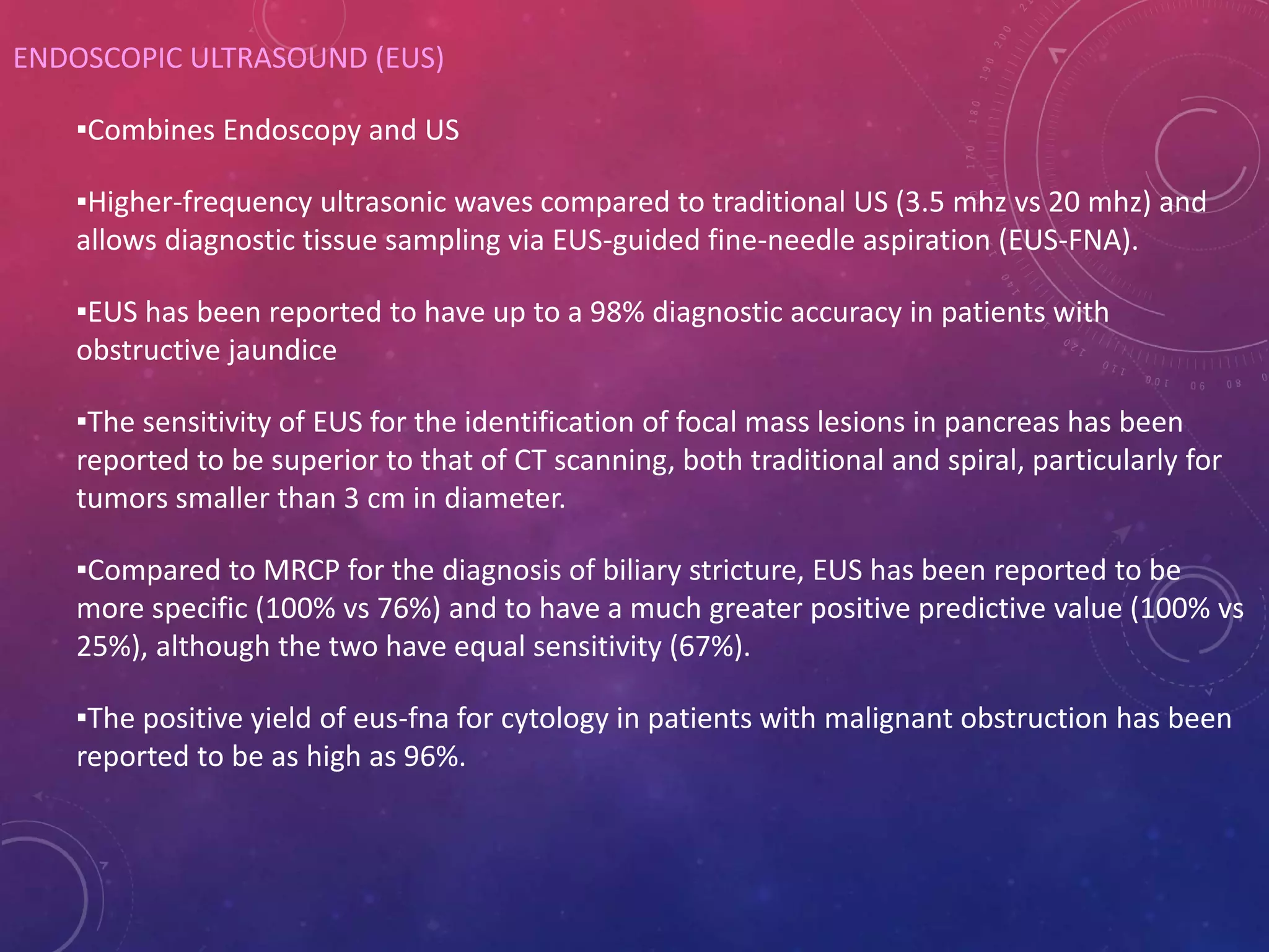
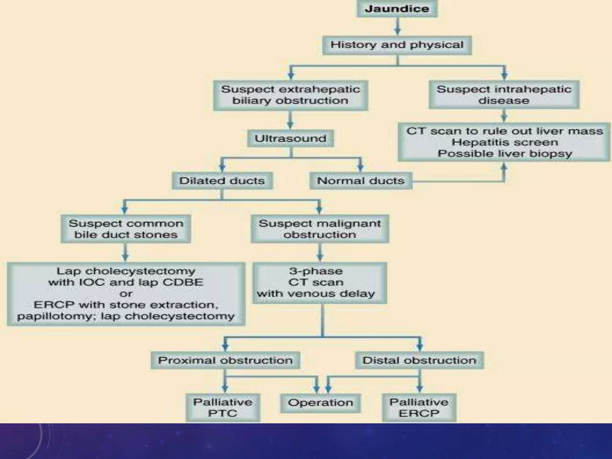
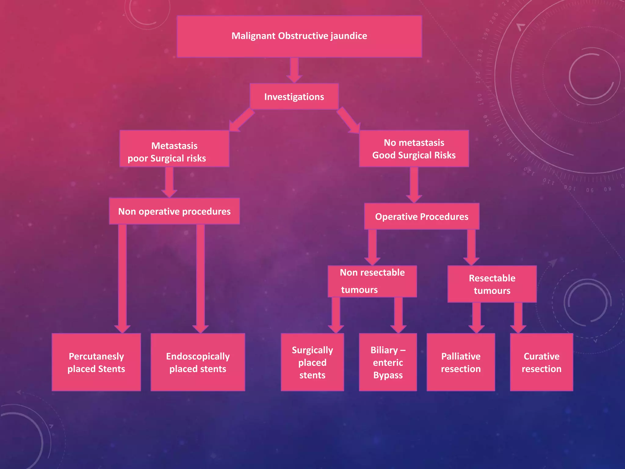
2. DEFINITION
Failure of normal amount of bile
to reach intestine
due to mechanical obstruction of the extra hepatic
biliary tree or within the porta hepatis
3. JAUNDICE
âȘJaundice (derived from French word âjauneâ for yellow) or icterus (Latin word for
Jaundice)
âȘYellowing of sclera at 3 mg%
âȘBilirubin has got high affinity for elastin and sclera has high elastin content
âȘYellowing of skin and mucous membrane at 6 mg%
âȘD/D beta carotenemia,Quinacrine therapy malingering with picric acid
âȘThey doesnât stain mucous membrane and sclera
âȘBilirubin level rise upto three weeks than stabilise
5. PHYSIOLOGICAL FACTS
âȘTotal bile flow-600ml/day(500-1000ml/day)
âȘHepatocyte component is -450ml/day
âȘCholangiocyte component-150ml/day
âȘIt depends on secretin stimulation
âȘTotal serum bilirubin is 0.3-1.2 mg/dl
âȘWith conjugated bilirubin<15 %
6. PHYSIOLOGY OF OBSTRUCTION
âȘNormal secretory pressure of bile is 15-25 cm of water
âȘAt 35 cm of water there is suppression of bile flow
âȘHigh pressure leads to cholangiovenous and cholangiolymphatic reflux of bile
âȘDilatation of bile duct and intra hepatic biliary radicals(IHBR)
âȘIHBR dilatation may be absent if there is secondary hepatic fibrosis or cirrhosis
7. PATHOPHYSIOLOGY
âȘIncrease in biliary pressure leads to
âȘDisruption of tight junctions between hepatocytes and bile duct cells with
increased permeability
âȘReflux of bile contents in liver sinusoids
âȘNeutrophil infiltration,increased fibrinogenesis and deposition of reticulin fiberes
in portal triad
âȘReticulin fibers gets converted in to type 1 collagen
âȘLaying down of collagen fibers leads to hepatic fibrosis obstruction of sinusoids
and secondary biliary cirrhosis and portal hypertension
âȘFibrosis can also lead to atrophy of obstructed liver
9. CHANGES IN LIVER BLOOD FLOW
âȘAcute obstruction
âȘ increase in hepatic arterial blood flow
âȘNo change in portal venous blood flow
âȘChronic obstruction
âȘDecrease in total liver blood flow , dilatation of sinusoids and elevation of portal
pressure
10. CARDIOVASCULAR EFFECTS
âȘDecreased cardiac contractability
âȘReduced left ventricular pressure
âȘImpaired response to beta agonist drugs
âȘDecreased peripheral vascular resistance
âȘBradycardia due to direct effect of bile salts on SA node.
Net result
âȘHypotensive patient
âȘExaggerated hypotensive response to bleeding
âȘMore prone to postoperative shock
11. RENAL FAILURE
âȘ10 % incidence with 70 % mortality
âȘFactors responsible are
âȘDecresed cardic function
âȘIncreased levels of ANP resulting in hypovolemia
âȘDecreased effect of bile salts on kidney mediated by increased prostaglandin E2
âȘEndotoxemia
âȘResulting in
âȘRenal vasoconstriction
âȘShunting of blood from cortex
âȘActivation of complement system peritubular and glomerular fibrin deposition
leading to tubular and cortical necrosis
12. IMMUNE SYSTEM
âȘDefects in cellular immunity
âȘImpaired T cell proliferation
âȘDecreased neutophil chemotaxis
âȘDefective bacterial phagocytosis
âȘDepressed function of RE system ie Kupffer cells
13. WOUND HEALING
âȘDelayed wound healing
âȘHigh incidence of wound dehiscence
âȘDecresed activity of enzyme Propyl hydroxylase in the skin
âȘThis helps in incorporation of proline in collagen
âȘDefective synthesis of collagen
14. COAGULATION FACTOR DEFECTS
âȘProlongation of Prothrombin time
âȘLoss of calcium
âȘEndotoxin induced damage to factor XI ,XII ,platelets
âȘLow grade DIC with increased fibrin degradation products
âȘThrombocytopenia from hyperspleenism
âȘDecreased absroption of fat solube vitamins A,D,E,K
15. ITCHING
âȘRetained bile salts
âȘItching disappears in terminal liver failure but bile salt level
still increased
âȘOther theory
âȘDue to endogenous opiate peptides
âȘInducing opiod receptor mediated scratching activity
of central origin
16. BIOCHEMICAL EFFECTS
âȘBilirubin
âȘRise by 25-43 micromol/litre/day
âȘMechanism of hyperbilirubinemia
âȘBiliary venous & biliary regurgitation of conjugated bilirubin due to
disruption of tight intracellular junction
âȘTrans hepatocytic regurgitation due to reversal of the secretory polarity of
hepatocytes
âȘRupture of dilated canaliculi in to sinusoids due to necrosis of hepatocytes
18. Why Bilirubin levels plateau
âȘIncreased excretion of bile pigments by kidney by products other than bilirubin not
giving DIAZO reaction
âȘA portion get covalently bounded to serum albumin
âȘThis protein bound bilirubin-Delta bilirubin,is not measurable by routine technique
21. HISTORY
âȘPrevious dyspepsia, fat intolerance
âȘJaundice- onset, course, itching
âȘPain
âȘPyrexia
âȘWeight loss
âȘDark urine and clay coloured stools
âȘTravel to endemic area
âȘContact with jaundice patient
âȘHistory of upper abdominal operation
âȘDrug intake ie ATT
âȘHistory of injection in preceding six months
22. CLINICAL EXAMINATION
âȘAge
âȘAnaemia - hemolysis, cancer , cirrhosis
âȘGross weight loss-malignancy
âȘHunched up position-chronic pancreatitis or ca pancreas
âȘFetor, flapping tremors,personality changes-impending hepatic coma
âȘSkin changes-Bruising,purpuric spots,spider naevi,palmar erythema,white
nails,loss of secondary sexual characters
23. ABDOMINAL EXAMINATION
âȘ Dilated peri umbalical veins- cirrhosis & portal collateral
circulation
âȘAscitis-Cirrhosis or malignant disease
âȘNodular liver
âȘCourvoisierâs Law-palpable non tender gall bladder in jaundice
patient-malignant biliary obstruction
âȘExceptions
âȘDouble impaction of stones
âȘImpaction of pancreatic calculus at ampulla of vater
âȘMirizzi syndrome
25. ALKALINE PHOSPHATSE(ALP)
âȘALP levels are elevated in nearly 100 % of patients with extra hepatic
obstruction except in some cases of intermittent obstruction.
âȘValues usually greater than 3 times the upper limit of reference range, and in
most typical cases, they exceed 5 times the upper limit.
âȘ An elevation less than 3 times the upper limit is evidence against complete
extra hepatic obstruction.
26. AST and ALT
âȘSerum enzymes that provide evidence of hepato cellular damage.ALT found primarily
in the liver, where as AST also found in heart ,kidney, skeletal muscle and brain
âȘAST is less specific for liver function. The levels of AST and ALT should be done
simultaneously since ALT can confirm the hepatic origin of the less specific but more
sensitive AST.
âȘIn extra hepatic obstruction usually AST levels are not elevated(< 10 times the upper
reference limit)
27. GAMMA âGLUTAMYL TRANSPEPTIDASE(GGTP)
âȘCorrelates with ALP level
âȘMost sensitive indicator of biliary tract disease
âȘBetter indicator of obstruction in children â levels are independent of age
âȘHelpful in the diagnosis of acute biliary tract obstruction in contrast to ALP because
ALP requires synthesis of fresh ALP and hence lags behind the onset of obstruction
28. 5- NUCLEOTIDASE
âȘThe principal value is to confirm the hepatic origin of an elevated ALP
âȘThis is particularly helpful in children, pregnant women and patients who
may have bone disease resulting in rise of ALP
âȘIt is more useful than ALP/GGTP in detecting hepatic metastasis
29. OTHER LAB INVESTIGATIONS
âȘProthrobin time
âȘSerum albumin
âȘStool for occult blood
âȘPresence of occult blood in the stools of a patient with jaundice must raise the
suspicion of malignancy.
30. Obstructive jaundice Medical jaundice
Serum Bilirubun
âȘ conjugated
âȘ unconjugated
+++
+
+
+++
Urobilinogen â â
Urinary Bilirubin + 0
Urinary Bile salts + 0
Serum ALP â No change
Serum GGTP â No change
Serum 5-nucleotidase â No change
Transaminases Mildly raised Markedly raised
33. INTERMITTENT OBSTRUCTION
âȘ Symptoms and typical biochemical changes
âȘClinically jaundice may or may not be present
âȘCauses
âȘCBD stones
âȘPeriampullary tumours
âȘDuodenal diverticulum
âȘCholedochal cyst
âȘBiliary parasites
âȘhemobilia
34. CHRONIC INCOMPLETE OBSTRUCTION
âȘWith or without classical symptoms or biochemical changes
âȘPathological changes in bile ducts or liver
âȘCauses
âȘStrictures of CBD
âȘStenosis of biliary-enteric anastamosis
âȘChronic pancreatitis
âȘCystic fibrosis
âȘSphincter of oddi stenosis
35. SEGMENTAL OBSTRUCTION
âȘ one or more segment of intrahepatic biliary tract obstructed
âȘCauses
âȘTraumatic
âȘIntrahepatic stones
âȘSclerosing cholangitis
âȘCholangiocarcinoma
36. BILIARY OBSTRUCTION
INTRINSIC
âȘ Ductal calculi
Primary - Develop de novo in bile ducts
Secondary - Migrate from gall bladder
âȘ Acute Cholangitis
âȘ Biliary Strictures
Idiopathic
Iatrogenic
âȘ Sclerosing Cholangitis
âȘ Parasites
âȘ Haemobilia
âȘ Benign Biliary Tumours
âȘ Cholangiocarcinoma
âȘ Carcinoma of ampulla of vater and Periampullary tumours
âȘ Intraductal secondary tumour seeding
37. BILIARY OBSTRUCTION
EXTRINSIC
âȘ Mirizzi syndrome
âȘ Pancreatitis- acute and chronic
âȘ Pancreatic pseudocyst
âȘ Carcinoma of gall bladder
âȘ Carcinoma of pancreas
âȘ Cystic tumours of pancreas
âȘ Metastatic carcinoma
âȘ Hepatocellular carcinoma
38. BILIARY OBSTRUCTION
CONGENITAL AND GENETIC DISORDERS
âȘ Biliary atresia
âȘ Choledocal cyst
âȘ Caroliâs disease
âȘ Progressive familial intra hepatic cholestasis
âȘ Primary biliary cirrhosis
âȘ Alpha 1 antitrypsin defeciency
âȘ Tyrosinemia
âȘ Neonatal hepatitis
âȘ Wilson disease
âȘ Others - dyskinesia of sphincter of odi
39. IMAGING GOALS
âȘTo confirm the presence of an extrahepatic obstruction
âȘTo determine the level of the obstruction, to identify the specific cause of the
obstruction
âȘTo provide complementary information relating to the underlying diagnosis (eg.,
Staging information in cases of malignancy).
40. Ultrasonography
Ultrasound of the abdomen is an extremely useful and accurate method for identifying
gallstones and pathologic changes in the gallbladder consistent with acute cholecystitis.
Abdominal ultrasound, if performed by an experienced operator, should be part of the routine
evaluation of patients suspected of having gallstone disease, given the high specificity (>98%)
and sensitivity (>95%) of this test for the diagnosis of cholelithiasis[1] ( Table 54-1 ). In addition
to identifying gallstones, ultrasound can also detail signs of cholecystitis such as thickening of
the gallbladder wall, pericholecystic fluid, and impacted stone in the neck of the gallbladder. It
is often the initial screening test for patients with suspected extrahepatic biliary obstruction (
Fig. 54-7 ). Dilation of the extrahepatic (>10 mm) or intrahepatic (>4 mm) bile ducts suggests
biliary obstruction. Intraoperative ultrasound is now used frequently to further evaluate
intrahepatic lesions, assess resectability, and determine involvement of vascular structures
41.
42.
43. .
MAGNETIC RESONANCE CHOLANGIOPANCREATOGRAPHY (MRCP)
âąNoninvasive test to visualize the hepato biliary tree
âąNo contrast
âąFluid found in the biliary tree is hyper intense on T2-weighted images. Surrounding structures
do not enhance and can be suppressed during image analysis.
âąSensitive in detecting biliary and pancreatic duct stones, strictures, or dilatations within the
biliary system.
âąMRCP combined with conventional MR imaging of the abdomen can provide information
about surrounding structures (eg, pseudocysts, masses).
âą ERCP and MRCP similarly effective in detecting malignant hilar and perihilar obstruction
âą MRCP is better able to determine the extent and type of tumor as compared to ERCP
44. Absolute contraindications
âȘcardiac pacemaker
âȘcerebral aneurysm clips
âȘocular or cochlear implants
âȘFluid stasis in the adjacent duodenum or ascitic fluid may produce image artifacts on
MRCP, making it difficult to clearly visualize the biliary tree.
45. ENDOSCOPIC ULTRASOUND (EUS)
âȘCombines Endoscopy and US
âȘHigher-frequency ultrasonic waves compared to traditional US (3.5 mhz vs 20 mhz) and
allows diagnostic tissue sampling via EUS-guided fine-needle aspiration (EUS-FNA).
âȘEUS has been reported to have up to a 98% diagnostic accuracy in patients with
obstructive jaundice
âȘThe sensitivity of EUS for the identification of focal mass lesions in pancreas has been
reported to be superior to that of CT scanning, both traditional and spiral, particularly for
tumors smaller than 3 cm in diameter.
âȘCompared to MRCP for the diagnosis of biliary stricture, EUS has been reported to be
more specific (100% vs 76%) and to have a much greater positive predictive value (100% vs
25%), although the two have equal sensitivity (67%).
âȘThe positive yield of eus-fna for cytology in patients with malignant obstruction has been
reported to be as high as 96%.
49. WORKUP AND MANAGEMENT OF POSTHEPATIC JAUNDICE
three possible clinical scenarios:DUCTAL OBSTRUCTION
SUSPECTED CHOLANGITIS
SUSPECTED CHOLEDOCHOLITHIASIS
WITHOUT CHOLANGITIS
SUSPECTED LESION OTHER THAN
CHOLEDOCHOLITHIASIS
50. SUSPECTED CHOLANGITIS
âȘA clinical picture compatible with acute suppurative cholangitis (charcot's triad or raynaud's
pentad) the most likely diagnosis is choledocholithiasis.
âȘAppropriate resuscitation, correction of any coagulopathies if present, and administration of
antibiotics
âȘERCP is indicated for diagnosis and treatment
âȘIf ERCP is unavailable or is not feasible (e.g., Because of previous roux-en-y reconstruction),
transhepatic drainage or surgery may be necessary
âȘMainstay of treatment of severe cholangitis is not just the administration of appropriate
antibiotics but rather the establishment of adequate biliary drainage.
51. SUSPECTED CHOLEDOCHOLITHIASIS WITHOUT CHOLANGITIS
âȘCholedocholithiasis is the most common cause of biliary obstruction.
âȘStrongly suspected if the jaundice is episodic or painful or if usg has shown presence of
gallstones or bile duct stones.
âȘPatients with suspected cbd stones should be referred for lap cholecystectomy with either
preoperative ERCP, intra operative cholangiography
âȘPreoperative ERCP in this setting of jaundice is prefered
âȘDiagnostic yield is high
âȘ Therapeutic-clearing the CBD of stones in 95% of cases.
âȘMany authors, however favor fully laparoscopic approach, in which CBD stone is detected in
the OR by means of intraoperative cholangiography and laparoscopic biliary clearance is
performed when choledocholithiasis is confirmed.
âȘ The optimal approach in a particular setting should be dictated by local expertise.
52. SUSPECTED LESION OTHER THAN CHOLEDOCHOLITHIASIS
âȘNo gallstones are seen
âȘClinical presentation is less acute (e.g., constant abdominal or back pain)
âȘAssociated constitutional symptoms (e.g., weight loss, fatigue, and long-standing anorexia)
âȘPossible causes of may be classified into three categories depending on the location of the
obstructing lesion
âȘthe upper third of the biliary tree
âȘthe middle third
âȘ the lower (distal) third
53. Upper-third obstruction
âȘ Polycystic liver disease
âȘ Caroli diseas
âȘ HCC
âȘ Oriental cholangiohepatitis
âȘ Hemobilia(e.g.,afterbiliarymani
pulation)
âȘ Iatrogenic bile duct injury
âȘ Cholangiocarcinoma
(Klatskin'stumor)
âȘ Sclerosing cholangitis
âȘ Papillomas of the bile duct
Mid-third obstruction
âȘCholangiocarcinoma
âȘSclerosing cholangitis
âȘPapillomas of the bile duct
âȘGallbladder cancer
âȘCholedochal cyst
âȘIntrabiliary parasites
âȘMirizzi syndrome
âȘExtrinsic nodal compression (e.g.,
lymphoma)
âȘIatrogenic bile duct injury
âȘCystic fibrosis
âȘBenign idiopathic bile duct stricture
Lower-third obstruction
âȘCholangiocarcinoma
âȘSclerosing cholangitis
âȘPapillomas of the bile duct
âȘPancreatic tumors
âȘAmpullary tumors
âȘChronic pancreatitis
âȘSphincter of Oddi dysfunction
âȘPapillary stenosis
âȘDuodenal diverticula
âȘPenetrating duodenal ulcer
âȘRetroduodenal adenopathy (e.g.,
lymphoma, carcinoid
ETIOLOGY
54. DIAGNOSIS AND ASSESSMENT OF RESECTABILITY
âȘInvolvement of the SUPERIOR MESENTERIC VEIN, THE PORTAL VEIN, THE SUPERIOR
MESENTERIC ARTERY, and the PORTA HEPATIS and on whether there is evidence of
significant LOCAL ADENOPATHY or EXTRAPANCREATIC EXTENSION OF TUMOR indicates
UNRESECTABILITY
âȘThe majority of lesions will be clearly unresectable, either because of tumor
extension or because of the presence of hepatic or peritoneal metastases
56. âȘIn the majority of patients with malignant obstructions, treatment is palliative rather than
curative.
âȘCholangiography and decompression of obstructed biliary system
âȘIn the absence of preexisting or concomitant hepatocellular dysfunction, drainage of one half
of the liver is generally sufficient for resolution of jaundice
57. âȘRoutine preoperative drainage of an obstructed biliary system does not benefit patients who
will soon undergo resection.1,2
âȘThere is evidence suggesting that in patients with either pancreatic 3,4 or hepatic malignancies,
routine preoperative direct cholangiography with decompression is associated with a higher
incidence of postoperative complications when tumor resection is ultimately carried out.
1. Pitt HA, Gomes AS, Lois JF: Does preoperative percutaneous biliary drainage reduce operative risk or
increase hospital cost? Ann Surg 201:545, 1985
2. McPherson GA, Benjamin IS, Hodgson HJ, et al: Preoperative percutaneous transhepatic biliary drainage:
results of a controlled trial. Br J Surg 71:371, 1984
3. Povoski SP, Karpeh MS Jr, Conlon KC, et al: Preoperative biliary drainage: impact on intraoperative bile
cultures and infectious morbidity and mortality after pancreaticoduodenectomy. J Gastrointest Surg 3:496,
1999
4. Sohn TA, Yeo CJ, Cameron JL, et al: Do preoperative biliary stents increase postpancreaticoduodenectomy
complications? J Gastrointest Surg 4:258, 2000
59. UPPER-THIRD OBSTRUCTION
PALLIATION.
âȘBecause the left hepatic duct has a long extrahepatic segment and is more accessible,
the preferred bypass -is a left hepaticojejunostomy
âȘ Laparoscopic bypass techniques that make use of segment 3 have been developed,
but their performance has yet to be formally assessed
60. RESECTION FOR CURE
âȘThe hilar plate is taken down to lengthen the hepatic duct segment available for subsequent
anastomosis.
âȘA formal hepatectomy or segmentectomy is required to ensure an adequate proximal margin of
resection
âȘ If the resection is carried out proximal to the hepatic duct bifurcation, several
cholangiojejunostomies have to be done to anastomose individual hepatic biliary branches.
âȘThe results of aggressive hilar tumor resections that included as much liver tissue as was
necessary to obtain a negative margin appear to justify this approach.
âȘ In cases of left hepatic involvement, resection of the caudate lobe is indicated as well.
61. MIDDLE-THIRD OBSTRUCTION
Palliation.
âȘSurgical bypass of middle-third lesions is technically simpler
âȘ Hepaticojejunostomy is done distal to the hepatic duct bifurcation,
âȘ Exposure of the hilar plate or the intrahepatic ducts is unnecessary.
Resection for cure.
âȘTumors in this part usually quite amenable to resection along with the lymphatic chains in the
porta hepatis.
âȘMirizzi syndrome -extrinsic obstruction of the CBD, either by causing inflammation of the
gallbladder wall or via direct impingement.
âȘTreatment of this syndrome - Hepaticojejunostomy
62. Lower-third obstruction
Palliation
âȘ The preferred bypass technique for lower-third lesions is a Roux-en-Y choledochojejunostomy.
âȘ Cholecystojejunostomy carries a higher risk of complications and subsequent development of
jaundice
Resection for cure.
âȘ Resection of a lower-third lesion usually involves a pancreaticoduodenectomy though
transduodenal ampullary resection may be an acceptable alternative for a small adenoma of the
ampulla
âȘFor optimal results, pancreaticoduodenectomy is best performed in specialized centers.
âȘpostoperative adjuvant therapy may improve the prognosis after resection of a pancreatic
adenocarcinoma
63. PALLIATION IN PATIENTS WITH ADVANCED MALIGNANT DISEASE
âȘWhen a patient has advanced malignant disease, drainage of the biliary system for palliation is
not routinely indicated, because the risk of complications related to the procedure may
outweigh the potential benefit
âȘThe best treatment for a patient with asymptomatic obstructive jaundice and liver metastases
may be supportive care alone.
âȘBiliary decompression is indicated if cholangitis or severe pruritus interferes with quality of life.
âȘStent placed with ercp to be the palliative modality of choice for advanced disease,
âȘUpper-third lesions may be managed most easily through the initial placement of an
internal/external catheter at the time of ptc.
64. âȘMetal expandable stents remain patent longer than large conventional plastic stents
âȘRCTs suggest that surgical biliary bypass should be reserved for patients who are expected to
survive for 6 months or longer because bypass is associated with more prolonged palliation at
the cost of greater initial morbidity.
âȘThe role of prophylactic gastric drainage at the time of operative biliary drainage remains
controversial,101,102
âȘ RCTs demonstrated a reduced incidence of subsequent clinical gastric outlet obstruction
when this measure was employed.
âȘ When a pancreatic malignancy is present, intraoperative celiac ganglion injection should be
performed for either prophylactic or therapeutic pain
65. POSTOPERATIVE JAUNDICE
âȘThe development of jaundice in the postoperative setting is approximately 1% of all surgical
patients after operation.120
âȘWhen jaundice occurs after a hepatobiliary procedure,
âȘAttributable to specific biliary causes,
âȘRetained cbd stones,
âȘPostoperative biliary leakage (through reabsorption of bile leaking into the peritoneum)
âȘ injury to the cbd
âȘDevelopment of biliary strictures
66. JAUNDICE WITHIN 48 HOURS OF THE OPERATION
âȘBreakdown of rbc âdue to multiple blood transfusions ,
âȘThe resorption of a large hematoma,
âȘTransfusion reaction.
âȘKnown underlying hemolytic anemia and may be precipitated specific drugs (e.G., Sulfa
drugs in a patient who has G6PD deficiency).
âȘGilbert syndrome may first manifest itself early in the postoperative period.
âȘIntraoperative hypotension or hypoxemia or the early development of heart failure can
lead to hyperbilirubinemia within 5 to 10 days after operation.
âȘThe hyperbilirubinemia may be associated with other end-organ damage (e.G., Acute
tubular necrosis).
âȘImpairment of renal function causes a decrease in bilirubin excretion and can be
responsible for a mild hyperbilirubinemia.
67. Jaundice developing 7 to 10 days after operation
âȘ In association with a medication-induced hepatitis attributable to an anesthetic agent.
âȘIncidence of 1/10,000 after an initial exposure.
âȘAdministration of antibiotics or other medications used in the perioperative setting
âȘJaundice associated with intrahepatic cholestasis is often a manifestation of a sepsis,
particularly in patients with mods
âȘ Jaundice may occur in as many as 30% of patients receiving total parenteral nutrition (tpn).
âȘIt may be due to steatosis, particularly with formulas containing large amounts of
carbohydrates.
âȘDecreased export of bilirubin from the hepatocytes may lead to cholestasis
âȘAcalculous cholecystitis or even ductal obstruction may develop as a result of sludge in the
gallbladder and the cbd.
68. âȘUnsuspected hepatic or post-hepatic causes (e.G., Occult cirrhosis, choledocholithiasis,
or cholecystitis)
âȘA rare cause of postoperative jaundice is the development of thyrotoxicosis.
âȘA diagnosis of exclusion- is so-called benign postoperative cholestasis, a primarily
cholestatic, self-limited process with no clearly demonstrable cause that typically arises
within 2 to 10 days after operation.
âȘMechanism-combination of an increased pigment load, impaired liver function, and
decreased renal bilirubin excretion caused by varying degrees of tubular necrosis.
âȘ The predominantly conjugated hyperbilirubinemia may reach 40 mg/dl and remain
elevated for as long as 3 weeks.