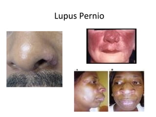
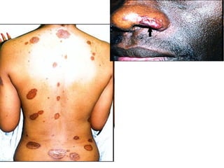
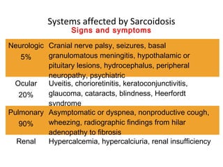
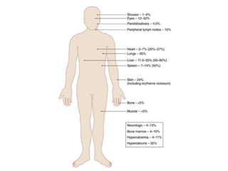
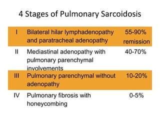
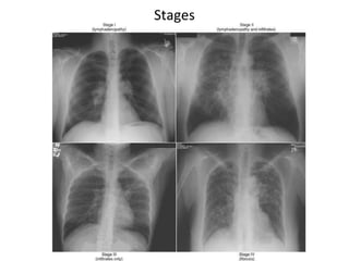
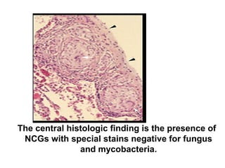
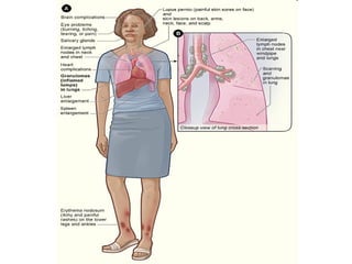
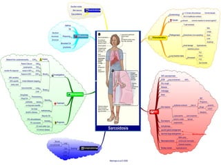
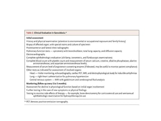
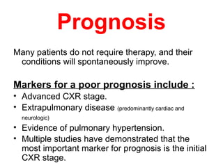
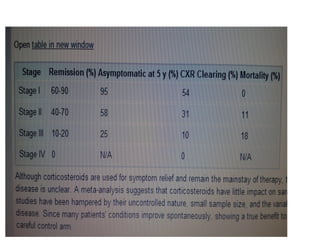
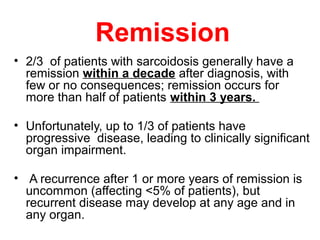
Sarcoidosis
Dr. Angelo Smith M.D
WHPL
Sarcoidosis
• Multisystem disorder of unknown etiology
that most commonly affects the lungs, but
can also affect other organs.
• Beethoven is thought to have been the first
person described with this condition.
Sarcoidosis is
manifested by the
presence of
noncaseating
granulomas (NCGs) in
affected organ tissues.
History of sarcoidosis
• In 1899, the pioneering Norwegian
dermatologist Caesar Boeck describe skin
nodules characterized by compact,
sharply defined foci of "epithelioid cells
with large pale nuclei and also a few giant
cells .
• Thinking this resembled sarcoma, he
called the condition "multiple benign
sarcoid of the skin.
Epidemiology
• All racial .
• All ethnic groups.
– More prevalent in Swedes, Danes, and US
blacks
• All ages (with the incidence peaking at 20 to 39 years).
• M-F ratio 2:1.
• Affects siblings of first- or second- degree
relatives in 15% of patients with
sarcoidosis.
Etiology and Pathogenesis
• Cause is unknown, although both genetic and
environmental factors suspected.
• Theory that disease develops in genetically
predetermined hosts who are exposed to certain
environmental agents that trigger an
exaggerated inflammatory immune response
leading to granuloma formation.
• Hallmark is noncaseating granulomas,
composed of a central core of epithelioid
histocytes and multinucleated giant cells.
• Activated T cells and macrophages
accumulate at site of inflammation.
• Release chemo attractants and GF’s lead
to cellular proliferation and granuloma
formation.
• Progressive granulomatous inflammation
leads to injury, dysfunction, and
destruction of the affected organs.
T cells, Macrophages
Chemoattractants
Growth Factors
Cellular proliferation
Granuloma
Fibrosi
s
Pathogenesis
The following have been suggested as
possible candidates that might play a
role in causing sarcoidosis:
• Mycobacteria, such as Mycobacterium tuberculosis, and
atypical pathogens have been suggested.
• Fungi and viruses, particularly Mycoplasma, Chlamydia,
and Epstein-Barr virus, have been unconvincingly
implicated.
Environmental Causes
• Some of the earliest studies of sarcoidosis reported
associations with exposures to irritants found in rural
settings, such as emissions from wood-burning stoves
and tree pollen.
• More recently, associations with sarcoidosis and
exposure to inorganic particles, insecticides, and
moldy environments have been reported.
• Occupational studies have shown positive associations
with service in the U.S. Navy, metalworking,
firefighting, and the handling of building supplies.
Common Clinical Features
Clinical Presentation
• 30-50% of patients are asymptomatic and are
diagnosed on routine CXR.
• One third have non-specific symptoms of fever,
fatigue, weight loss and malaise.
• A clinical variant of sarcoidosis, Lofgren’s
syndrome, includes constellation of erythema
nodosum, polyarthritis, and BHL. Remission
occurs in 80%.
Clinical Presentation
• Onset of sarcoidosis in white patients is usually
asymptomatic.
• African Americans tend to present with an earlier
onset and a more aggressive and severe clinical
course.
• Chronic pulmonary sarcoidosis and the
disfiguring cutaneous lesions of lupus pernio are
also more common in African Americans.
Clinical Presentation
• A progressive course is more likely in:
– Age of onset > 40 yrs
– Black race
– Cardiac or renal involvement
– Lupus pernio
– Chronic uveitis
– Hypercalcemia
– Nasal mucosal involvement
– Cystic bone lesions
– Neurosarcoidosis
– Pulmonary fibrosis
Systems affected by Sarcoidosis
Cardiac
30%
Palpitations, syncope, dizziness, chest pain,
arrhythmia, sudden death
Cutaneous
25%
Erythema nodosum, lupus pernio, plaques,
subcutaneous nodules, maculopapular eruption,
alopecia, hyper/hypopigmentation
Endocrin
e
Hypo/hyperthyroidism, adrenal insufficiency
Exocrine Painless swelling of parotid gland, keratocon-junctivis
sicca
Hepatic
50-80%
Asymptomatic or abdominal pain, abnormal LFT’s,
hepatomegaly
Lymphatic Extrapulmonary lymphadenopathy, splenomegaly
Signs and symptoms
Erythema Nodosum
Lupus Pernio
Systems affected by Sarcoidosis
Neurologic
5%
Cranial nerve palsy, seizures, basal
granulomatous meningitis, hypothalamic or
pituitary lesions, hydrocephalus, peripheral
neuropathy, psychiatric
Ocular
20%
Uveitis, chorioretinitis, keratoconjunctivitis,
glaucoma, cataracts, blindness, Heerfordt
syndrome
Pulmonary
90%
Asymptomatic or dyspnea, nonproductive cough,
wheezing, radiographic findings from hilar
adenopathy to fibrosis
Renal Hypercalcemia, hypercalciuria, renal insufficiency
Signs and symptoms
4 Stages of Pulmonary Sarcoidosis
I Bilateral hilar lymphadenopathy
and paratracheal adenopathy
55-90%
remission
II Mediastinal adenopathy with
pulmonary parenchymal
involvements
40-70%
III Pulmonary parenchymal without
adenopathy
10-20%
IV Pulmonary fibrosis with
honeycombing
0-5%
Stages
Löfgren's syndrome, an acute presentation
consisting of:
• Fever.
• Arthralgia.
• erythema nodosum.
• bilateral hilar adenopathy.
• occurs in 9 to 34% of patients.
Heerford's syndrome :
• Anterior Uveitis
• Fever
• Parotid enlargment
• Facial palsy
Laboratory Studies
• Routine lab evaluation often is
unrevealing.
• Hypercalcemia or hypercalciuria may
occur (NCGs secrete 1,25 vitamin D).
• Hypercalcemia is seen in about 10-13%
of patients, whereas hypercalciuria is 3
times more common.
• An elevated alkaline phosphatase level
suggests hepatic involvement.
• Angiotensin converting enzyme (ACE)
levels may be elevated.
• NCGs secrete ACE, which may function
as a cytokine.
• Serum ACE levels are elevated in 60%
of patients at the time of diagnosis.
• Levels may be increased in fluid from
bronchoalveolar lavage or in CSF.
• Sensitivity and specificity as a
diagnostic test is limited (60 and 70%,
respectively).
• There is no clear prognostic value.
• Serum ACE levels may decline in
response to therapy.
• Decisions on treatment should not be
based on the ACE level alone.
Imaging Studies
• A chest radiograph is central to
evaluation.
• Routine chest CT scan adds little.
• HRCT of the chest may be helpful.
Biopsy specimen
• A biopsy specimen should be obtained
from the involved organ that is most
easily accessed, such as the skin,
peripheral LN, lacrimal glands, or
conjunctiva.
• If diagnosis requires pulmonary tissue,
transbronchial biopsy by means of
bronchoscopy has a diagnostic yield of at
least 85% when multiple lung segments
are sampled.
The central histologic finding is the presence of
NCGs with special stains negative for fungus
and mycobacteria.
• Sarcoidal granulomas have no unique histologic
features to differentiate them from other granulomas.
• Special stains for acid-fast bacilli and fungi, as well
as cultures of such organisms, are essential.
• If the results of lung biopsy with bronchoscopy are
negative and other organs are not obviously involved,
biopsy of intrathoracic lymph nodes, which are often
enlarged in patients with sarcoidosis, may be
necessary to confirm the diagnosis.
Differential Diagnosis of Noncaseating
Granulomas
• TB
• Fungal infections
• Lymphoma
• Epithelioid tumors of the breast
• Lung cancer
Treatment
• Observation
• Initiating corticosteroid therapy when
appropriate
• Monitoring response to therapy
• Discontinuing corticosteroids when clinically or
physiologically indicated.
Treatment
• Topical therapy for cutaneous or ophthalmic
disease.
• Systemic corticosteroids for patients with
unresponsive ophthalmic manifestations,
cardiac, neurologic and progressive pulmonary
involvement.
• Systemic therapy for patients with
hypercalcemia.
Treatment
• Prednisone, 20 to 40 mg/d in divided doses or
alternate-day dosing is used for organ
involvement that is not life threatening.
• Higher dosage is used off-label for potentially life
threatening disease.
• High-dose inhaled corticosteroids may be useful
in patients with symptomatic pulmonary disease.
Treatment
• Clinical improvement should be assessed after 3
months of corticosteroids.
• If no improvement is found, further treatment is
unlikely to be beneficial.
• Long term adverse affects of therapy include
weight gain, mood swings, cataracts, GERD,
osteoporosis
Prognosis
Many patients do not require therapy, and their
conditions will spontaneously improve.
Markers for a poor prognosis include :
• Advanced CXR stage.
• Extrapulmonary disease (predominantly cardiac and
neurologic)
• Evidence of pulmonary hypertension.
• Multiple studies have demonstrated that the
most important marker for prognosis is the initial
CXR stage.
Remission
• 2/3 of patients with sarcoidosis generally have a
remission within a decade after diagnosis, with
few or no consequences; remission occurs for
more than half of patients within 3 years.
• Unfortunately, up to 1/3 of patients have
progressive disease, leading to clinically significant
organ impairment.
• A recurrence after 1 or more years of remission is
uncommon (affecting <5% of patients), but
recurrent disease may develop at any age and in
any organ.
Death
• Less than 5% of patients die from
sarcoidosis.
• death is usually the result of pulmonary
fibrosis with respiratory failure or of
cardiac or neurologic involvement.

Sarcoidosis

  • 1.
  • 2.
    Sarcoidosis • Multisystem disorderof unknown etiology that most commonly affects the lungs, but can also affect other organs. • Beethoven is thought to have been the first person described with this condition.
  • 3.
    Sarcoidosis is manifested bythe presence of noncaseating granulomas (NCGs) in affected organ tissues.
  • 4.
    History of sarcoidosis •In 1899, the pioneering Norwegian dermatologist Caesar Boeck describe skin nodules characterized by compact, sharply defined foci of "epithelioid cells with large pale nuclei and also a few giant cells . • Thinking this resembled sarcoma, he called the condition "multiple benign sarcoid of the skin.
  • 5.
    Epidemiology • All racial. • All ethnic groups. – More prevalent in Swedes, Danes, and US blacks • All ages (with the incidence peaking at 20 to 39 years). • M-F ratio 2:1. • Affects siblings of first- or second- degree relatives in 15% of patients with sarcoidosis.
  • 6.
    Etiology and Pathogenesis •Cause is unknown, although both genetic and environmental factors suspected. • Theory that disease develops in genetically predetermined hosts who are exposed to certain environmental agents that trigger an exaggerated inflammatory immune response leading to granuloma formation.
  • 7.
    • Hallmark isnoncaseating granulomas, composed of a central core of epithelioid histocytes and multinucleated giant cells. • Activated T cells and macrophages accumulate at site of inflammation. • Release chemo attractants and GF’s lead to cellular proliferation and granuloma formation. • Progressive granulomatous inflammation leads to injury, dysfunction, and destruction of the affected organs.
  • 8.
    T cells, Macrophages Chemoattractants GrowthFactors Cellular proliferation Granuloma Fibrosi s Pathogenesis
  • 10.
    The following havebeen suggested as possible candidates that might play a role in causing sarcoidosis: • Mycobacteria, such as Mycobacterium tuberculosis, and atypical pathogens have been suggested. • Fungi and viruses, particularly Mycoplasma, Chlamydia, and Epstein-Barr virus, have been unconvincingly implicated.
  • 11.
    Environmental Causes • Someof the earliest studies of sarcoidosis reported associations with exposures to irritants found in rural settings, such as emissions from wood-burning stoves and tree pollen. • More recently, associations with sarcoidosis and exposure to inorganic particles, insecticides, and moldy environments have been reported. • Occupational studies have shown positive associations with service in the U.S. Navy, metalworking, firefighting, and the handling of building supplies.
  • 12.
  • 13.
    Clinical Presentation • 30-50%of patients are asymptomatic and are diagnosed on routine CXR. • One third have non-specific symptoms of fever, fatigue, weight loss and malaise. • A clinical variant of sarcoidosis, Lofgren’s syndrome, includes constellation of erythema nodosum, polyarthritis, and BHL. Remission occurs in 80%.
  • 14.
    Clinical Presentation • Onsetof sarcoidosis in white patients is usually asymptomatic. • African Americans tend to present with an earlier onset and a more aggressive and severe clinical course. • Chronic pulmonary sarcoidosis and the disfiguring cutaneous lesions of lupus pernio are also more common in African Americans.
  • 15.
    Clinical Presentation • Aprogressive course is more likely in: – Age of onset > 40 yrs – Black race – Cardiac or renal involvement – Lupus pernio – Chronic uveitis – Hypercalcemia – Nasal mucosal involvement – Cystic bone lesions – Neurosarcoidosis – Pulmonary fibrosis
  • 17.
    Systems affected bySarcoidosis Cardiac 30% Palpitations, syncope, dizziness, chest pain, arrhythmia, sudden death Cutaneous 25% Erythema nodosum, lupus pernio, plaques, subcutaneous nodules, maculopapular eruption, alopecia, hyper/hypopigmentation Endocrin e Hypo/hyperthyroidism, adrenal insufficiency Exocrine Painless swelling of parotid gland, keratocon-junctivis sicca Hepatic 50-80% Asymptomatic or abdominal pain, abnormal LFT’s, hepatomegaly Lymphatic Extrapulmonary lymphadenopathy, splenomegaly Signs and symptoms
  • 18.
  • 19.
  • 21.
    Systems affected bySarcoidosis Neurologic 5% Cranial nerve palsy, seizures, basal granulomatous meningitis, hypothalamic or pituitary lesions, hydrocephalus, peripheral neuropathy, psychiatric Ocular 20% Uveitis, chorioretinitis, keratoconjunctivitis, glaucoma, cataracts, blindness, Heerfordt syndrome Pulmonary 90% Asymptomatic or dyspnea, nonproductive cough, wheezing, radiographic findings from hilar adenopathy to fibrosis Renal Hypercalcemia, hypercalciuria, renal insufficiency Signs and symptoms
  • 23.
    4 Stages ofPulmonary Sarcoidosis I Bilateral hilar lymphadenopathy and paratracheal adenopathy 55-90% remission II Mediastinal adenopathy with pulmonary parenchymal involvements 40-70% III Pulmonary parenchymal without adenopathy 10-20% IV Pulmonary fibrosis with honeycombing 0-5%
  • 24.
  • 25.
    Löfgren's syndrome, anacute presentation consisting of: • Fever. • Arthralgia. • erythema nodosum. • bilateral hilar adenopathy. • occurs in 9 to 34% of patients. Heerford's syndrome : • Anterior Uveitis • Fever • Parotid enlargment • Facial palsy
  • 26.
    Laboratory Studies • Routinelab evaluation often is unrevealing. • Hypercalcemia or hypercalciuria may occur (NCGs secrete 1,25 vitamin D). • Hypercalcemia is seen in about 10-13% of patients, whereas hypercalciuria is 3 times more common. • An elevated alkaline phosphatase level suggests hepatic involvement. • Angiotensin converting enzyme (ACE) levels may be elevated.
  • 27.
    • NCGs secreteACE, which may function as a cytokine. • Serum ACE levels are elevated in 60% of patients at the time of diagnosis. • Levels may be increased in fluid from bronchoalveolar lavage or in CSF. • Sensitivity and specificity as a diagnostic test is limited (60 and 70%, respectively). • There is no clear prognostic value. • Serum ACE levels may decline in response to therapy. • Decisions on treatment should not be based on the ACE level alone.
  • 28.
    Imaging Studies • Achest radiograph is central to evaluation. • Routine chest CT scan adds little. • HRCT of the chest may be helpful.
  • 29.
    Biopsy specimen • Abiopsy specimen should be obtained from the involved organ that is most easily accessed, such as the skin, peripheral LN, lacrimal glands, or conjunctiva. • If diagnosis requires pulmonary tissue, transbronchial biopsy by means of bronchoscopy has a diagnostic yield of at least 85% when multiple lung segments are sampled.
  • 30.
    The central histologicfinding is the presence of NCGs with special stains negative for fungus and mycobacteria.
  • 31.
    • Sarcoidal granulomashave no unique histologic features to differentiate them from other granulomas. • Special stains for acid-fast bacilli and fungi, as well as cultures of such organisms, are essential. • If the results of lung biopsy with bronchoscopy are negative and other organs are not obviously involved, biopsy of intrathoracic lymph nodes, which are often enlarged in patients with sarcoidosis, may be necessary to confirm the diagnosis.
  • 32.
    Differential Diagnosis ofNoncaseating Granulomas • TB • Fungal infections • Lymphoma • Epithelioid tumors of the breast • Lung cancer
  • 33.
    Treatment • Observation • Initiatingcorticosteroid therapy when appropriate • Monitoring response to therapy • Discontinuing corticosteroids when clinically or physiologically indicated.
  • 34.
    Treatment • Topical therapyfor cutaneous or ophthalmic disease. • Systemic corticosteroids for patients with unresponsive ophthalmic manifestations, cardiac, neurologic and progressive pulmonary involvement. • Systemic therapy for patients with hypercalcemia.
  • 35.
    Treatment • Prednisone, 20to 40 mg/d in divided doses or alternate-day dosing is used for organ involvement that is not life threatening. • Higher dosage is used off-label for potentially life threatening disease. • High-dose inhaled corticosteroids may be useful in patients with symptomatic pulmonary disease.
  • 36.
    Treatment • Clinical improvementshould be assessed after 3 months of corticosteroids. • If no improvement is found, further treatment is unlikely to be beneficial. • Long term adverse affects of therapy include weight gain, mood swings, cataracts, GERD, osteoporosis
  • 40.
    Prognosis Many patients donot require therapy, and their conditions will spontaneously improve. Markers for a poor prognosis include : • Advanced CXR stage. • Extrapulmonary disease (predominantly cardiac and neurologic) • Evidence of pulmonary hypertension. • Multiple studies have demonstrated that the most important marker for prognosis is the initial CXR stage.
  • 42.
    Remission • 2/3 ofpatients with sarcoidosis generally have a remission within a decade after diagnosis, with few or no consequences; remission occurs for more than half of patients within 3 years. • Unfortunately, up to 1/3 of patients have progressive disease, leading to clinically significant organ impairment. • A recurrence after 1 or more years of remission is uncommon (affecting <5% of patients), but recurrent disease may develop at any age and in any organ.
  • 43.
    Death • Less than5% of patients die from sarcoidosis. • death is usually the result of pulmonary fibrosis with respiratory failure or of cardiac or neurologic involvement.