Sarcoidosis is a systemic granulomatous disease of unknown cause that most commonly involves the lungs and intrathoracic lymph nodes. It can affect any organ and presents variably from asymptomatic to acute symptoms like fever and weight loss. Lung involvement is common and is staged based on chest x-ray findings. Skin, eye, and musculoskeletal involvement also occur. The diagnosis involves ruling out other causes through biopsy showing non-caseating granulomas and clinical/radiological findings. Treatment focuses on symptomatic cases using steroids or immunosuppressants for severe or organ-threatening disease.
Polymyalgia rheumatica is a syndrome characterized by symmetrical pain and stiffness in the shoulder and pelvic girdle muscles, most commonly affecting those over 50 years old. It is important to rule out giant cell arteritis when diagnosing PMR. Blood tests like ESR and CRP will often be elevated. Treatment involves corticosteroids, which provide rapid relief, and exercise. PMR has a generally good prognosis but relapses can occur if steroids are tapered too quickly.
This document discusses the approach to interstitial lung diseases (ILD) and diffuse parenchymal lung diseases (DPLD). It begins by reviewing the spectrum of ILD and DPLD, identifying clues from clinical presentation to make a diagnosis, and reviewing common radiographic findings. Key points include that ILD involves the pulmonary interstitium located between the epithelial and endothelial basement membranes. Clinical presentation of DPLD/ILD often involves dyspnea, cough, and abnormal chest imaging. Diagnosis involves considering history, physical exam, pulmonary function tests, imaging like chest radiographs and CT, and tissue sampling. Management depends on the specific diagnosis but may include treatments like corticosteroids, immunosuppressants, anti
The document discusses several myths and realities regarding systemic lupus erythematosus (SLE). It notes that while early deaths were once caused by active SLE, later deaths are now primarily due to cardiovascular disease. It also clarifies that fatigue and pain are often due to other conditions like fibromyalgia rather than active SLE. Additionally, the document states that ANA-negative lupus can still exist and SLE can develop after age 50, contradicting common misconceptions.
This document provides an overview of atrial fibrillation (AF). It begins with the basic electrophysiology of the heart and defines AF. It describes the classification, causes, pathophysiology and epidemiology of AF. It discusses the risks of stroke and methods for assessing stroke risk, including various risk scores. The document outlines the guidelines for managing AF, including treatment options and newer oral anticoagulants. It provides details on evaluating a patient with AF through history, physical exam, ECG and echocardiogram.
This document discusses interstitial lung disease (ILD) associated with connective tissue diseases (CTDs). It begins by providing background on ILD and defining common presentations. It then discusses the classification of ILD and patterns associated with different CTDs. Common CTDs that can cause ILD are described such as systemic sclerosis, rheumatoid arthritis, and polymyositis/dermatomyositis. Risk factors, pathophysiology, epidemiology, clinical presentation, investigations including radiography and antibodies, and biomarkers for ILD associated with CTDs are summarized. Specific CT features that can help differentiate CTD-ILD from idiopathic pulmonary fibrosis are also outlined.
This document provides an overview of interstitial lung disease (ILD), also known as diffuse parenchymal lung disease. It discusses the epidemiology, diagnostic approach, classification, and treatment of ILD. The diagnostic approach involves obtaining a thorough history, physical exam, pulmonary function tests, imaging like chest X-rays and HRCT, and tissue sampling via bronchoscopy or surgical lung biopsy. ILDs can be classified as idiopathic interstitial pneumonias, granulomatous diseases like sarcoidosis, connective tissue disease-associated, and those related to occupational or environmental exposures. Treatment depends on the underlying cause but may include immunosuppression, antifibrotic drugs, managing comorbid
This document provides an overview of connective tissue disease (CTD)-associated interstitial lung disease (ILD). Some key points:
- ILD is a common pulmonary complication in patients with CTDs like systemic sclerosis (SSc), rheumatoid arthritis (RA), and systemic lupus erythematosus (SLE). It can occur concurrently with or after diagnosis of the CTD.
- The pathogenesis involves autoimmune mechanisms, genetic factors, environmental exposures, and inflammatory cytokines that cause lung inflammation and fibrosis.
- SSc has the highest rate of ILD of all CTDs, affecting 40-80% of patients. Antibodies to topoisomerase I are associated with increased
1. Chronic coronary syndromes (CCS) refer to conditions involving atherosclerotic plaque buildup in the coronary arteries that can cause various clinical presentations depending on the dynamic nature of the disease process.
2. The most common clinical scenarios in patients with suspected or established CCS involve those with stable angina symptoms, new onset of heart failure, recent acute coronary syndrome, or asymptomatic patients more than 1 year after initial diagnosis or revascularization.
3. Evaluation and management of patients with suspected CCS involves assessing symptoms, risk factors and comorbidities, performing basic testing, estimating pre-test probability of CAD, selecting appropriate non-invasive testing to confirm diagnosis when needed, calculating risk, and determining long-
This document provides an overview of interstitial lung disease (ILD). ILD encompasses over 200 lung disorders that involve scarring or damage to the lungs' interstitium. Progressive fibrosis can occur in some ILDs and is associated with worse outcomes. Idiopathic pulmonary fibrosis is the most common progressive ILD and is characterized by lung scarring. Progressive-fibrosing ILD describes patients with fibrotic ILDs that may deteriorate despite treatment. Diagnosis involves evaluating symptoms, imaging, pulmonary function tests, biopsies and labs to identify the specific ILD and develop a treatment plan which may include immunosuppressants or removing environmental exposures.
This document discusses interstitial lung disease (ILD), which refers to a group of lung conditions that involve scarring or damage to the lungs' interstitium. ILD has many potential causes and is not a single disease. The document covers classification of ILD, common symptoms, diagnostic testing including HRCT scans and pulmonary function tests, management through immunosuppression, antifibrotic drugs, oxygen therapy, and potentially lung transplantation in advanced cases. Key idiopathic forms of ILD discussed are idiopathic pulmonary fibrosis and nonspecific interstitial pneumonia. Occupational and environmental exposures are important to consider in ILD evaluation.
The document discusses various COPD phenotypes including:
1) Asthma-COPD overlap phenotype characterized by incompletely reversible airway obstruction and asthma-like features.
2) Frequent exacerbator phenotype defined as 2 or more exacerbations per year which increases health risks.
3) Upper lobe-predominant emphysema phenotype where surgical lung volume reduction may help.
4) Infrequent exacerbator phenotype experiencing less than two exacerbations per year requiring only bronchodilators.
5) Alpha-1 antitrypsin deficiency phenotype which is a genetic cause of panlobular emphysema.
Giant cell arteritis is a disease that affects medium and large arteries, often causing ischemia. It typically affects older adults and presents with symptoms of temporal headache, jaw claudication, and scalp tenderness. Laboratory tests often show elevated ESR and CRP levels. Diagnosis is made through temporal artery biopsy or response to treatment with corticosteroids like prednisone. Treatment involves high doses of prednisone tapered over two years to prevent vision loss and other complications.
This document discusses diffuse parenchymal lung diseases (DPLD), also known as interstitial lung diseases. It describes the different categories and subtypes of DPLD, including idiopathic interstitial pneumonias (IIP) such as idiopathic pulmonary fibrosis (IPF). IPF is the most important subtype of IIP, with a poor prognosis. The document outlines approaches to diagnosing and treating IPF.
La esclerosis múltiple es una enfermedad inflamatoria crónica del sistema nervioso central caracterizada por áreas de desmielinización en el encéfalo y la médula espinal. Se presenta más comúnmente en mujeres jóvenes entre 20-40 años y su diagnóstico requiere hallazgos clínicos y de resonancia magnética compatibles. El tratamiento incluye terapias inmunomoduladoras para reducir los brotes y síntomas, así como rehabilitación multidisciplinaria para mejorar la calidad de vida.
This document provides information on sarcoidosis, a multisystem chronic inflammatory disease characterized by non-caseating granulomas. It discusses the epidemiology, etiology, clinical presentation, pathology, diagnosis and systems involvement of the disease. Sarcoidosis most commonly affects the lungs and lymph nodes, presenting as bilateral hilar lymphadenopathy. The cause is unknown but believed to involve a disordered immune response in genetically predisposed individuals. Diagnosis is based on clinical features along with identification of non-caseating granulomas in biopsy specimens.
Interstitial lung disease (ILD) is a group of disorders causing scarring of the lungs. Idiopathic pulmonary fibrosis is the most common ILD, accounting for around half of cases. It generally affects older adults and smokers and causes shortness of breath, cough, and lung function decline. Diagnosis involves imaging, pulmonary function tests, and biopsy. Approved treatments are pirfenidone and nintedanib, which can slow disease progression, but prognosis remains poor with median survival of 3-4 years. Nonspecific interstitial pneumonia is another ILD that may be idiopathic or associated with connective tissue diseases.
This document discusses asthma-COPD overlap syndrome (ACOS). It defines asthma and COPD, noting their differences and similarities. Both are chronic inflammatory airway diseases but COPD is characterized by persistent airflow limitation and progressive lung function decline while asthma is often reversible. The document then discusses clinical features that can help distinguish asthma from COPD. It notes that some patients have features of both diseases, termed ACOS. Spirometry, biomarkers, imaging and response to treatment are discussed to help identify ACOS. The inflammatory patterns in asthma and COPD are compared, showing that eosinophilic inflammation is more prominent in asthma while neutrophilic inflammation dominates in COPD.
skin is an organ where internal disorders are manifested. some are early signs, some are late signs, some may be the only manifestation. they can result in diagnostic dilemma.
This document discusses chronic coronary syndrome (CCS), previously known as stable coronary artery disease. It defines CCS and outlines the six entities it can be classified into. It discusses the natural history, pathophysiology, risk factors, diagnosis and management of CCS. Key changes from 2013 guidelines include exercise stress testing now recommended for assessing symptoms and risk rather than diagnosis, and several new second-line treatment options for angina added based on heart rate, blood pressure and tolerance. Invasive testing guidelines were also updated.
recent advances in the pathophysiology of psoriasisMikhin Thomas
This document discusses recent advances in the pathophysiology of psoriasis. It begins by providing background on psoriasis as a chronic inflammatory skin condition influenced by both genetic and environmental factors. It then covers the genetic factors and risk factors involved, including specific HLA alleles. The pathophysiology involves both the innate and adaptive immune system, with an overview of the cellular components like T cells, dendritic cells, and cytokines involved. It discusses recent concepts around the interplay between genetic susceptibility, skin barrier defects, and immune dysregulation. Key genes associated with both adaptive and innate immunity as well as skin barrier function are highlighted.
The document discusses interstitial lung disease (ILD), including its common features, types, causes, diagnostic approach and treatment. It describes various ILD types such as idiopathic pulmonary fibrosis and sarcoidosis. Imaging and biopsy are used to diagnose ILD and determine prognosis. Treatment involves identifying and removing environmental causes, suppressing inflammation, and managing complications like right heart failure.
Allergic rhinitis and asthma are linked airway diseases that share common inflammatory pathways. Both conditions involve inflammation of the respiratory mucosa and similar inflammatory cells and mediators. Up to 88% of asthma patients also have allergic rhinitis, and allergic rhinitis is a risk factor for the development of asthma. Symptoms of allergic rhinitis and asthma correlate with the early and late phase inflammatory responses in both conditions. Treatment of rhinitis may help control asthma symptoms and reduce exacerbations.
This document summarizes various cutaneous manifestations that can occur in patients with diabetes mellitus. It classifies them into vascular, metabolic, necrobiotic, bullous, infection, neuropathic, treatment related and miscellaneous categories. Some of the most common manifestations discussed include diabetic dermopathy, acanthosis nigricans, necrobiosis lipoidica, bullous diabeticorum, bacterial and fungal infections, lipodystrophy and various cutaneous complications related to diabetes treatment. Evaluation and management of these cutaneous signs are important for diabetes care and control.
Scleroderma is a group of autoimmune diseases that may result in changes to the skin, blood vessels, muscles, and internal organs.
The disease can be either localized to the skin or involve other organs in addition to the skin.
Symptoms may include areas of thickened skin, stiffness, feeling tired, and poor blood flow to the fingers or toes with cold exposure.
Scleroderma is an autoimmune connective tissue disease that causes hardening of the skin and internal organs. It is classified into limited and diffuse subtypes based on the extent of skin involvement. Raynaud's phenomenon, skin thickening, and pulmonary and gastrointestinal issues are common clinical manifestations. The underlying pathogenesis involves vascular dysfunction, immune dysregulation, and fibrosis. Management focuses on treating individual organ system complications. Prognosis depends on the specific organ systems affected and can range from relatively mild to severe with significant morbidity and mortality.
References
Fisherman's Pulmonary Diseases & Disorders 5th ed
Murray & Nadel's Textbook of Respiratory Medicine 6th ed
Croatian & Douglas Respiratory Medicine 5th ed
Harrison's Principle of Internal Medicine 19th edition
NEJM Article
Sarcoidosis is a multisystem granulomatous disorder of unknown etiology characterized by non-caseating granulomas. It most commonly affects the lungs presenting as bilateral hilar lymphadenopathy and pulmonary infiltrates. Extrapulmonary involvement can include the skin, eyes, liver and musculoskeletal system. Diagnosis involves clinical features and exclusion of other causes. Treatment involves corticosteroids while prognosis depends on organ involvement with lung fibrosis carrying the worst prognosis.
This document provides information on mixed connective tissue disease (MCTD). It discusses the definition, etiology, pathophysiology, diagnosis, treatment and prognosis of MCTD. MCTD is a rare autoimmune disease with overlapping features of at least two connective tissue diseases like SLE, SSc, PM and DM. It is characterized by the presence of anti-U1 RNP antibodies. Symptoms can affect multiple organ systems. Diagnosis involves assessing clinical features and antibody levels. Treatment aims to control symptoms and is tailored based on organ involvement. Prognosis varies, with some patients experiencing complete resolution while others face life-threatening complications like pulmonary hypertension.
Sarcoidosis is a systemic granulomatous disease of unknown cause that commonly involves the lungs. It occurs worldwide and is characterized by the formation of non-caseating granulomas in affected organs. While the cause is unknown, it is believed to involve a dysregulated immune response in genetically susceptible individuals. Clinical manifestations vary depending on the organs involved but commonly include respiratory symptoms as well as skin and eye lesions. Diagnosis involves clinical and radiological evidence of granulomatous inflammation along with exclusion of other potential causes. Prognosis is generally good with many patients experiencing resolution of symptoms within a few years.
Sarcoidosis is a multisystem disorder characterized by noncaseating granulomas in affected tissues. It most commonly involves the lungs but can affect other organs. The cause is unknown but genetic and environmental factors are thought to play a role. Diagnosis is made through biopsy showing granulomas and excluding other causes. Treatment involves corticosteroids for organ involvement. Prognosis is generally good with remission occurring within 3 years for over half of patients.
Sarcoidosis is a multisystem disorder characterized by noncaseating granulomatous inflammation that can affect any organ but most commonly involves the lungs and intrathoracic lymph nodes. It has an unknown etiology but is thought to be triggered by exposure to microbial agents in genetically susceptible individuals. The pathology shows noncaseating epithelioid cell granulomas. It has variable presentation based on ethnicity and geography. Acute sarcoidosis may present with erythema nodosum while chronic sarcoidosis can lead to pulmonary fibrosis over two years.
Scleroderma is a chronic connective tissue disorder characterized by fibrosis of the skin and internal organs. There are two main types - diffuse, which is more severe and affects 20% of patients, and limited, which affects 80% of patients. The pathophysiology involves autoimmunity, excessive collagen deposition by fibroblasts in response to cytokines, and vasculopathy. Clinical features include Raynaud's phenomenon, skin thickening and tightening, gastrointestinal issues like reflux, pulmonary fibrosis or hypertension, and renal crisis. Diagnosis involves tests for autoantibodies and imaging/pulmonary function tests. Treatment focuses on symptoms like reflux, Raynaud's, and organ involvement using medications, though there is no
Rheumatoid arthritis is a chronic inflammatory disease that commonly results in joint damage and physical disability. It is characterized by a symmetric, peripheral polyarthritis of unknown etiology that most frequently involves the small joints of the hands and feet. While the disease primarily affects the joints, it can also result in a variety of systemic manifestations involving other organ systems. The risk of rheumatoid arthritis is genetically influenced and increases with certain HLA-DRB1 alleles.
Systemic sclerosis is a chronic autoimmune disease characterized by fibrosis of the skin and internal organs. It is more common in females and there are three subtypes: limited cutaneous, diffuse cutaneous, and morphea. Complications can include pulmonary hypertension, interstitial lung disease, gastrointestinal issues, and renal crisis. Treatment involves managing symptoms, slowing disease progression, and treating complications aggressively. Scleroderma renal crisis, in particular, requires strict blood pressure control using ACE inhibitors.
This document provides an overview of sarcoidosis, including:
- It is a multisystem granulomatous disorder of unknown cause that commonly affects the lungs, skin and eyes.
- Risk factors include genetics and environmental exposures, and it has the highest rates in the United States and Sweden.
- Clinical presentation varies from asymptomatic to involvement of multiple organ systems. Lung involvement is most common and is staged based on chest x-ray findings.
- Diagnosis involves ruling out other causes and may include biopsy showing non-caseating granulomas. Treatment involves corticosteroids and prognosis is generally good with many experiencing remission.
Syphilis is caused by the bacterium Treponema pallidum and is transmitted sexually or from mother to fetus. It progresses through primary, secondary, latent, and tertiary stages if left untreated. Primary syphilis causes a chancre at the infection site. Secondary syphilis symptoms may include a rash, fever, and lymphadenopathy. Latent syphilis is asymptomatic but seropositive. Tertiary syphilis can cause damage to the heart, brain, and other organs through conditions like tabes dorsalis, general paresis, and gummas. Syphilis also increases the risk of HIV transmission. Proper treatment can cure syphilis at any stage.
Sarcoidosis is a multisystem inflammatory disease characterized by the formation of non-caseating granulomas in multiple organs. Ocular involvement occurs in 25-60% of sarcoidosis patients and most commonly manifests as chronic anterior uveitis. The document outlines the definition, epidemiology, pathogenesis, clinical features involving the eyes and other organ systems, diagnostic criteria, treatment, and complications of ocular sarcoidosis. Investigations like lab tests, imaging, and biopsy are used to diagnose sarcoidosis and rule out other differentials when the clinical features are present.
Sarcoidosis is a multisystem inflammatory disease characterized by the formation of non-caseating granulomas in multiple organs. Ocular involvement occurs in 25-60% of sarcoidosis patients and most commonly manifests as chronic anterior uveitis. Other ocular features include conjunctival and lacrimal gland nodules, keratitis, optic neuropathy, and posterior uveitis. Systemic features commonly involve the lungs, skin, and lymph nodes. Investigations include blood tests, imaging, and tissue biopsy demonstrating granulomas. Treatment involves topical or oral corticosteroids and immunosuppressants. Complications include cataracts, glaucoma, cystoid macular edema,
Sjögren's syndrome is an autoimmune disease that causes inflammation of the exocrine glands, most commonly the salivary and lacrimal glands, leading to dry eyes and dry mouth. It exists as either a primary form that occurs alone or a secondary form associated with other connective tissue diseases like rheumatoid arthritis. Diagnosis involves evaluating symptoms of dry eyes and dry mouth along with tests like salivary gland biopsy, salivary flow tests, and lab tests for autoantibodies. Treatment focuses on managing symptoms while complications can involve other organ systems.
1) Systemic sclerosis is a disorder of connective tissue that causes hardening and tightening of the skin. It occurs more often in females and peaks between ages 40-50.
2) There are two main types: limited cutaneous which mainly affects the skin, and diffuse cutaneous which has more severe internal organ involvement.
3) Symptoms include thickened skin, especially on the hands, as well as Raynaud's phenomenon and potential lung, heart, kidney, or gastrointestinal complications. Management focuses on treating specific organ involvement and symptoms.
The vertebral canal contains the spinal cord, spinal nerve roots, spinal meninges, and neurovascular structures. The spinal cord begins in the foramen magnum and ends between L1-L2, protected by vertebrae and other structures. It has 31 segments and two enlargements. Spinal nerve roots exit through intervertebral foramina. The spinal meninges (dura mater, arachnoid mater, pia mater) cover and protect the spinal cord, suspended in cerebrospinal fluid. The spinal cord receives blood supply from the anterior/posterior spinal arteries and segmental medullary arteries.
The contents of the vertebral canal include the spinal cord, spinal nerve roots, spinal meninges, and neurovascular structures. The spinal cord has 31 segments and extends from the foramen magnum to the L1/L2 vertebrae. It is protected by vertebrae, muscles, ligaments, meninges, and cerebrospinal fluid. The spinal meninges consist of the dura mater, arachnoid mater, and pia mater, and contain cerebrospinal fluid. The spinal cord receives its blood supply from the anterior and posterior spinal arteries as well as segmental medullary arteries.
Phenylketonuria (PKU) is a genetic disorder caused by a mutation in the PAH gene which results in deficiency of the enzyme phenylalanine hydroxylase. This prevents the breakdown of the amino acid phenylalanine, causing its toxic buildup in the blood and tissues. Left untreated, high phenylalanine levels can lead to intellectual disabilities and developmental delays. Treatment involves maintaining low phenylalanine levels through a phenylalanine-restricted diet.
Cancer is caused by uncontrolled cell growth due to mutations in genes that regulate the cell cycle. There are several types of cancer named according to the tissue of origin, including carcinomas of epithelial tissues, sarcomas of bone and soft tissues, leukemias of blood cells, and lymphomas of immune cells. Cancer development is driven by alterations in two main classes of cancer genes: tumor suppressor genes, which limit cell growth and are inactivated in cancer, and oncogenes, which promote cell growth and are activated. Cancer is diagnosed through tumor markers, biopsies, imaging tests, and pathology to identify malignant cells.
This document discusses several medical conditions related to blood flow and clotting: thrombosis, embolism, infarction, coagulation, and shock. Thrombosis is the formation of a blood clot within the vascular system and can be caused by factors like damaged blood vessels, slowed blood flow, and hypercoagulability. Embolism occurs when a blood clot or air bubble breaks off and travels through the bloodstream, potentially blocking a vessel. Infarction is tissue death caused by inadequate blood supply due to a blocked artery or vein. Coagulation disorders like disseminated intravascular coagulation can lead to abnormal clotting throughout the blood vessels. The document provides classifications, causes, treatments and complications
The antidiuretic hormone (ADH), also known as vasopressin, is produced in the hypothalamus and secreted by the neurohypophysis. It acts on the kidneys to increase water reabsorption in the collecting ducts. Specifically, ADH binds to vasopressin receptors on principal cells in the collecting duct, activating a G protein that stimulates adenylate cyclase to produce cAMP. cAMP activates protein kinase A, which causes aquaporin-2 water channels to insert into the cell membrane. This allows water to move from the collecting duct into the blood, increasing blood volume and pressure while decreasing plasma osmolality. ADH secretion is stimulated by low blood
Atrial natriuretic peptide (ANP) is a protein hormone secreted by heart muscle cells that acts as a powerful vasodilator. It is involved in homeostatic control of body water, sodium, potassium and fat levels. ANP binds to specific receptors and its effects include reducing blood pressure by decreasing blood volume and cardiac output through increased sodium excretion and water diuresis, as well as increasing lipolysis. The overall effect of ANP is to counteract increases in blood pressure and volume caused by the renin-angiotensin system.
There are two major groups of muscles in the back - extrinsic and intrinsic muscles. The extrinsic muscles are further divided into superficial and intermediate muscles. The intrinsic muscles have superficial, intermediate and deep layers. Some of the key muscles include the trapezius, latissimus dorsi, erector spinae group and multifidus. Together these muscles work to extend, flex, rotate and laterally bend the vertebral column and ribs. Common back issues include sprains from ligament injuries and strains from overcontraction of muscles like the erector spinae.
Sympathomimetic drugs directly bind to and stimulate adrenergic receptors like alpha, beta, and dopamine receptors. They act on the sympathetic nervous system to activate the body's fight or flight response. The main types are alpha and beta agonists which exert their effects through G-protein coupled receptors and can cause effects like vasoconstriction, increased heart rate, bronchodilation, and more. These drugs are typically administered parenterally due to their short duration of action and lack of oral effectiveness.
The neck joins the head to the trunk and contains many important structures like the trachea, esophagus, blood vessels and nerves. The skeleton of the neck consists of 7 cervical vertebrae and other bones like the hyoid bone. The neck region is divided into anterior, posterior and lateral areas defined by muscles and boundaries. It contains many lymph nodes, blood vessels, nerves and the larynx. Common issues of the neck include pain, fractures and injuries to the structures within the deep and superficial fascia layers that surround muscles and organs of the neck.
Plasma contains circulating nutrients like glucose, fatty acids, and amino acids that tissues use for energy production through cell respiration. After a meal, the primary energy reserves synthesized are glycogen and fat through glycogenesis and lipogenesis, while proteins act secondarily as reserves. The formation and use of these reserves are regulated by hormones. Between meals, energy substrates in the bloodstream are produced through glycogenolysis, lipolysis, gluconeogenesis, and ketogenesis. Different organs prefer different energy sources depending on their enzyme contents, such as the brain needing glucose and resting muscles using fatty acids. There are absorptive and postabsorptive states that provide energy and maintain blood glucose levels, with the absorptive state occurring after a
Neonatal respiratory distress syndrome is a breathing disorder that mainly affects premature newborns. It occurs due to immature lungs that cannot produce enough surfactant, a substance that keeps the lungs from collapsing. Without sufficient surfactant, the lungs collapse and the infant must work hard to breathe, risking organ damage from lack of oxygen if untreated. The case study describes a premature newborn, Kristin, exhibiting several clinical signs of respiratory distress syndrome, including a fast respiratory rate, expiratory grunting, and cyanosis around the mouth. Her blood tests also indicate low oxygen levels incompatible with life.
This document summarizes key concepts in pharmacokinetics and metabolism. It describes how drugs are absorbed, distributed, metabolized and eliminated from the body. Key terms discussed include volume of distribution, clearance, half-life, bioavailability, routes of administration including enteral and parenteral, and factors that influence drug metabolism such as urine pH, the cytochrome P450 system, and drug interactions. Drug dosing considerations like maintenance doses, loading doses, and adjusting for bioavailability are also covered.
This document discusses key concepts in pharmacology including:
- Pharmacodynamics is the study of what drugs do to the body, how they act on receptors and their downstream effects. Pharmacokinetics describes how the body affects drugs through absorption, distribution, metabolism and excretion.
- Dose-response curves can be graded, showing the change in effect with increasing dose on a linear scale, or quantal, showing the percentage of a population exhibiting an effect.
- The therapeutic window is the difference between the effective dose (ED50) and the toxic (TD50) or lethal dose (LD50). Understanding dose-response helps optimize drug safety and efficacy.
The document summarizes the different regions of the vertebral column and vertebrae. It discusses the 7 cervical, 12 thoracic, 5 lumbar, 5 sacral, and 4 coccygeal vertebrae. Each region has distinguishing characteristics, such as the cervical vertebrae being smaller and allowing the most movement including flexion, extension, and lateral flexion. The thoracic vertebrae are located in the upper and middle back and facilitate rotation. The lumbar vertebrae are the largest and help bear the weight of the upper body through flexion, extension, and lateral flexion. The sacrum consists of 5 fused vertebrae that stabilize the pelvis.
The neck contains four layers of deep fascia that divide it into compartments. The sides of the neck are divided into anterior and posterior triangles by the sternocleidomastoid muscle. The anterior triangle contains the submandibular gland and carotid arteries, while the posterior triangle contains nerves of the brachial plexus and subclavian vessels. Major structures in the neck include the pharynx, which is divided into nasopharynx, oropharynx, and laryngopharynx, and the larynx, which is a cartilaginous structure that acts as an air valve and aids in voice production. The thyroid gland lies in the neck anterior to the trachea.
This document summarizes several chromosomal disorders including trisomies, deletions, and sex chromosome abnormalities. It describes the karyotypes, causes, and typical symptoms of several conditions including Down syndrome, Patau syndrome, Edwards syndrome, DiGeorge syndrome, Klinefelter syndrome, and Turner syndrome. Structural abnormalities of chromosomes can result in an abnormal number of chromosomes or deletions/insertions of genetic material, leading to developmental delays and physical abnormalities.
This document discusses several genetic concepts including pseudoautosomal inheritance, mosaicism, parent-of-origin effects, dynamic mutations involving unstable repeat expansions like those seen in Fragile X syndrome and Huntington disease, mitochondrial inheritance of disorders, and correlating genotype and phenotype. Pseudoautosomal regions allow for genes on the X and Y chromosomes to be inherited like autosomal genes. Mosaicism results from genetic changes occurring after fertilization leading to the presence of multiple cell lines. Parent-of-origin effects influence inheritance patterns of disorders like Prader-Willi and Angelman syndromes. Unstable repeat expansions can cause diseases when the repeat number expands above a critical threshold as seen in Fragile X, Friedreich
This document summarizes carbohydrate digestion. It explains that carbohydrates are broken down into monosaccharides like glucose and fructose through digestion by salivary amylase in the mouth and pancreatic amylase in the pancreas. The monosaccharides are then absorbed into the bloodstream through the small intestine, either through facilitated diffusion down a concentration gradient or active transport against a gradient, powered by sodium-glucose cotransporters.
Promoting Wellbeing - Applied Social Psychology - Psychology SuperNotesPsychoTech Services
A proprietary approach developed by bringing together the best of learning theories from Psychology, design principles from the world of visualization, and pedagogical methods from over a decade of training experience, that enables you to: Learn better, faster!
TEST BANK For Community Health Nursing A Canadian Perspective, 5th Edition by...Donc Test
TEST BANK For Community Health Nursing A Canadian Perspective, 5th Edition by Stamler, Verified Chapters 1 - 33, Complete Newest Version Community Health Nursing A Canadian Perspective, 5th Edition by Stamler, Verified Chapters 1 - 33, Complete Newest Version Community Health Nursing A Canadian Perspective, 5th Edition by Stamler Community Health Nursing A Canadian Perspective, 5th Edition TEST BANK by Stamler Test Bank For Community Health Nursing A Canadian Perspective, 5th Edition Pdf Chapters Download Test Bank For Community Health Nursing A Canadian Perspective, 5th Edition Pdf Download Stuvia Test Bank For Community Health Nursing A Canadian Perspective, 5th Edition Study Guide Test Bank For Community Health Nursing A Canadian Perspective, 5th Edition Ebook Download Stuvia Test Bank For Community Health Nursing A Canadian Perspective, 5th Edition Questions and Answers Quizlet Test Bank For Community Health Nursing A Canadian Perspective, 5th Edition Studocu Test Bank For Community Health Nursing A Canadian Perspective, 5th Edition Quizlet Test Bank For Community Health Nursing A Canadian Perspective, 5th Edition Stuvia Community Health Nursing A Canadian Perspective, 5th Edition Pdf Chapters Download Community Health Nursing A Canadian Perspective, 5th Edition Pdf Download Course Hero Community Health Nursing A Canadian Perspective, 5th Edition Answers Quizlet Community Health Nursing A Canadian Perspective, 5th Edition Ebook Download Course hero Community Health Nursing A Canadian Perspective, 5th Edition Questions and Answers Community Health Nursing A Canadian Perspective, 5th Edition Studocu Community Health Nursing A Canadian Perspective, 5th Edition Quizlet Community Health Nursing A Canadian Perspective, 5th Edition Stuvia Community Health Nursing A Canadian Perspective, 5th Edition Test Bank Pdf Chapters Download Community Health Nursing A Canadian Perspective, 5th Edition Test Bank Pdf Download Stuvia Community Health Nursing A Canadian Perspective, 5th Edition Test Bank Study Guide Questions and Answers Community Health Nursing A Canadian Perspective, 5th Edition Test Bank Ebook Download Stuvia Community Health Nursing A Canadian Perspective, 5th Edition Test Bank Questions Quizlet Community Health Nursing A Canadian Perspective, 5th Edition Test Bank Studocu Community Health Nursing A Canadian Perspective, 5th Edition Test Bank Quizlet Community Health Nursing A Canadian Perspective, 5th Edition Test Bank Stuvia
8 Surprising Reasons To Meditate 40 Minutes A Day That Can Change Your Life.pptxHolistified Wellness
We’re talking about Vedic Meditation, a form of meditation that has been around for at least 5,000 years. Back then, the people who lived in the Indus Valley, now known as India and Pakistan, practised meditation as a fundamental part of daily life. This knowledge that has given us yoga and Ayurveda, was known as Veda, hence the name Vedic. And though there are some written records, the practice has been passed down verbally from generation to generation.
Basavarajeeyam is a Sreshta Sangraha grantha (Compiled book ), written by Neelkanta kotturu Basavaraja Virachita. It contains 25 Prakaranas, First 24 Chapters related to Rogas& 25th to Rasadravyas.
Adhd Medication Shortage Uk - trinexpharmacy.comreignlana06
The UK is currently facing a Adhd Medication Shortage Uk, which has left many patients and their families grappling with uncertainty and frustration. ADHD, or Attention Deficit Hyperactivity Disorder, is a chronic condition that requires consistent medication to manage effectively. This shortage has highlighted the critical role these medications play in the daily lives of those affected by ADHD. Contact : +1 (747) 209 – 3649 E-mail : sales@trinexpharmacy.com
Osteoporosis - Definition , Evaluation and Management .pdfJim Jacob Roy
Osteoporosis is an increasing cause of morbidity among the elderly.
In this document , a brief outline of osteoporosis is given , including the risk factors of osteoporosis fractures , the indications for testing bone mineral density and the management of osteoporosis
Does Over-Masturbation Contribute to Chronic Prostatitis.pptxwalterHu5
In some case, your chronic prostatitis may be related to over-masturbation. Generally, natural medicine Diuretic and Anti-inflammatory Pill can help mee get a cure.
share - Lions, tigers, AI and health misinformation, oh my!.pptxTina Purnat
• Pitfalls and pivots needed to use AI effectively in public health
• Evidence-based strategies to address health misinformation effectively
• Building trust with communities online and offline
• Equipping health professionals to address questions, concerns and health misinformation
• Assessing risk and mitigating harm from adverse health narratives in communities, health workforce and health system
Local Advanced Lung Cancer: Artificial Intelligence, Synergetics, Complex Sys...Oleg Kshivets
Overall life span (LS) was 1671.7±1721.6 days and cumulative 5YS reached 62.4%, 10 years – 50.4%, 20 years – 44.6%. 94 LCP lived more than 5 years without cancer (LS=2958.6±1723.6 days), 22 – more than 10 years (LS=5571±1841.8 days). 67 LCP died because of LC (LS=471.9±344 days). AT significantly improved 5YS (68% vs. 53.7%) (P=0.028 by log-rank test). Cox modeling displayed that 5YS of LCP significantly depended on: N0-N12, T3-4, blood cell circuit, cell ratio factors (ratio between cancer cells-CC and blood cells subpopulations), LC cell dynamics, recalcification time, heparin tolerance, prothrombin index, protein, AT, procedure type (P=0.000-0.031). Neural networks, genetic algorithm selection and bootstrap simulation revealed relationships between 5YS and N0-12 (rank=1), thrombocytes/CC (rank=2), segmented neutrophils/CC (3), eosinophils/CC (4), erythrocytes/CC (5), healthy cells/CC (6), lymphocytes/CC (7), stick neutrophils/CC (8), leucocytes/CC (9), monocytes/CC (10). Correct prediction of 5YS was 100% by neural networks computing (error=0.000; area under ROC curve=1.0).
TEST BANK For An Introduction to Brain and Behavior, 7th Edition by Bryan Kol...rightmanforbloodline
TEST BANK For An Introduction to Brain and Behavior, 7th Edition by Bryan Kolb, Ian Q. Whishaw, Verified Chapters 1 - 16, Complete Newest Versio
TEST BANK For An Introduction to Brain and Behavior, 7th Edition by Bryan Kolb, Ian Q. Whishaw, Verified Chapters 1 - 16, Complete Newest Version
Integrating Ayurveda into Parkinson’s Management: A Holistic ApproachAyurveda ForAll
Explore the benefits of combining Ayurveda with conventional Parkinson's treatments. Learn how a holistic approach can manage symptoms, enhance well-being, and balance body energies. Discover the steps to safely integrate Ayurvedic practices into your Parkinson’s care plan, including expert guidance on diet, herbal remedies, and lifestyle modifications.
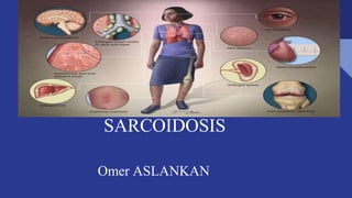
2. SARCOİDOSİS
• Sarcoidosis is a systemic granulomatous and
inflammatory disease of unknown cause.
• The most involved structures are intrathoracic
lymph nodes and lungs, but any organ can be
involved.
3. SARCOIDOSIS EPIDEMIOLOGY
• There is no clear information about the incidence and prevalence
of sarcoidosis.
• Sarcoidosis can be seen all over the world, in both sexes and in all
age groups.
• However, there are societies and age groups where it is more
common.
• Although it is seen in all age groups, it is mostly a disease of
young adults.
• The most common populations are Scandinavians and African
Americans.
• The course of the disease and the forms of involvement vary
according to races.
• 75% of the cases are non-smokers.
4. SARCOİDOSİS ETİOLOGY
• The cause of sarcoidosis is unknown.
• It is accepted that genetic predisposition plays a role in the
development of sarcoidosis.
• Regarding the genetics of sarcoidosis, a study was conducted on the
HLA Gene.
• Some genetic features have been associated with the frequency of
sarcoidosis, the mode of presentation or prognosis of sarcoidosis.
• It is accepted that environmental factors play a role in the
development of sarcoidosis.
• The most important of the environmental factors is the infectious
agents, Mycobacterium tuberculosis.
• The development of sarcoidosis has been reported in relation to
drugs that affect the immune system (such as interferon, anti-TNF
agents, “highly activated retroviral therapy” used in HIV+ patients...)
5. SARCOİDOSİS PATHOGENESİS
• The main event in the development of sarcoidosis is the development of granuloma.
• The antigen that triggers sarcoidosis is presented to CD4+ T lymphocytes by antigen-
presenting cells.
• Activated CD4+ T lymphocytes (Th0) transform into T helper type 1 (Th1) effector
cells with the effect of interleukin (IL) 12 and 18.
• Continuation of antigen presentation by alveolar macrophages to effector Th1 cells
and production of many cytokines, chemokines; It provides granuloma formation
through migration, collection and local proliferation of cells (especially T lymphocytes,
monocytes/macrophages).
• In later periods, in some cases, granulomas disappear with spontaneous resolution,
while in others they persist and go to chronic disease.
• In a very small group of patients, granulomas are replaced by fibrotic changes, and if
this is progressive, end-stage fibrosis may develop.
6. SARCOİDOSİS PATHOLOGY
• Typical histopathological lesion of sarcoidosis is compact epithelioid cell granulomas
without caseification necrosis.
• Granulomas contain epithelioid cells, giant cells and lymphocytes.
• Giant cells may contain cytoplasmic inclusions such as asteroid bodies and
Schaumann bodies.
• Occasionally, focal coagulation necrosis may be found in granulomas.
• In sarcoid granulomas, fibrotic changes may develop, starting from the periphery and
progressing to the centre, resulting in complete fibrosis and/or hyalinization.
• Granulomas may disappear or progress to fibrosis.
• Most of the granulomas in the lung are located in the connective tissue sheaths around
the bronchioles, in the subpleural or perilobular areas.
7. SARCOİDOSİS CLİNİC
• The manifestation of sarcoidosis is very variable.
• It may start with an acute noisy picture and
manifest itself with an insidious picture.
• Some patients with sarcoidosis present with non-
specific constitutional symptoms such as fever,
fatigue, fatigue, and weight loss.
• Weight loss is usually around 2-6 kg in the last 2-3
months.
• Weakness and fatigue are common
8. SARCOİDOSİS CLİNİC (LUNG SARCOİDOSİS)
• As the lungs are the most common site of involvement, respiratory system symptoms
are present in one third or half of the cases.
• In the ACCESS study, lung involvement was detected in 95% of the cases.
• The most common respiratory symptoms are shortness of breath, cough and chest
pain.
• Sarcoidosis is staged according to lung radiography.
• Stage 0: normal chest X-ray
• Stage 1: bilateral hilar lymphadenopathy
• Stage 2: bilateral hilar adenopathy and parenchymal infiltrates
• Stage 3: parenchymal infiltrates only
• Stage 4: fibrosis
9. SARCOİDOSİS CLİNİC (LUNG SARCOİDOSİS)
• The radiology of the vast majority of cases is compatible with stage 1 or 2.
• Stage 3 or 4 radiology was observed in only 15% of the cases in the
ACCESS study.
• Restrictive defects in pulmonary function tests are detected in more than 20-
30% of patients with sarcoidosis during the initial diagnosis period.
• There may be airway involvement in sarcoidosis, it is most commonly
observed as nodular swellings on the mucosa, and there may be the
appearance of a mass lesion from time to time.
• Pleural involvement is rare in sarcoidosis. Pleural fluid, pneumothorax,
pleural thickening and nodules, hydropneumorax, chylothorax may develop.
10. SARCOİDOSİS CLİNİC (SKİN SARCOİDOSİS)
• There is skin involvement in 25-35% of cases.
• Skin involvement may be in the form of specific lesions with granulomas on
biopsy or non-specific lesions without granulomatous inflammation on biopsy.
• Erythema nodosum is the most common, easily recognized non-specific
lesion.
• Some of the patients with sarcoidosis present with erythema nodosum,
fever, arthralgia, and bilateral hilar adenopathy (parenchymal infiltrates can
also be found) on chest X-ray.
• This picture, called Löfgren's syndrome, strongly suggests sarcoidosis,
• The prognosis of this group of patients is generally very good. Erythema
nodosum often resolves within 3 weeks.
• Another important skin involvement that is easily recognized in sarcoidosis
is lupus pernio.
• It is often a sign of chronic sarcoidosis, accompanying bone cysts and lung
fibrosis.
11. SARCOİDOSİS CLİNİC (EYE INVOLVEMENT)
• The frequency of eye involvement is reported to vary
between 10-80%.
• Every patient with sarcoidosis should be referred for
routine eye consultation.
• Although every layer of the eye can be involved,
uveitis is the most common.
• Chronic uveitis can lead to glaucoma, cataract and
blindness.
• Conjunctival follicles, retinal vasculitis, lacrimal gland
enlargement, dacryocystitis, keratoconjunctivitis sicca
can also be seen in sarcoidosis.
12. SARCOİDOSİS CLİNİC (MUSCULOSKELETAL INVOLVEMENT)
• Bone, muscle and joint involvement may occur in patients with sarcoidosis.
• Symptomatic muscle involvement is rare.
• When chronic myopathy is present, it is important to differentiate it from
corticosteroid-induced myopathy.
• The frequency of bone lesions due to sarcoidosis is between 3-13%, most
commonly the bones of the hands and feet are involved.
• Lytic lesions (bone cysts), reticular appearance, destructive changes may
occur in the bone.
• Acute sarcoid arthritis often accompanies Löfgren's syndrome and has a
good prognosis.
13. LİVER GASTROINTESTINAL SYSTEM INVOLVEMENT
• Granuloma can be detected frequently in liver biopsies; but most of the
patients do not have any complaints.
• Liver enzymes may be elevated in 10% of patients with sarcoidosis.
• Although hepatic sarcoidosis is often silent, itching, jaundice, liver failure
and portal hypertension may develop rarely due to liver involvement.
• Liver failure, hepatopulmonary syndrome, portal hypertension with varicose
bleeding is seen in less than 1% of cases.
• Gastrointestinal system involvement is very rare, gastric sarcoidosis can be
found in less than 10% of cases.
• Gastrointestinal involvement usually does not cause clinical symptoms.
14. SARCOİDOSİS CLİNİC ( HEART INVOLVEMENT)
• In sarcoidosis, the heart can be affected both by lung involvement and
directly by disease involvement.
• Clinically detected cardiac involvement is around 5%; however, higher
rates are reported in autopsy series.
• Cardiac sarcoidosis is a rare but life-threatening form of involvement.
• Sudden death may occur in cases with cardiac sarcoidosis if the diagnosis is
not considered.
• Cardiac involvement can occur at any time during the course of sarcoidosis,
before other organ involvement, simultaneously with others or after other
involvements.
• Sarcoid granulomas can affect every layer of the heart, myocardium is most
commonly affected.
• The clinical picture is related to the localization and prevalence of
granulomas.
• Major clinical findings occur with infiltration of myocardium and
conduction system.
• Benign arrhythmias, conduction delays, blocks, heart failure, and even
sudden cardiac death may occur with this infiltration.
• Diagnosis is difficult; Diagnostic difficulties and delayed diagnosis
negatively affect the prognosis.
• Endomyocardial biopsy, which is a definitive diagnostic method, is both
difficult and has low diagnostic value.
• Therefore, in a case with granulomas in a non-cardiac organ, a diagnosis of
cardiac sarcoidosis can be made by clinical signs and symptoms, ECG and
other non-invasive cardiac imaging methods.
• Electrocardiography, 24-hour Holter monitoring, echocardiography,
myocardial perfusion scintigraphy with thallium, gallium scintigraphy are
auxiliary tests in the diagnosis.
• In recent years, cardiac magnetic resonance imaging (MRI) and 18F-fluoro-
2-deoxyglucose positron emission tomography (FDG-PET) have been the
most emphasized methods in the diagnosis of cardiac sarcoidosis.
15. SARCOİDOSİS CLİNİC ( NEUROSARCOIDOSIS)
• Clinically detectable nervous system involvement is below 10%, but up to
25% nervous system involvement is reported in autopsies.
• Common forms of neurosarcoidosis are cranial nerve involvement,
especially facial paralysis, hypothalamic and hypopituitary lesions; Less
frequently, space-occupying masses, peripheral nerve involvement,
lymphocytic meningitis may be seen.
• Cranial nerve paralysis, headache, ataxia, cognitive dysfunction, loss of
strength and convulsions may be seen in the clinical picture.
• In 62-74% of patients with neurosarcoidosis, neurological symptoms are the
initial manifestations of the disease; in these cases, other systemic
manifestations occur later.
• The clinical picture can be acute, subacute or chronic, insidious.
• Angiotensin converting enzyme (ACE) level in cerebrospinal fluid (CSF)
and cranial MR are helpful in diagnosis.
• Paralysis of the seventh cranial pair may accompany uveo-parotid fever
(Herfort syndrome-uveitis, parotid swelling, 7th nerve paralysis).
16. SARCOİDOSİS CLİNİC ( HEMATOLOGICAL PROBLEMS)
• Not very serious anemia, leukopenia may be found.
• Hematological disorders may be related to splenomegaly or bone
marrow involvement.
• Leucomoid reaction, eosinophilia, thrombocytopenia are rare.
• Spleen enlargement is usually mild and asymptomatic; but
sometimes it can get too big and cause compression symptoms and
hyperseplenism.
• Hypercalcemia is found in 2-10% of cases, hypercalciuria is more
common; Detection and correction of hypercalcemia and
hypercalciuria may cause kidney stones, nephrocalcinosis, and kidney
failure.
• Therefore, blood calcium and 24-hour urine calcium should be
measured in all patients diagnosed with sarcoidosis.
• Endocrine findings such as diabetes insipidus, hypothyroidism,
hyperthyroidism, and adrenal suppression are very rare. There may be
swelling, painful enlargement of the parotid glands.
• Rarely, kidneys, reproductive organs, breast tissue may be involved.
• Involvement of oral cavity, larynx, tonsils, nasopharynx can be seen
rarely in sarcoidosis; hoarseness, sore throat, acute respiratory failure
and obstructive sleep apnea may develop.
17. SARCOİDOSİS DIAGNOSIS
• The diagnosis of sarcoidosis is made by excluding other causes that may cause these pictures in the presence of a compatible clinical,
radiological and histopathological picture.
• Presence of granuloma alone does not make the diagnosis of sarcoidosis.
• Tissue biopsy is required for histopathological demonstration of granulomas that do not contain caseification necrosis.
• However, in some cases, the patient may be considered sarcoidosis without a tissue diagnosis.
• These are bilateral hilar lymphadenopathy, Löfgren's syndrome, Heerford's syndrome, involvement of the lacrimal and parotid glands
on Gallium 67 scintigraphy (panda sign) and right paratracheal and bilateral hilar involvement (lambda sign) in an asymptomatic
patient.
• If the clinical picture is not so typical as to be called sarcoidosis without tissue diagnosis, biopsy should be performed.
• Other possibilities should be excluded when biopsy shows granulomatous inflammation..
18. SARCOİDOSİS DIAGNOSIS
• Detection of excess organ involvement is helpful in differential diagnosis.
• For example, sarcoidosis can be confused with lymphoma and tuberculosis; however, if uveitis is detected, it is an auxiliary finding in the
diagnosis of sarcoidosis, since uveitis is very rare in these diseases.
• Multiple sclerosis, like sarcoidosis, can cause optic neuritis and uveitis, but these patients do not have mediastinal, hilar lymphadenopathy.
• In the initial evaluation of sarcoidosis, after the history and physical examination, chest X-ray, pulmonary function tests (spirometry, diffusion
test), whole blood, complete urinalysis, all biochemistry (liver, kidney functions, angiotensin converting enzyme-ACE level), 24-hour urine calcium,
ECG, PPD should be done, all patients should be sent for routine eye consultation and fiberoptic bronchoscopy, bronchial mucosa and transbronchial
lung biopsy, BAL examination should be planned..
• Biopsy can be performed from any organ involved in the patient with sarcoidosis.
• Bronchoscopy can be performed to obtain bronchoalveolar lavage, bronchial mucosal biopsy, transbronchial needle aspiration, and transbronchial
biopsy.
19. SARCOİDOSİS DIAGNOSIS
• Although EBUS prolongs the bronchoscopy time a little, it greatly increases the chance of diagnosis.
• If chest X-ray findings are atypical, and if the chest X-ray is normal even though the disease is considered, CT, especially high-resolution CT (HRCT), is
helpful in the diagnosis.
• Generally, nodules with bronchovascular and subpleural distribution, thickening of the interlobular septum, structural deterioration, and conglomerate
masses are observed on CT.
• Sarcoidosis sometimes progresses with single or multiple nodules or mass lesions on chest X-ray and CT.
• Exclusion of malignant diseases is important in these cases, bronchoscopy and transbronchial biopsy are valuable in diagnosis.
• High serum ACE level is helpful in diagnosis, reflecting the total granuloma burden in sarcoidosis.
• Its sensitivity is low, therefore it is not diagnostic. Its sensitivity was found to be 57% and specificity around 90%.
• PPD is negative in approximately 85% of patients.
20. SARCOİDOSİS TREATMENT
• Spontaneous remission develops in a significant portion of patients with
sarcoidosis.
• Therefore, treatment should only be considered for symptomatic cases with
impaired organ functions.
• Steroids are used in treatment.
• In appropriate cases, treatment with local steroids should be tried.
• Steroid pomades in skin lesions, intralesional steroid injections, steroid eye
drops in eye involvement, steroid inhalers in cough complaints can control the
disease or symptoms.
• Nonsteroidal anti-inflammatories may be useful in acute conditions such as
erythema nodosum, arthralgia, and arthritis.
• Antimalarial drugs can be used instead of steroids in common skin lesions,
hypercalcemia, and hyperlaxia.
• Cardiac, neurological involvement, eye involvement that does not respond
to local treatment, severe hypercalcemia are absolute steroid treatment
indications.
21. SARCOİDOSİS TREATMENT
• In lung and other organ involvement, treatment decision should be made according to the patient's symptoms and organ functions.
• Stage 1, asymptomatic cases should be followed without treatment.
• If stage 2 or stage 3 cases have mild or moderate symptoms, the decision to treat can be left at the end of 6-12 months with a close follow-up.
• In these cases, it is appropriate to check every 2-3 months and to start treatment if the disease worsens.
• Symptomatic cases with impaired pulmonary function tests and diffuse infiltration should be treated.
• Only persistent radiological infiltrates and progressive loss of lung functions should bring up treatment in an asymptomatic patient ..
• Spontaneous remission is common in patients with acute sarcoidosis.
• Spontaneous remission usually occurs in the first 6 months, but it can last up to 2-5 years.
• Chronic cases usually have progressive or persistent disease that requires treatment.
• Patients whose disease lasts longer than two years and whose respiratory functions are found to be impaired in the last three months should be treated.
• Stage 4 cases may not respond to steroid/immunosuppressive therapy.
22. SARCOİDOSİS TREATMENT
• However, treatment may be attempted for a while to assess whether symptomatic or functional improvement will occur.
• This group of cases will require more supportive treatment.
• In cases requiring treatment, 20-40 mg of prednisone or its equivalent per day is started and a 12-24 month treatment is applied.
• Treatment may not be discontinued for a long time in a group of chronic cases.
• There are drugs that reduce the need for steroids in patients who need steroids for a long time:
• Methotrexate: 5-15mg once a week.
• 1mg/day folic acid is added to reduce the toxicity of methotrexate.
• Azathioprine: 50-200mg/day
• Leflunomide: 10-20mg/day Mycophenolate: 2-3x500mg
• Anti-TNF agents are recommended in chronic refractory cases..
23. SARCOİDOSİS RESOURCES
1-MD.. Özlem Özdemir Kumbasar Ankara University Faculty of Medicine Department of Chest Diseases
2- Musellim B, Kumbasar OO, Ongen G, Cetinkaya E, et al. Epidemiological features of Turkish patients with sarcoidosis. Respir Med 2009;
103:907-912.
3-de Boer S, Wilsher M. Sarcoidosis. Chronic Resp Dis 2010; 7:247-258.
4-Baughman RP, Teirstein AS, Judson MA, et al. Clinical Characteristics of Patients in a Case Control Study of Sarcoidosis. Am J Respir Crit Care
Med 2001;164: 1885-1889. 5-Grutter JC, Drent M, van den Bosch JMM. sarcoidosis. Eur Respir Mon 2009;46:126-154. 6- Judson MA.
Sarcoidosis: Clinical presentation, diagnosis and approach to treatment Am J Med Sci 2008; 335:26-33.
7--Ianuzzi MC, Rybicki BA, Teirstein AS. Sarcoidosis N Engl J Med 2007: 2153-65.
8- Lynch JP, Ma YL, Koss MN, White ES. pulmonary sarcoidosis. Semin Respir Crit Care Med 2007; 28:53-74.