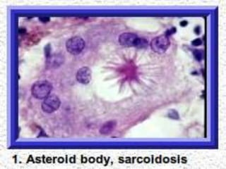
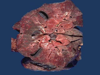
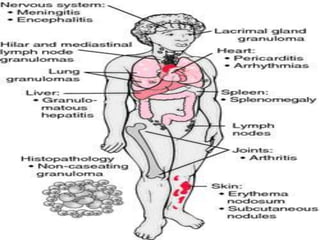
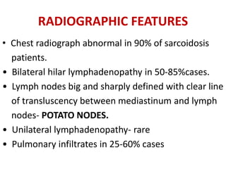
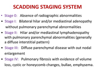
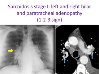
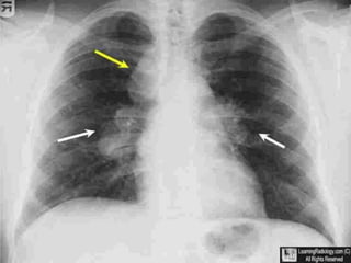
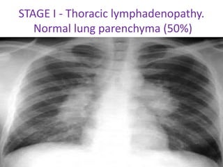
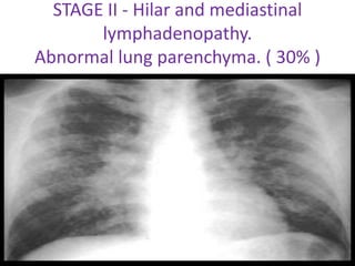
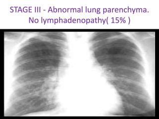
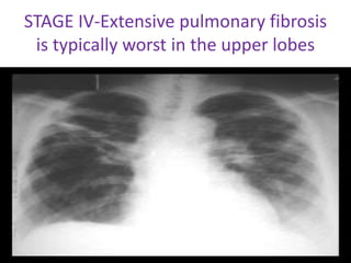
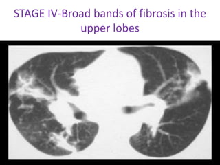
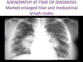
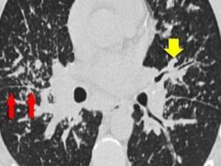
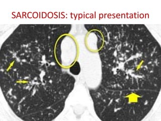
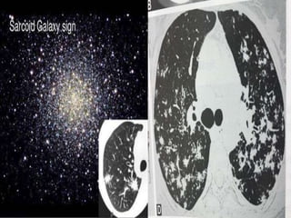
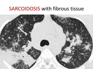
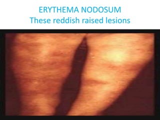
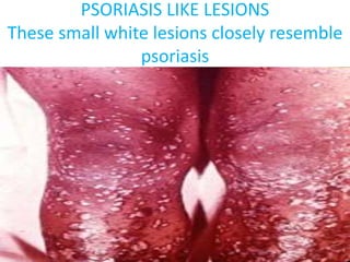
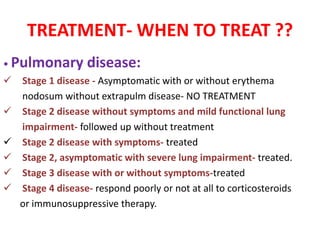
SlideShare a Scribd company logo
SARCOIDOSIS
PRESENTOR
• Dr PRITI
CONTENTS
• HISTORY AND EPIDEMIOLOGY
• RISK FACTORS
• PATHOLOGY
• CLINICAL FEATURES
• DIAGNOSIS
• TREATMENT
• PROGNOSIS AND MORTALITY
SYNONYMS
Besnier Boeck Disease
Schaumann's syndrome
INTRODUCTION
• Multisystem non-caseous granulomatous disorder
of unknown aetiology
• Characterized by depression of cutaneous delayed
type hypersenstivity and heightened Th1 immune
response in affected organs.
• Most commonly affecting young adults
• Presenting most frequently with bilateral hilar
lymphadenopathy, pulmonary infiltration and skin
or eye lesions.
EPIDEMIOLOGY
• Occurs worldwide
• Highest incidence - United States and
Sweden.
• Lower - Asian
• Prevalence - 10 and 40 cases per 100,000
• Mortality - 1 to 5 percent
• Environmental exposures - winter and early
spring months
• Geographic variation and time–space clustering
• Occupational association- health care
professionals,firefighters, military personnel.
• Age : 20-40 yr
• Sex : female ; bimodal
• 16 times more common in blacks
• Genetic influence
monozygotic : dizygotic twins (13: 1)
AETIOLOGY
• Unknown
• Exposure to pine pollen or beryllium
• Infection with Mycobacterium, viruses and
fungi
• protoplast or L form of the tubercle bacillus
may be a cause of sarcoidosis
ACCESS STUDY
• A Case Control Etiologic Study of Sarcoidosis
• 706 subjects
• Absence of occupational and environmental
association
• Weak assoc-insecticide;mold;musty odour
• No association-berylium;wood ;rural; smoking
• No single dominant factor
• Gene environmental factors
INFECTIONS
Mycobacterium DNA
• DNA studies :0-80%
• Control :0-30%
• Propionibacterium acne
• 80-98%
• Control 0-60%
• Borrelia
• Chlamydiae
• Rickettsiae
• Viruses : EBV,CMV,herpes 6
• High titres of these viruses may be reflect
generalised B cell activation in sarcoidosis
AUTOIMMUNITY
ANA,RF,hypergammaglobulinaemia
Molecular mimicry
BIOCHEMICAL EVIDENCE
• mKatG (mycobacterial catalase peroxidase
protein) -50% of patients
GENETICS
• STRONG EVIDENCE
• ACCESS STUDY
• Siblings are higher risk than parents
• More of mother-child than father-child
associations.
ROLE OF HLA
• HLAB 8 – most consistently associated.
• HLA-B1,B8, DR3- also associated with abnormal
immune responsiveness.
• DR1, DR 4 – protective.
• HLA-B8, DR3, Cw7 associated with good
prognosis in caucasians.
• Other alleles are associated with chronic disease,
favorable outcomes.
Non-HLA genes:
• TNF genes(polymorphisms associated
with 1.5 fold increased risk)
• CC chemokine receptors.
• ACE complement receptor 1.
• German study – Chromosome 6
• SAGA study – Chromosome 5
Non-HLA Candidate Genes Evaluated
in Sarcoidosis
PATHOGENESIS
• IL-12 contributes to proliferation of activated T
cells in early disease.
• Increased levels of IL-12 and IL-18 stimulate
IFN-gamma production.
• TGF-beta inhibits IL-12 and IFN-
gammadownregulation of granulomatous
inflammation in sarcoidosis.
• Elevated levels of IL-6 and IL-8 in BAL in active
sarcoidosis.
HISTOPATHOLOGY
• The pathologic hallmark is presence of non
caseating
epitheloid cell granuloma.
• Active sarcoidosis has nodular collections of large
closely packed, pale-staining histiocytes -
epithelioid cells with giant cells.
• Necrosis doesn’t occur.
• Electron microscopy showed that epitheloid cells
tend to be central and macrophages peripheral.
• As lesion ages, reticulin fibres between epitheloid
cells ramify and converted to collagen.
The cytoplasm of giant cells have inclusion bodies
1) Schaumann bodies- round or oval and vary in size from
that of a leucocyte to about 100μm in diameter.The larger
of these bodies ( conchoid bodies) seem to be formed of
basophilic concentric lamellae that appear to contain
calcium and iron
2) Doubly refractile crystalline inclusion bodies are 1–20 μm
in diameter and may take up stains for calcium and iron.
3) Asteroid bodies consist of a central mass 2.3–3μm in
diameter with radiating straight or centred spinous
projections, the whole being 5–25μm in diameter
4) Hamazaki wesenberg bodies.
DIFFERENCES BETWEEN GRANULOMA
OF TB and SARCOIDOSIS
Histological features of sarcoid lesionare
not specific. Can also be seen in
• tuberculosis
• leprosy
• tertiary syphilis
• brucellosis
• primary biliary cirrhosis
• hypogammaglobulinemia
• Brucellosis
• fungal infections.
CLINICAL PRESENTATION
Acute Sarcoidosis
• Lofgren's syndrome - acute erythema nodosum
with
bilateral hilar lymphadenopathy, fever, and
polyarthritis, non granulomatous uveitis
• Symptoms are typically abrupt in onset and have a
transient course.
• A vesicular or maculopapular rash, acute iritis,
conjunctivitis, and Bell's palsy may also be features
of acute disease
CHRONIC SARCOIDOSIS
• More gradual, insidious onset, is more likely to
develop chronic disease.
• Other risk factors for chronic disease include the
presence of lupus pernio, which is a persistent,
disfiguring, violaceous rash over the nose, cheeks,
and ears, and the presence of multiorgan involvement
at the time of diagnosis.
• Chronic eye involvement includes chronic uveitis,
cataracts, glaucoma, or keratoconjunctivitis sicca,which
can be confused with Sjogren's syndrome
Major Clinical Manifestations
LUNGS
First site involved
• Begins with alveolitis involving small bronchi
and small blood vessels
• Alveolitis either clears up spontaneously or
leads to granuloma and fibrosis.
Around 50% patients asymptomatic.
• Dry cough and dyspnoea
• Chest pain-rare
• Wheezing secondary to endobronchial disease,
extrinsic compression by lymphadenopathy or
bronchial distrortion secondary to fibrosis.
• Productive cough- traction bronchiectasis
• Hemoptysis- bronchiectasis or aspergilloma
RADIOGRAPHIC FEATURES
• Chest radiograph abnormal in 90% of sarcoidosis
patients.
• Bilateral hilar lymphadenopathy in 50-85%cases.
• Lymph nodes big and sharply defined with clear line
of transluscency between mediastinum and lymph
nodes- POTATO NODES.
• Unilateral lymphadenopathy- rare
• Pulmonary infiltrates in 25-60% cases
SCADDING STAGING SYSTEM
• Stage 0: Absence of radiographic abnormalities
• Stage I: Bilateral hilar and/or mediastinal adenopathy
without pulmonary parenchymal abnormalities
• Stage II: Hilar and/or mediastinal lymphadenopathy
with pulmonary parenchymal abnormalities (generally
a diffuse interstitial pattern)
• Stage III: Diffuse parenchymal disease with out nodal
enlargement
• Stage IV: Pulmonary fibrosis with evidence of volume
loss, cystic or honeycomb changes, bullae, emphysema.
• Radiographic staging of intrathoracic
sarcoidosis
• Staging also helps in prognosis.
Sarcoidosis stage I: left and right hilar
and paratracheal adenopathy
(1-2-3 sign)
STAGE I - Thoracic lymphadenopathy.
Normal lung parenchyma (50%)
STAGE II - Hilar and mediastinal
lymphadenopathy.
Abnormal lung parenchyma. ( 30% )
STAGE III - Abnormal lung parenchyma.
No lymphadenopathy( 15% )
Differential diagnosis of sarcoidosis
with pulmonary infiltrates
• Extrinsic Allergic alveolitis
• Fibrosing alveolitis
• Carcinomatous infiltration
• Pneumoconiosis
• Miliary tuberculosis
• Metastatic malignancy
• Alveolar cell carcinoma
• Honey comb lung.
STAGE IV-Extensive pulmonary fibrosis
is typically worst in the upper lobes
STAGE IV-Broad bands of fibrosis in the
upper lobes
ADENOPATHY AT TIME OF DIAGNOSIS
Marked enlarged hilar and mediastinal
lymph nodes
Radiographic abnormalities on chest x ray.
• 1) disseminated miliary lesions
• 2) disseminated nodular lesions
• 3) linear type of infiltration extending fan-wise from the hilum
• 4) diffuse and confluent patchy shadows;
• 5) diffuse fibrosis
• 6) diffuse fibrosis with cavitation
• 7) diffuse ground-glass shadowing
• 8) changes similar to chronic tuberculosis as regards location
and distribution
• 9) bilateral confluent massive opacities resembling areas of
pneumonia
• 10) atelectasis.
UNUSUAL
• “Egg shell” calcification of hilar nodes
• Pleural effusions
• Cavitations
• Atelectasis
• Pulmonary hypertension
• Pneumothorax
• Cardiomegaly
HRCT findings in Sarcoidosis.
• Common findings:
– Small nodules in a perilymphatic distribution
(i.e. along subpleural surface and fissures, along
interlobular septa and the peribronchovascular
bundle).
– Upper and middle zone predominance.
– Lymphadenopathy in left hilus, right hilus and
paratracheal (1-2-3 sign). Often with
calcifications.
SARCOIDOSIS: typical presentation
Uncommon findings:
– Conglomerate masses in a perihilar location.
– Larger nodules (> 1cm in diameter, in < 20%)
– Grouped nodules or coalescent nodules
surrounded by multiple satellite nodules (sarcoid
galaxy sign)
• Nodules so small and dense that they appear as
ground glass or even as consolidations (alveolar
sarcoidosis)
• Reverse halo sign
SARCOIDOSIS with fibrous tissue
GALLIUM 67 SCAN
• Gallium 67 concentrated in metabolically and mitotically
active tissues.
• Intravenous injection of 11X107 Bq of gallium 67 citrate,
simultaneous anterior and posterior scans performed 3 days
later.
• Close correlations between gallium uptake and total number
of lymphocytes recovered in BAL.
• Not specific for sarcoidosis.
• Expensive and radiation exposure.
the parotid, salivary and lacrimal gland
region (panda sign) is pathognomic for
sarcoidosis
Gallium scan
showing increased
uptake in the
lacrimal and parotid
glands and
pulmonary regions
in a patient with
active sarcoidosis
Other Intrathoracic Manifestations
Pleural invovlment in 5-10% cases.
( effusions and pleural thickening)
• Aspergilloma
• Bronchiectasis
• Necrotising sarcoid angitis
• Superior venacaval syndrome
• Mediastinal lymph node calcification
• Mediastinal fibrosis
• Vanishing lung syndrome (giant bulla)
Functional abnormalties
• Seen in 20%-40% in patients with normal chest x
ray and 50%-70% when x ray is visibly abnormal.
• Abnormalities in vital capacity, diffusion
capacity, PaO2 at rest, PaO2 at exercise and lung
compliance.
• Usually restrictive defect noted on PFT but
obstructive defect in endobronchial
involvement leading to airflow obstruction, also
from airway distortion secondary to lung fibrosis
EXTRAPULMONARY SARCOIDOSIS
UPPER RESPIRATORY TRACT
Uncommon but disabling.
• Nasal mucosa- crusting,obstruction, discharge. The
mucosa erythematous and granular with polypoid
hypertrophy.
• Laryngeal and pharyngeal mucosa- hoarseness,cough,
dysphagia,dypsnoea
• Patients with sarcoidosis of upper respiratory tract
have 50% chance of developing lupus pernio
• Nasal septal and palatal perforations in untreated
cases.
• d/d tuberculosis, wegeners granulomatosis, leprosy
KRESPI STAGING SYSTEM
• Stage I : mild nasal disease without paranasal sinus
disease. - saline nasal spray, nasal irrigation, and topical
nasal steroids.
• Stage II : moderate disease, with involvement of both
nasal and paranasal sinuses; it is typically treated with
both stage I therapy and intralesional steroids.
• Stage III : severe, often irreversible, nasal and sinus
disease that usually requires the therapeutic
interventions of stages I and II, as well as systemic
therapy.
LYMPHATIC SYSTEM
• Hilar and Mediastinal lymph nodes (>90% of
patients)
• Peripheral lymphadenopathy (5%–30%).
• Cervical, axillary, epitrochlear, and inguinal
regions.
• Non tender, mobile
• Of superficial nodes, right scalene group most
common
EYES
• Upto 25% of sarcoidosis patients ant uveitis most
common manifestation.
• Anterior uveitis - acutely, with pain, photophobia,
lacrimation, and redness, self limiting
• Posterior uveitis is typically gradual in onset and is
more likely to result in visual morbidity, chronic form of
disease.
• Chronic uveitis leads to glaucoma, cataracts and
blindness ,Keratoconjunctivitis sicca Papilledema
• 10% of patients with sarcoid-associated uveitis
develop blindness in at least one eye
CONJUNCTIVITIS
PAPILLEDEMA - Often associated with
7th nerve facial palsy
Multifocal choroiditis & candle waxy spots
Ocular Involvement - KPs as ‘mutton fat’ -
large and greasy facial palsy
SKIN
• Most common manifestation- erythema nodosum.
• Erythema nodosum typically presents as raised, tender, red
nodules, 1 to 2 cm in diameter, on the anterior surface of the
lower legs.
• Lupus pernio is a rare lesion,most severe dermatological
manifesation - purplish plaques typically found over the
nose, cheeks, lips, and ears. Associated with poor prognosis
and severe pulmonary disease.
• Skin abnormalities include red-brown to orange macules and
papules, keloids, and hyper- or hypopigmentation.
LOFGREN'S SYNDROME; acute triad of
erythema nodosum,polyartropathy, bilateral
hilar adenopathy and non granulomatous
uveitis
LUPUS PERNIO
LUPUS PERNIO
Indurated and violaceous range from a few
small lesions to large lesions
ERYTHEMA NODOSUM
These reddish raised lesions
PSORIASIS LIKE LESIONS
These small white lesions closely resemble
psoriasis
LIVER
• 33% have hepatomegaly or biochemical
evidence of disease
• Symptoms usually absent
• Cholestasis, fibrosis, cirrhosis, portal
hypertension, and the Budd-Chiari syndrome
have been seen
MUSCULOSKELETAL
• Bone involvement most commonly affects terminal
phalanges of hands and feet and proximal bones in
severe cases.
• Three types of bony lesions:
 Lytic
 Permeative
 Destructive
• Bone lesions not affected by corticosteroids
PUNCHED OUT LYTIC LESIONS
Focal osteolytic lesions in the fingers are
most common abnormality
SCLEROTIC LESIONS,NONSPECIFIC
Focal sclerosis (arrows) of distal phalanges
is unusual
• Subcutaneous swellings also noted along with
digit abnormalities but they respond to
corticosteroid therapy.
• Skeletal granulomas commonly affecting
pectoral,shoulder, arm and calf muscles
NERVOUS SYSTEM
• Peripheral neuropathy or mononeuritis multiplex
• Cranial nerve palsy- 7th nerve facial palsy is most
common (sarcoidosis is most common cause of
bilateral facial nerve palsy)
 Acute, transient, and can be unilateral or bilateral
 HEERFORDT'S SYNDROME: facial palsy accompanied
by fever, uveitis, and enlargement of the parotid gland
• Lymphocytic meningitis
• Meningoencephalitis
• SOLs
• Epilepsy
• Brain stem and spinal cord syndromes
• Hematopoietic system: splenomegaly common
• Genitourinary system: nephrocalcinosis due to
unexplained increase in senstivity to vitamin D
leading to increased absorption of calcium from
gut.
• Rarely glomerulonephritis and sarcoidosis of
epididymis.
CARDIAC
• Extensive pulmonary fibrosis leading to cor
pulmonale.
• Involvement of myocardium leading to dysrhythmias,
conduction disorders, heart failure and sudden death.
• PAH may occur due to parenchymal fibrosis distorting
the pulmonary vasculature, granulomatous
inflammation of pulmonary vasculature,hypoxic
pulm arterial vasoconstriction and from elevated left
ventricular diastolic pressure of cardiac involvement
ATS criteria diagnosis of
pulmonary sarcoidosis
(1) Presence of a consistent clinical and
radiographic picture
(2) Demonstration of noncaseating granulomas
on biopsy
(3) Exclusion of other conditions that can
produce granulomatous inflammation
Recommended Test for all patients :
• Complete blood count with differential count
• Chest radiograph
• PFT-spirometry, diffusion capacity, lung volumes
• Ophthalmologic examination
• Complete metabolic profile
• ECG
• PPD skin test. (<10mm reaction to 5TU has 100%
sensitivity but not specific. About 2/3rd of patients
with active disease fail to react to 100TU and 1/4th
to 100TU and less than 1/10th to 10TU.)
• Serum & Urine Calcium
Organ Specific Test :
Cardiac-
• ECG
• Holter monitoring
• Thalium or sestamibi myocardial scan
• Cardiac MRI
• Cardiac PET.
Neurologic-
• Brain or spine MRI with gadolinium enhancement
• CSF examination
• EMG or nerve conduction studies.
URT :
Flow-volume loop
ENT evaluation.
X-Ray PNS
CT Scan PNS
Endocrine :
Pituitary function test
Thyroid profile.
Biopsy :
Tissue biopsy is essential.
Biopsy almost always +ve if skin, lymphnodes,
conjunctiva involved accessible sites.
• Transbronchial lung biopsy is usually performed
because high yield and relative safe.
• The diagnostic yield of TBLB is ranges 40-90% if
atleast 4 biopsies are taken.
• Higher yield if pulmonary infiltrates an CXR or CT
scan shows.
• TBLB in advanced fibrocystic sarcoidosis has a
low yield.
• TBNA has diagnostic sensitivity of 100%
• Combining EBUS with TBNA increases the
sensitivity
BAL Findings
• Increased lymphocytes.
• Raised CD4: CD8 ratio >3.5 to 4.0 supports the diagnosis of
sarcoidosis.
• When FOB is non diagnostic – Mediastionscopy, VATS
• Open lung biopsy establish the diagnosis at >90% diagnostic yield.
• For organs that are difficult to biopsy image techniques such as
gallium 67 scan or more recently 18F – fluoro deoxyglucose position
emmison tomography (FDG-PET) may help.
• F-methyltyrosine-PET uses amino acids that is preferentially taken up
and expressed on tumor cells and is negative in sarcoidosis
• Tissues from mediastinoscopy +ve in 95%.
• Scalene lymph node biopsy positive in 80
Serum ACE levels (SACE levels) :
Libermann first to report raised ACE in sarcoidosis
• Produced by epitheloid cells.
• Elevated in 30 to 80% of patients.
• Elevated levels are seen in infections , graulomatous
disease, lymphoma, hepatitis, DM, thyroid disease.
Thus SACE is not recommended as a diagnostic test.
• Neither sensitive nor specific to be used as a
diagnostic tool.
• Provides good monitor of disease activity
Kveim-stiltzbach test :
Intradermal injection of homogenised tissue of organs
involved with sarcoidosis causes delayed cutaneous
reaction in 4 – 6 weeks.
Method of testing
• The test is performed by injecting intradermally 0.1- 0.2
ml of suspension of human sarcoid tissue , usually
obtained from cervical gland.
• Rarely , splenectomy for splenomegaly due to
sarcoidosis allows the preparation of large quantities of
test substance.
Within 2-3 wks positive test shows purplish red
nodule at the site of injection.
• Biopsy at 4- 6 wks reveals sarcoid tissue on
histological examination.
• Value of the test : false positive reactions are rare,
only 1 – 2% .
• Positive test can be regarded as a virtual proof of
active sarcoidosis.
• Positive results are obtained in 75% of patients with
clinical evidence of disease.
FDG PET SCAN
Assessment of disease activity
Best is by clinical assessment by worsening or
persistence of symptoms, with skin lesions and
changes in chest radiograph and PFTs.
• Serum ACE levels
• Others:
blood( lysozyme, neopterin,soluble IL-2 receptor)
BAL fluid(high lymphocytes, CD4/CD8 ratio, TNFalpha,
collagenase etc.,)
Differential Diagnosis
Diagnostic criteria for diagnosis of
tuberculous sarcoidosis
• Age: 25-45 years
• Gender: No gender predisposition
• History of previous tuberculosis infection which was
adequately treated with ATT with adequate drug
combinations, dosages and duration.
• H/O close contact with pt’s having tuberculosis
infection or family
• H/O tubeculosis or association with type II DM.
• Onset: Asymtomatic or with mild fever, anorexia
and loss of weight
• Important symtoms: Chronic cough, brethlessness
on exertion.
• Important signs: Involvement of multiple nodes, Eg-
Scalene or
• cervical group of lymph nodes, at Multiple
locations.
• B/L bibasilar end inspiratory velcrow crackles.
• The Marshall Protocol is a medical treatment
• While other treatments for chronic disease
use palliative medications in an effort to cover
up symptoms, the Marshall Protocol is a
curative treatment, which strives to address
the root cause of the disease process
TREATMENT- WHEN TO TREAT ??
• Pulmonary disease:
 Stage 1 disease - Asymptomatic with or without erythema
nodosum without extrapulm disease- NO TREATMENT
 Stage 2 disease without symptoms and mild functional lung
impairment- followed up without treatment
 Stage 2 disease with symptoms- treated
 Stage 2, asymptomatic with severe lung impairment- treated.
 Stage 3 disease with or without symptoms-treated
 Stage 4 disease- respond poorly or not at all to corticosteroids
or immunosuppressive therapy.
• Extrapulmonary disease:
 Ocular, cardiac, neurological, upper airway
involvement-treated with higher doses(40-60mg/day)
 Glandular, splenic, parotid, cutaneous lesions
treated with modest doses (20-30mg/day)
 Hepatic no treatment required but regular follow
up required.
Choice of drug
Corticosteroids drug of choice.
Act by gene transcription leading to inactivation of
nuclear Factor-kappa B which inhibits synthesis of most
known cytokines.
• Prednisolone 40mg daily maximum dose recommended
for acute pulmonary sarcoidosis which is tapered after
3-6months to maintainance dose of 5-10mg/day in
patients responding to therapy with close monitoring
for relapse and treatment continued for 12months.
• Insufficient data to recommend inhalational
corticosteroids in pulmonary disease
Major complication of corticosteroids therapy is
osteoporosis for which calcium and vitamin D
considered after ruling out hypercalcemia and
hypercalciuria.
• Use of bisphosphonates recommended in patients
with >5mg prednisolone for >3 months.
• Others: hypertension, diabetes, increased
susceptibility to infection, mood changes, skin
thinning, cataracts, weight gain etc
Alternative drugs
CYTOTOXIC DRUGS:
Rationale use of cytotoxic drugs with low doses of
corticosteroids enhances efficacy and reduces toxicity.
• Methotrexate preferred 2nd line drug. 10-15mg
once a week given often in combination with
steroid therapy.
• Others: azathioprine, chlorambucil,
cyclophosphamide
ANTI MALARIAL DRUGS:
• Good response in upper airway, sarcoid related
hypercalcemia and neurosarcoid.
• Chloroquine and hydroxychloroquine
• Hydroxychloroquine 200-400mg daily.
• Side effects of irreversible retinopathy and
blindness. (more with chloroquine)
• Rare side effects- agranulocytosis and myopathy
Reports clinical improvement in refractory
neurosarcoidosis, cardiac sarcoid, upper airway
sarcoid and skin lesions.
Drugs: infliximab and adalimumab
Infliximab- 3-5mg/kg intravenous infusion on weeks
0 and 2 with repeat dose every 4-8 weeks thereafter.
Concern is reactivation of latent tuberculosis and
other opportunistic infections. And also lymphomas,
new onset and worsening of congestive cardiac
failure and demyelinating diseases
• Other rare drugs include:
• leflunomide, mycophenolate,colchicine,
• pentoxyfylline, cyclosporin A
Treat how long??
• Duration individualised based on symptoms, organ
involved and response.
• Mostly for 1 year.
• Some from 6 months to even 10 years.
• If needed for longer periods, alternate day regimen to
minimise side effects.
• Recently showed that in acute exacerbations of
pulmonary sarcoidosis a 20mg prednsiolone for 21days
improved spirometry back to baseline and improved
clinical symptoms.
Treatment of complications
• Sarcoidosis associated fatigue - dexmethylphenidate
hydrochloride
• Bronchiectasis - antibiotic therapy, postural drainage,
anti inflammatory therapy
• Extensive lung fibrosis - long term oxygen therapy
• Sarcoid cardiomyopathy - decongestive therapy
• Refractory neurosarcoidosis - whole brain irradiation
• Bronchostenosis following sarcoidosis- bronchoscopic
balloon dilatation with topical mitomycin C
• Pulmonary HTN- sildenafil and bosentan. Candidates
for lung transplantation
PROGNOSIS AND MORTALITY
Prognosis related to severity of disease.
• Mortality rates of 1-6%
• Fibrosis in lung and forced vital capacity of less than
1.5litres are predictive of death due to respiratory
failure caused by sarcoidosis.
• In survival analysis, sarcoidosis has better prognosis
at 5 years (91.6% survival) compared to other
diffuse interstitial lung diseases
THANK YOU….

More Related Content

What's hot

Sarcoidosis
SarcoidosisSarcoidosis
Sarcoidosis
DR.SHARIF AHSAN
 
Sarcoidosis
SarcoidosisSarcoidosis
Sarcoidosis
Aswin Rm
 
Sarcoidosis
SarcoidosisSarcoidosis
Sarcoidosis
Thomas Kurian
 
Sarcoidosis
SarcoidosisSarcoidosis
Sarcoidosis
drangelosmith
 
DIFFUSE ALVEOLAR HAEMORRHAGE
DIFFUSE ALVEOLAR HAEMORRHAGEDIFFUSE ALVEOLAR HAEMORRHAGE
DIFFUSE ALVEOLAR HAEMORRHAGE
Ashraf Hefny
 
CTD ILDs.
CTD ILDs.CTD ILDs.
Pulmonary eosinophilic infiltrates
Pulmonary eosinophilic infiltratesPulmonary eosinophilic infiltrates
Pulmonary eosinophilic infiltrates
Firoz Hakkim
 
Sarcoidosis
SarcoidosisSarcoidosis
Sarcoidosis
tareq chowdhury
 
Cavitatoy lung lesions
Cavitatoy lung lesionsCavitatoy lung lesions
Cavitatoy lung lesions
Sumiya Arshad
 
Lymphangioleiomyomatosis
LymphangioleiomyomatosisLymphangioleiomyomatosis
Lymphangioleiomyomatosis
Vaishnavi S Nair
 
Idiopathic pulmonary fibrosis copy
Idiopathic pulmonary fibrosis   copyIdiopathic pulmonary fibrosis   copy
Idiopathic pulmonary fibrosis copy
Adetunji Adesegun
 
Sarcoidosis and IgG4
Sarcoidosis and IgG4Sarcoidosis and IgG4
Sarcoidosis and IgG4
ikramdr01
 
Sarcoidosis sagar gandhi
Sarcoidosis sagar gandhiSarcoidosis sagar gandhi
Sarcoidosis sagar gandhi
Sagar Gandhi
 
Lung in pancreatitis
Lung in pancreatitisLung in pancreatitis
Lung in pancreatitis
Rikin Hasnani
 
Approach to interstitial lung disease
Approach to interstitial lung diseaseApproach to interstitial lung disease
Approach to interstitial lung disease
Nahid Sherbini
 
Hypersensitivity Pneumonitis
Hypersensitivity  PneumonitisHypersensitivity  Pneumonitis
Sarcoidosis
SarcoidosisSarcoidosis
Sarcoidosis
MilDoc
 
Idiopathic interstitial pneumonias
Idiopathic interstitial pneumoniasIdiopathic interstitial pneumonias
Idiopathic interstitial pneumonias
Arvind Ghongane
 
Pulmonary alveolar proteinosis
Pulmonary alveolar proteinosisPulmonary alveolar proteinosis
Pulmonary alveolar proteinosis
Vijay Sal
 
Pulmonary sequestration
Pulmonary sequestrationPulmonary sequestration
Pulmonary sequestration
Drhammad Rehman
 

What's hot (20)

Sarcoidosis
SarcoidosisSarcoidosis
Sarcoidosis
 
Sarcoidosis
SarcoidosisSarcoidosis
Sarcoidosis
 
Sarcoidosis
SarcoidosisSarcoidosis
Sarcoidosis
 
Sarcoidosis
SarcoidosisSarcoidosis
Sarcoidosis
 
DIFFUSE ALVEOLAR HAEMORRHAGE
DIFFUSE ALVEOLAR HAEMORRHAGEDIFFUSE ALVEOLAR HAEMORRHAGE
DIFFUSE ALVEOLAR HAEMORRHAGE
 
CTD ILDs.
CTD ILDs.CTD ILDs.
CTD ILDs.
 
Pulmonary eosinophilic infiltrates
Pulmonary eosinophilic infiltratesPulmonary eosinophilic infiltrates
Pulmonary eosinophilic infiltrates
 
Sarcoidosis
SarcoidosisSarcoidosis
Sarcoidosis
 
Cavitatoy lung lesions
Cavitatoy lung lesionsCavitatoy lung lesions
Cavitatoy lung lesions
 
Lymphangioleiomyomatosis
LymphangioleiomyomatosisLymphangioleiomyomatosis
Lymphangioleiomyomatosis
 
Idiopathic pulmonary fibrosis copy
Idiopathic pulmonary fibrosis   copyIdiopathic pulmonary fibrosis   copy
Idiopathic pulmonary fibrosis copy
 
Sarcoidosis and IgG4
Sarcoidosis and IgG4Sarcoidosis and IgG4
Sarcoidosis and IgG4
 
Sarcoidosis sagar gandhi
Sarcoidosis sagar gandhiSarcoidosis sagar gandhi
Sarcoidosis sagar gandhi
 
Lung in pancreatitis
Lung in pancreatitisLung in pancreatitis
Lung in pancreatitis
 
Approach to interstitial lung disease
Approach to interstitial lung diseaseApproach to interstitial lung disease
Approach to interstitial lung disease
 
Hypersensitivity Pneumonitis
Hypersensitivity  PneumonitisHypersensitivity  Pneumonitis
Hypersensitivity Pneumonitis
 
Sarcoidosis
SarcoidosisSarcoidosis
Sarcoidosis
 
Idiopathic interstitial pneumonias
Idiopathic interstitial pneumoniasIdiopathic interstitial pneumonias
Idiopathic interstitial pneumonias
 
Pulmonary alveolar proteinosis
Pulmonary alveolar proteinosisPulmonary alveolar proteinosis
Pulmonary alveolar proteinosis
 
Pulmonary sequestration
Pulmonary sequestrationPulmonary sequestration
Pulmonary sequestration
 

Similar to Sarcoidosis

Sarcoidosis .pptx
Sarcoidosis .pptxSarcoidosis .pptx
Sarcoidosis .pptx
Ömer aslankan
 
Sle and lupus pneumonitis
Sle and lupus pneumonitis Sle and lupus pneumonitis
Sle and lupus pneumonitis
Sagar Gandhi
 
Sexually transmitted infections comp
Sexually transmitted infections compSexually transmitted infections comp
Sexually transmitted infections comp
pgijeff
 
Sarcoidosis
SarcoidosisSarcoidosis
Sarcoidosis
Opeyemi Muyiwa
 
Interstitial lung disease
Interstitial lung diseaseInterstitial lung disease
Interstitial lung disease
Dr. Akash Bharti
 
A RARE CASE OF THE GREAT IMITATOR IN DERMATOLOGY-SARCOIDOSIS.pptx
A RARE CASE OF THE GREAT IMITATOR IN DERMATOLOGY-SARCOIDOSIS.pptxA RARE CASE OF THE GREAT IMITATOR IN DERMATOLOGY-SARCOIDOSIS.pptx
A RARE CASE OF THE GREAT IMITATOR IN DERMATOLOGY-SARCOIDOSIS.pptx
SujataDora2
 
Ild diagnosis
Ild diagnosis Ild diagnosis
Ild diagnosis
Anusha Jahagirdar
 
REACTIVE ARTHRITIS.pptx
REACTIVE ARTHRITIS.pptxREACTIVE ARTHRITIS.pptx
REACTIVE ARTHRITIS.pptx
AnupamAnand60
 
Inflammation(3)
Inflammation(3)Inflammation(3)
Inflammation(3)
SECULAR HARYANA
 
Sjogren syndrome by aseem
Sjogren syndrome by aseemSjogren syndrome by aseem
Sjogren syndrome by aseem
Dr. Aseem Sharma
 
Hiv manifestations in ent
Hiv manifestations in entHiv manifestations in ent
Hiv manifestations in ent
Nayeem Baig
 
Neuroblastoma
NeuroblastomaNeuroblastoma
Neuroblastoma
shockarz
 
syphilis.ppt
syphilis.pptsyphilis.ppt
syphilis.ppt
Abhishek Singhai
 
Sjögren syndrome
Sjögren syndromeSjögren syndrome
Sjögren syndrome
dattasrisaila
 
8-Guideline for elaborate SLE management.ppt
8-Guideline for elaborate  SLE management.ppt8-Guideline for elaborate  SLE management.ppt
8-Guideline for elaborate SLE management.ppt
Bosan Khalid
 
Sarcoidosis
SarcoidosisSarcoidosis
4 ECZEMA 2023.pptx
4 ECZEMA 2023.pptx4 ECZEMA 2023.pptx
4 ECZEMA 2023.pptx
LuisVelarde40
 
Sarcoidosis agreat mimic
Sarcoidosis agreat mimicSarcoidosis agreat mimic
Sarcoidosis agreat mimic
hythemhashim
 
GR 11 NEPHROTIC AND NEPHRITIC SYNDROME.pptx
GR 11 NEPHROTIC AND NEPHRITIC SYNDROME.pptxGR 11 NEPHROTIC AND NEPHRITIC SYNDROME.pptx
GR 11 NEPHROTIC AND NEPHRITIC SYNDROME.pptx
KelfalaHassanDawoh
 
Systemic Sclerosis 2017
Systemic Sclerosis 2017Systemic Sclerosis 2017
Systemic Sclerosis 2017
singlamanik
 

Similar to Sarcoidosis (20)

Sarcoidosis .pptx
Sarcoidosis .pptxSarcoidosis .pptx
Sarcoidosis .pptx
 
Sle and lupus pneumonitis
Sle and lupus pneumonitis Sle and lupus pneumonitis
Sle and lupus pneumonitis
 
Sexually transmitted infections comp
Sexually transmitted infections compSexually transmitted infections comp
Sexually transmitted infections comp
 
Sarcoidosis
SarcoidosisSarcoidosis
Sarcoidosis
 
Interstitial lung disease
Interstitial lung diseaseInterstitial lung disease
Interstitial lung disease
 
A RARE CASE OF THE GREAT IMITATOR IN DERMATOLOGY-SARCOIDOSIS.pptx
A RARE CASE OF THE GREAT IMITATOR IN DERMATOLOGY-SARCOIDOSIS.pptxA RARE CASE OF THE GREAT IMITATOR IN DERMATOLOGY-SARCOIDOSIS.pptx
A RARE CASE OF THE GREAT IMITATOR IN DERMATOLOGY-SARCOIDOSIS.pptx
 
Ild diagnosis
Ild diagnosis Ild diagnosis
Ild diagnosis
 
REACTIVE ARTHRITIS.pptx
REACTIVE ARTHRITIS.pptxREACTIVE ARTHRITIS.pptx
REACTIVE ARTHRITIS.pptx
 
Inflammation(3)
Inflammation(3)Inflammation(3)
Inflammation(3)
 
Sjogren syndrome by aseem
Sjogren syndrome by aseemSjogren syndrome by aseem
Sjogren syndrome by aseem
 
Hiv manifestations in ent
Hiv manifestations in entHiv manifestations in ent
Hiv manifestations in ent
 
Neuroblastoma
NeuroblastomaNeuroblastoma
Neuroblastoma
 
syphilis.ppt
syphilis.pptsyphilis.ppt
syphilis.ppt
 
Sjögren syndrome
Sjögren syndromeSjögren syndrome
Sjögren syndrome
 
8-Guideline for elaborate SLE management.ppt
8-Guideline for elaborate  SLE management.ppt8-Guideline for elaborate  SLE management.ppt
8-Guideline for elaborate SLE management.ppt
 
Sarcoidosis
SarcoidosisSarcoidosis
Sarcoidosis
 
4 ECZEMA 2023.pptx
4 ECZEMA 2023.pptx4 ECZEMA 2023.pptx
4 ECZEMA 2023.pptx
 
Sarcoidosis agreat mimic
Sarcoidosis agreat mimicSarcoidosis agreat mimic
Sarcoidosis agreat mimic
 
GR 11 NEPHROTIC AND NEPHRITIC SYNDROME.pptx
GR 11 NEPHROTIC AND NEPHRITIC SYNDROME.pptxGR 11 NEPHROTIC AND NEPHRITIC SYNDROME.pptx
GR 11 NEPHROTIC AND NEPHRITIC SYNDROME.pptx
 
Systemic Sclerosis 2017
Systemic Sclerosis 2017Systemic Sclerosis 2017
Systemic Sclerosis 2017
 

Recently uploaded

Computer in pharmaceutical research and development-Mpharm(Pharmaceutics)
Computer in pharmaceutical research and development-Mpharm(Pharmaceutics)Computer in pharmaceutical research and development-Mpharm(Pharmaceutics)
Computer in pharmaceutical research and development-Mpharm(Pharmaceutics)
MuskanShingari
 
June 2024 Oncology Cartoons By Dr Kanhu Charan Patro
June 2024 Oncology Cartoons By Dr Kanhu Charan PatroJune 2024 Oncology Cartoons By Dr Kanhu Charan Patro
June 2024 Oncology Cartoons By Dr Kanhu Charan Patro
Kanhu Charan
 
Test bank for karp s cell and molecular biology 9th edition by gerald karp.pdf
Test bank for karp s cell and molecular biology 9th edition by gerald karp.pdfTest bank for karp s cell and molecular biology 9th edition by gerald karp.pdf
Test bank for karp s cell and molecular biology 9th edition by gerald karp.pdf
rightmanforbloodline
 
Recent advances on Cervical cancer .pptx
Recent advances on Cervical cancer .pptxRecent advances on Cervical cancer .pptx
Recent advances on Cervical cancer .pptx
DrGirishJHoogar
 
Medical Quiz ( Online Quiz for API Meet 2024 ).pdf
Medical Quiz ( Online Quiz for API Meet 2024 ).pdfMedical Quiz ( Online Quiz for API Meet 2024 ).pdf
Medical Quiz ( Online Quiz for API Meet 2024 ).pdf
Jim Jacob Roy
 
Case presentation On Urinary tract infection
Case presentation On Urinary tract infectionCase presentation On Urinary tract infection
Case presentation On Urinary tract infection
KarthyAks1
 
Lecture 6 -- Memory 2015.pptlearning occurs when a stimulus (unconditioned st...
Lecture 6 -- Memory 2015.pptlearning occurs when a stimulus (unconditioned st...Lecture 6 -- Memory 2015.pptlearning occurs when a stimulus (unconditioned st...
Lecture 6 -- Memory 2015.pptlearning occurs when a stimulus (unconditioned st...
AyushGadhvi1
 
Nano-gold for Cancer Therapy chemistry investigatory project
Nano-gold for Cancer Therapy chemistry investigatory projectNano-gold for Cancer Therapy chemistry investigatory project
Nano-gold for Cancer Therapy chemistry investigatory project
SIVAVINAYAKPK
 
acne vulgaris -Mpharm (2nd semester) Cosmetics and cosmeceuticals
acne vulgaris -Mpharm (2nd semester) Cosmetics and cosmeceuticalsacne vulgaris -Mpharm (2nd semester) Cosmetics and cosmeceuticals
acne vulgaris -Mpharm (2nd semester) Cosmetics and cosmeceuticals
MuskanShingari
 
STUDIES IN SUPPORT OF SPECIAL POPULATIONS: GERIATRICS E7
STUDIES IN SUPPORT OF SPECIAL POPULATIONS: GERIATRICS E7STUDIES IN SUPPORT OF SPECIAL POPULATIONS: GERIATRICS E7
STUDIES IN SUPPORT OF SPECIAL POPULATIONS: GERIATRICS E7
shruti jagirdar
 
pharmacology for dummies free pdf download.pdf
pharmacology for dummies free pdf download.pdfpharmacology for dummies free pdf download.pdf
pharmacology for dummies free pdf download.pdf
KerlynIgnacio
 
How to Control Your Asthma Tips by gokuldas hospital.
How to Control Your Asthma Tips by gokuldas hospital.How to Control Your Asthma Tips by gokuldas hospital.
How to Control Your Asthma Tips by gokuldas hospital.
Gokuldas Hospital
 
Promoting Wellbeing - Applied Social Psychology - Psychology SuperNotes
Promoting Wellbeing - Applied Social Psychology - Psychology SuperNotesPromoting Wellbeing - Applied Social Psychology - Psychology SuperNotes
Promoting Wellbeing - Applied Social Psychology - Psychology SuperNotes
PsychoTech Services
 
SENSORY NEEDS B.SC. NURSING SEMESTER II.
SENSORY NEEDS B.SC. NURSING SEMESTER II.SENSORY NEEDS B.SC. NURSING SEMESTER II.
SENSORY NEEDS B.SC. NURSING SEMESTER II.
KULDEEP VYAS
 
DECLARATION OF HELSINKI - History and principles
DECLARATION OF HELSINKI - History and principlesDECLARATION OF HELSINKI - History and principles
DECLARATION OF HELSINKI - History and principles
anaghabharat01
 
Top Travel Vaccinations in Manchester
Top Travel Vaccinations in ManchesterTop Travel Vaccinations in Manchester
Top Travel Vaccinations in Manchester
NX Healthcare
 
Osvaldo Bernardo Muchanga-GASTROINTESTINAL INFECTIONS AND GASTRITIS-2024.pdf
Osvaldo Bernardo Muchanga-GASTROINTESTINAL INFECTIONS AND GASTRITIS-2024.pdfOsvaldo Bernardo Muchanga-GASTROINTESTINAL INFECTIONS AND GASTRITIS-2024.pdf
Osvaldo Bernardo Muchanga-GASTROINTESTINAL INFECTIONS AND GASTRITIS-2024.pdf
Osvaldo Bernardo Muchanga
 
K CỔ TỬ CUNG.pdf tự ghi chép, chữ hơi xấu
K CỔ TỬ CUNG.pdf tự ghi chép, chữ hơi xấuK CỔ TỬ CUNG.pdf tự ghi chép, chữ hơi xấu
K CỔ TỬ CUNG.pdf tự ghi chép, chữ hơi xấu
HongBiThi1
 
Artificial Intelligence Symposium (THAIS)
Artificial Intelligence Symposium (THAIS)Artificial Intelligence Symposium (THAIS)
Artificial Intelligence Symposium (THAIS)
Josep Vidal-Alaball
 
The Nervous and Chemical Regulation of Respiration
The Nervous and Chemical Regulation of RespirationThe Nervous and Chemical Regulation of Respiration
The Nervous and Chemical Regulation of Respiration
MedicoseAcademics
 

Recently uploaded (20)

Computer in pharmaceutical research and development-Mpharm(Pharmaceutics)
Computer in pharmaceutical research and development-Mpharm(Pharmaceutics)Computer in pharmaceutical research and development-Mpharm(Pharmaceutics)
Computer in pharmaceutical research and development-Mpharm(Pharmaceutics)
 
June 2024 Oncology Cartoons By Dr Kanhu Charan Patro
June 2024 Oncology Cartoons By Dr Kanhu Charan PatroJune 2024 Oncology Cartoons By Dr Kanhu Charan Patro
June 2024 Oncology Cartoons By Dr Kanhu Charan Patro
 
Test bank for karp s cell and molecular biology 9th edition by gerald karp.pdf
Test bank for karp s cell and molecular biology 9th edition by gerald karp.pdfTest bank for karp s cell and molecular biology 9th edition by gerald karp.pdf
Test bank for karp s cell and molecular biology 9th edition by gerald karp.pdf
 
Recent advances on Cervical cancer .pptx
Recent advances on Cervical cancer .pptxRecent advances on Cervical cancer .pptx
Recent advances on Cervical cancer .pptx
 
Medical Quiz ( Online Quiz for API Meet 2024 ).pdf
Medical Quiz ( Online Quiz for API Meet 2024 ).pdfMedical Quiz ( Online Quiz for API Meet 2024 ).pdf
Medical Quiz ( Online Quiz for API Meet 2024 ).pdf
 
Case presentation On Urinary tract infection
Case presentation On Urinary tract infectionCase presentation On Urinary tract infection
Case presentation On Urinary tract infection
 
Lecture 6 -- Memory 2015.pptlearning occurs when a stimulus (unconditioned st...
Lecture 6 -- Memory 2015.pptlearning occurs when a stimulus (unconditioned st...Lecture 6 -- Memory 2015.pptlearning occurs when a stimulus (unconditioned st...
Lecture 6 -- Memory 2015.pptlearning occurs when a stimulus (unconditioned st...
 
Nano-gold for Cancer Therapy chemistry investigatory project
Nano-gold for Cancer Therapy chemistry investigatory projectNano-gold for Cancer Therapy chemistry investigatory project
Nano-gold for Cancer Therapy chemistry investigatory project
 
acne vulgaris -Mpharm (2nd semester) Cosmetics and cosmeceuticals
acne vulgaris -Mpharm (2nd semester) Cosmetics and cosmeceuticalsacne vulgaris -Mpharm (2nd semester) Cosmetics and cosmeceuticals
acne vulgaris -Mpharm (2nd semester) Cosmetics and cosmeceuticals
 
STUDIES IN SUPPORT OF SPECIAL POPULATIONS: GERIATRICS E7
STUDIES IN SUPPORT OF SPECIAL POPULATIONS: GERIATRICS E7STUDIES IN SUPPORT OF SPECIAL POPULATIONS: GERIATRICS E7
STUDIES IN SUPPORT OF SPECIAL POPULATIONS: GERIATRICS E7
 
pharmacology for dummies free pdf download.pdf
pharmacology for dummies free pdf download.pdfpharmacology for dummies free pdf download.pdf
pharmacology for dummies free pdf download.pdf
 
How to Control Your Asthma Tips by gokuldas hospital.
How to Control Your Asthma Tips by gokuldas hospital.How to Control Your Asthma Tips by gokuldas hospital.
How to Control Your Asthma Tips by gokuldas hospital.
 
Promoting Wellbeing - Applied Social Psychology - Psychology SuperNotes
Promoting Wellbeing - Applied Social Psychology - Psychology SuperNotesPromoting Wellbeing - Applied Social Psychology - Psychology SuperNotes
Promoting Wellbeing - Applied Social Psychology - Psychology SuperNotes
 
SENSORY NEEDS B.SC. NURSING SEMESTER II.
SENSORY NEEDS B.SC. NURSING SEMESTER II.SENSORY NEEDS B.SC. NURSING SEMESTER II.
SENSORY NEEDS B.SC. NURSING SEMESTER II.
 
DECLARATION OF HELSINKI - History and principles
DECLARATION OF HELSINKI - History and principlesDECLARATION OF HELSINKI - History and principles
DECLARATION OF HELSINKI - History and principles
 
Top Travel Vaccinations in Manchester
Top Travel Vaccinations in ManchesterTop Travel Vaccinations in Manchester
Top Travel Vaccinations in Manchester
 
Osvaldo Bernardo Muchanga-GASTROINTESTINAL INFECTIONS AND GASTRITIS-2024.pdf
Osvaldo Bernardo Muchanga-GASTROINTESTINAL INFECTIONS AND GASTRITIS-2024.pdfOsvaldo Bernardo Muchanga-GASTROINTESTINAL INFECTIONS AND GASTRITIS-2024.pdf
Osvaldo Bernardo Muchanga-GASTROINTESTINAL INFECTIONS AND GASTRITIS-2024.pdf
 
K CỔ TỬ CUNG.pdf tự ghi chép, chữ hơi xấu
K CỔ TỬ CUNG.pdf tự ghi chép, chữ hơi xấuK CỔ TỬ CUNG.pdf tự ghi chép, chữ hơi xấu
K CỔ TỬ CUNG.pdf tự ghi chép, chữ hơi xấu
 
Artificial Intelligence Symposium (THAIS)
Artificial Intelligence Symposium (THAIS)Artificial Intelligence Symposium (THAIS)
Artificial Intelligence Symposium (THAIS)
 
The Nervous and Chemical Regulation of Respiration
The Nervous and Chemical Regulation of RespirationThe Nervous and Chemical Regulation of Respiration
The Nervous and Chemical Regulation of Respiration
 

Sarcoidosis

  • 2. CONTENTS • HISTORY AND EPIDEMIOLOGY • RISK FACTORS • PATHOLOGY • CLINICAL FEATURES • DIAGNOSIS • TREATMENT • PROGNOSIS AND MORTALITY
  • 4. INTRODUCTION • Multisystem non-caseous granulomatous disorder of unknown aetiology • Characterized by depression of cutaneous delayed type hypersenstivity and heightened Th1 immune response in affected organs. • Most commonly affecting young adults • Presenting most frequently with bilateral hilar lymphadenopathy, pulmonary infiltration and skin or eye lesions.
  • 5. EPIDEMIOLOGY • Occurs worldwide • Highest incidence - United States and Sweden. • Lower - Asian • Prevalence - 10 and 40 cases per 100,000 • Mortality - 1 to 5 percent
  • 6. • Environmental exposures - winter and early spring months • Geographic variation and time–space clustering • Occupational association- health care professionals,firefighters, military personnel.
  • 7. • Age : 20-40 yr • Sex : female ; bimodal • 16 times more common in blacks • Genetic influence monozygotic : dizygotic twins (13: 1)
  • 8. AETIOLOGY • Unknown • Exposure to pine pollen or beryllium • Infection with Mycobacterium, viruses and fungi • protoplast or L form of the tubercle bacillus may be a cause of sarcoidosis
  • 9. ACCESS STUDY • A Case Control Etiologic Study of Sarcoidosis • 706 subjects • Absence of occupational and environmental association • Weak assoc-insecticide;mold;musty odour • No association-berylium;wood ;rural; smoking • No single dominant factor • Gene environmental factors
  • 10. INFECTIONS Mycobacterium DNA • DNA studies :0-80% • Control :0-30% • Propionibacterium acne • 80-98% • Control 0-60%
  • 11. • Borrelia • Chlamydiae • Rickettsiae • Viruses : EBV,CMV,herpes 6 • High titres of these viruses may be reflect generalised B cell activation in sarcoidosis
  • 13. BIOCHEMICAL EVIDENCE • mKatG (mycobacterial catalase peroxidase protein) -50% of patients
  • 14. GENETICS • STRONG EVIDENCE • ACCESS STUDY • Siblings are higher risk than parents • More of mother-child than father-child associations.
  • 15. ROLE OF HLA • HLAB 8 – most consistently associated. • HLA-B1,B8, DR3- also associated with abnormal immune responsiveness. • DR1, DR 4 – protective. • HLA-B8, DR3, Cw7 associated with good prognosis in caucasians. • Other alleles are associated with chronic disease, favorable outcomes.
  • 16.
  • 17. Non-HLA genes: • TNF genes(polymorphisms associated with 1.5 fold increased risk) • CC chemokine receptors. • ACE complement receptor 1. • German study – Chromosome 6 • SAGA study – Chromosome 5
  • 18. Non-HLA Candidate Genes Evaluated in Sarcoidosis
  • 20.
  • 21. • IL-12 contributes to proliferation of activated T cells in early disease. • Increased levels of IL-12 and IL-18 stimulate IFN-gamma production. • TGF-beta inhibits IL-12 and IFN- gammadownregulation of granulomatous inflammation in sarcoidosis. • Elevated levels of IL-6 and IL-8 in BAL in active sarcoidosis.
  • 22. HISTOPATHOLOGY • The pathologic hallmark is presence of non caseating epitheloid cell granuloma. • Active sarcoidosis has nodular collections of large closely packed, pale-staining histiocytes - epithelioid cells with giant cells. • Necrosis doesn’t occur. • Electron microscopy showed that epitheloid cells tend to be central and macrophages peripheral. • As lesion ages, reticulin fibres between epitheloid cells ramify and converted to collagen.
  • 23. The cytoplasm of giant cells have inclusion bodies 1) Schaumann bodies- round or oval and vary in size from that of a leucocyte to about 100μm in diameter.The larger of these bodies ( conchoid bodies) seem to be formed of basophilic concentric lamellae that appear to contain calcium and iron 2) Doubly refractile crystalline inclusion bodies are 1–20 μm in diameter and may take up stains for calcium and iron. 3) Asteroid bodies consist of a central mass 2.3–3μm in diameter with radiating straight or centred spinous projections, the whole being 5–25μm in diameter 4) Hamazaki wesenberg bodies.
  • 24.
  • 25.
  • 26. DIFFERENCES BETWEEN GRANULOMA OF TB and SARCOIDOSIS
  • 27. Histological features of sarcoid lesionare not specific. Can also be seen in • tuberculosis • leprosy • tertiary syphilis • brucellosis • primary biliary cirrhosis • hypogammaglobulinemia • Brucellosis • fungal infections.
  • 28.
  • 29.
  • 30. CLINICAL PRESENTATION Acute Sarcoidosis • Lofgren's syndrome - acute erythema nodosum with bilateral hilar lymphadenopathy, fever, and polyarthritis, non granulomatous uveitis • Symptoms are typically abrupt in onset and have a transient course. • A vesicular or maculopapular rash, acute iritis, conjunctivitis, and Bell's palsy may also be features of acute disease
  • 31. CHRONIC SARCOIDOSIS • More gradual, insidious onset, is more likely to develop chronic disease. • Other risk factors for chronic disease include the presence of lupus pernio, which is a persistent, disfiguring, violaceous rash over the nose, cheeks, and ears, and the presence of multiorgan involvement at the time of diagnosis. • Chronic eye involvement includes chronic uveitis, cataracts, glaucoma, or keratoconjunctivitis sicca,which can be confused with Sjogren's syndrome
  • 32.
  • 34.
  • 35. LUNGS First site involved • Begins with alveolitis involving small bronchi and small blood vessels • Alveolitis either clears up spontaneously or leads to granuloma and fibrosis.
  • 36. Around 50% patients asymptomatic. • Dry cough and dyspnoea • Chest pain-rare • Wheezing secondary to endobronchial disease, extrinsic compression by lymphadenopathy or bronchial distrortion secondary to fibrosis. • Productive cough- traction bronchiectasis • Hemoptysis- bronchiectasis or aspergilloma
  • 37.
  • 38.
  • 39. RADIOGRAPHIC FEATURES • Chest radiograph abnormal in 90% of sarcoidosis patients. • Bilateral hilar lymphadenopathy in 50-85%cases. • Lymph nodes big and sharply defined with clear line of transluscency between mediastinum and lymph nodes- POTATO NODES. • Unilateral lymphadenopathy- rare • Pulmonary infiltrates in 25-60% cases
  • 40. SCADDING STAGING SYSTEM • Stage 0: Absence of radiographic abnormalities • Stage I: Bilateral hilar and/or mediastinal adenopathy without pulmonary parenchymal abnormalities • Stage II: Hilar and/or mediastinal lymphadenopathy with pulmonary parenchymal abnormalities (generally a diffuse interstitial pattern) • Stage III: Diffuse parenchymal disease with out nodal enlargement • Stage IV: Pulmonary fibrosis with evidence of volume loss, cystic or honeycomb changes, bullae, emphysema.
  • 41. • Radiographic staging of intrathoracic sarcoidosis • Staging also helps in prognosis.
  • 42.
  • 43. Sarcoidosis stage I: left and right hilar and paratracheal adenopathy (1-2-3 sign)
  • 44.
  • 45. STAGE I - Thoracic lymphadenopathy. Normal lung parenchyma (50%)
  • 46. STAGE II - Hilar and mediastinal lymphadenopathy. Abnormal lung parenchyma. ( 30% )
  • 47. STAGE III - Abnormal lung parenchyma. No lymphadenopathy( 15% )
  • 48. Differential diagnosis of sarcoidosis with pulmonary infiltrates • Extrinsic Allergic alveolitis • Fibrosing alveolitis • Carcinomatous infiltration • Pneumoconiosis • Miliary tuberculosis • Metastatic malignancy • Alveolar cell carcinoma • Honey comb lung.
  • 49. STAGE IV-Extensive pulmonary fibrosis is typically worst in the upper lobes
  • 50. STAGE IV-Broad bands of fibrosis in the upper lobes
  • 51. ADENOPATHY AT TIME OF DIAGNOSIS Marked enlarged hilar and mediastinal lymph nodes
  • 52. Radiographic abnormalities on chest x ray. • 1) disseminated miliary lesions • 2) disseminated nodular lesions • 3) linear type of infiltration extending fan-wise from the hilum • 4) diffuse and confluent patchy shadows; • 5) diffuse fibrosis • 6) diffuse fibrosis with cavitation • 7) diffuse ground-glass shadowing • 8) changes similar to chronic tuberculosis as regards location and distribution • 9) bilateral confluent massive opacities resembling areas of pneumonia • 10) atelectasis.
  • 53. UNUSUAL • “Egg shell” calcification of hilar nodes • Pleural effusions • Cavitations • Atelectasis • Pulmonary hypertension • Pneumothorax • Cardiomegaly
  • 54. HRCT findings in Sarcoidosis. • Common findings: – Small nodules in a perilymphatic distribution (i.e. along subpleural surface and fissures, along interlobular septa and the peribronchovascular bundle). – Upper and middle zone predominance. – Lymphadenopathy in left hilus, right hilus and paratracheal (1-2-3 sign). Often with calcifications.
  • 55.
  • 56.
  • 58. Uncommon findings: – Conglomerate masses in a perihilar location. – Larger nodules (> 1cm in diameter, in < 20%) – Grouped nodules or coalescent nodules surrounded by multiple satellite nodules (sarcoid galaxy sign) • Nodules so small and dense that they appear as ground glass or even as consolidations (alveolar sarcoidosis) • Reverse halo sign
  • 59.
  • 61. GALLIUM 67 SCAN • Gallium 67 concentrated in metabolically and mitotically active tissues. • Intravenous injection of 11X107 Bq of gallium 67 citrate, simultaneous anterior and posterior scans performed 3 days later. • Close correlations between gallium uptake and total number of lymphocytes recovered in BAL. • Not specific for sarcoidosis. • Expensive and radiation exposure.
  • 62. the parotid, salivary and lacrimal gland region (panda sign) is pathognomic for sarcoidosis
  • 63.
  • 64. Gallium scan showing increased uptake in the lacrimal and parotid glands and pulmonary regions in a patient with active sarcoidosis
  • 65. Other Intrathoracic Manifestations Pleural invovlment in 5-10% cases. ( effusions and pleural thickening) • Aspergilloma • Bronchiectasis • Necrotising sarcoid angitis • Superior venacaval syndrome • Mediastinal lymph node calcification • Mediastinal fibrosis • Vanishing lung syndrome (giant bulla)
  • 66. Functional abnormalties • Seen in 20%-40% in patients with normal chest x ray and 50%-70% when x ray is visibly abnormal. • Abnormalities in vital capacity, diffusion capacity, PaO2 at rest, PaO2 at exercise and lung compliance. • Usually restrictive defect noted on PFT but obstructive defect in endobronchial involvement leading to airflow obstruction, also from airway distortion secondary to lung fibrosis
  • 68. UPPER RESPIRATORY TRACT Uncommon but disabling. • Nasal mucosa- crusting,obstruction, discharge. The mucosa erythematous and granular with polypoid hypertrophy. • Laryngeal and pharyngeal mucosa- hoarseness,cough, dysphagia,dypsnoea • Patients with sarcoidosis of upper respiratory tract have 50% chance of developing lupus pernio • Nasal septal and palatal perforations in untreated cases. • d/d tuberculosis, wegeners granulomatosis, leprosy
  • 69. KRESPI STAGING SYSTEM • Stage I : mild nasal disease without paranasal sinus disease. - saline nasal spray, nasal irrigation, and topical nasal steroids. • Stage II : moderate disease, with involvement of both nasal and paranasal sinuses; it is typically treated with both stage I therapy and intralesional steroids. • Stage III : severe, often irreversible, nasal and sinus disease that usually requires the therapeutic interventions of stages I and II, as well as systemic therapy.
  • 70. LYMPHATIC SYSTEM • Hilar and Mediastinal lymph nodes (>90% of patients) • Peripheral lymphadenopathy (5%–30%). • Cervical, axillary, epitrochlear, and inguinal regions. • Non tender, mobile • Of superficial nodes, right scalene group most common
  • 71. EYES • Upto 25% of sarcoidosis patients ant uveitis most common manifestation. • Anterior uveitis - acutely, with pain, photophobia, lacrimation, and redness, self limiting • Posterior uveitis is typically gradual in onset and is more likely to result in visual morbidity, chronic form of disease. • Chronic uveitis leads to glaucoma, cataracts and blindness ,Keratoconjunctivitis sicca Papilledema • 10% of patients with sarcoid-associated uveitis develop blindness in at least one eye
  • 73. PAPILLEDEMA - Often associated with 7th nerve facial palsy
  • 74. Multifocal choroiditis & candle waxy spots
  • 75. Ocular Involvement - KPs as ‘mutton fat’ - large and greasy facial palsy
  • 76. SKIN • Most common manifestation- erythema nodosum. • Erythema nodosum typically presents as raised, tender, red nodules, 1 to 2 cm in diameter, on the anterior surface of the lower legs. • Lupus pernio is a rare lesion,most severe dermatological manifesation - purplish plaques typically found over the nose, cheeks, lips, and ears. Associated with poor prognosis and severe pulmonary disease. • Skin abnormalities include red-brown to orange macules and papules, keloids, and hyper- or hypopigmentation.
  • 77. LOFGREN'S SYNDROME; acute triad of erythema nodosum,polyartropathy, bilateral hilar adenopathy and non granulomatous uveitis
  • 79. LUPUS PERNIO Indurated and violaceous range from a few small lesions to large lesions
  • 81. PSORIASIS LIKE LESIONS These small white lesions closely resemble psoriasis
  • 82. LIVER • 33% have hepatomegaly or biochemical evidence of disease • Symptoms usually absent • Cholestasis, fibrosis, cirrhosis, portal hypertension, and the Budd-Chiari syndrome have been seen
  • 83. MUSCULOSKELETAL • Bone involvement most commonly affects terminal phalanges of hands and feet and proximal bones in severe cases. • Three types of bony lesions:  Lytic  Permeative  Destructive • Bone lesions not affected by corticosteroids
  • 84. PUNCHED OUT LYTIC LESIONS Focal osteolytic lesions in the fingers are most common abnormality
  • 85. SCLEROTIC LESIONS,NONSPECIFIC Focal sclerosis (arrows) of distal phalanges is unusual
  • 86. • Subcutaneous swellings also noted along with digit abnormalities but they respond to corticosteroid therapy. • Skeletal granulomas commonly affecting pectoral,shoulder, arm and calf muscles
  • 87. NERVOUS SYSTEM • Peripheral neuropathy or mononeuritis multiplex • Cranial nerve palsy- 7th nerve facial palsy is most common (sarcoidosis is most common cause of bilateral facial nerve palsy)  Acute, transient, and can be unilateral or bilateral  HEERFORDT'S SYNDROME: facial palsy accompanied by fever, uveitis, and enlargement of the parotid gland • Lymphocytic meningitis • Meningoencephalitis • SOLs • Epilepsy • Brain stem and spinal cord syndromes
  • 88. • Hematopoietic system: splenomegaly common • Genitourinary system: nephrocalcinosis due to unexplained increase in senstivity to vitamin D leading to increased absorption of calcium from gut. • Rarely glomerulonephritis and sarcoidosis of epididymis.
  • 89. CARDIAC • Extensive pulmonary fibrosis leading to cor pulmonale. • Involvement of myocardium leading to dysrhythmias, conduction disorders, heart failure and sudden death. • PAH may occur due to parenchymal fibrosis distorting the pulmonary vasculature, granulomatous inflammation of pulmonary vasculature,hypoxic pulm arterial vasoconstriction and from elevated left ventricular diastolic pressure of cardiac involvement
  • 90. ATS criteria diagnosis of pulmonary sarcoidosis (1) Presence of a consistent clinical and radiographic picture (2) Demonstration of noncaseating granulomas on biopsy (3) Exclusion of other conditions that can produce granulomatous inflammation
  • 91. Recommended Test for all patients : • Complete blood count with differential count • Chest radiograph • PFT-spirometry, diffusion capacity, lung volumes • Ophthalmologic examination • Complete metabolic profile • ECG • PPD skin test. (<10mm reaction to 5TU has 100% sensitivity but not specific. About 2/3rd of patients with active disease fail to react to 100TU and 1/4th to 100TU and less than 1/10th to 10TU.) • Serum & Urine Calcium
  • 92. Organ Specific Test : Cardiac- • ECG • Holter monitoring • Thalium or sestamibi myocardial scan • Cardiac MRI • Cardiac PET. Neurologic- • Brain or spine MRI with gadolinium enhancement • CSF examination • EMG or nerve conduction studies.
  • 93. URT : Flow-volume loop ENT evaluation. X-Ray PNS CT Scan PNS Endocrine : Pituitary function test Thyroid profile. Biopsy : Tissue biopsy is essential. Biopsy almost always +ve if skin, lymphnodes, conjunctiva involved accessible sites.
  • 94. • Transbronchial lung biopsy is usually performed because high yield and relative safe. • The diagnostic yield of TBLB is ranges 40-90% if atleast 4 biopsies are taken. • Higher yield if pulmonary infiltrates an CXR or CT scan shows. • TBLB in advanced fibrocystic sarcoidosis has a low yield. • TBNA has diagnostic sensitivity of 100% • Combining EBUS with TBNA increases the sensitivity
  • 95. BAL Findings • Increased lymphocytes. • Raised CD4: CD8 ratio >3.5 to 4.0 supports the diagnosis of sarcoidosis. • When FOB is non diagnostic – Mediastionscopy, VATS • Open lung biopsy establish the diagnosis at >90% diagnostic yield. • For organs that are difficult to biopsy image techniques such as gallium 67 scan or more recently 18F – fluoro deoxyglucose position emmison tomography (FDG-PET) may help. • F-methyltyrosine-PET uses amino acids that is preferentially taken up and expressed on tumor cells and is negative in sarcoidosis
  • 96. • Tissues from mediastinoscopy +ve in 95%. • Scalene lymph node biopsy positive in 80
  • 97. Serum ACE levels (SACE levels) : Libermann first to report raised ACE in sarcoidosis • Produced by epitheloid cells. • Elevated in 30 to 80% of patients. • Elevated levels are seen in infections , graulomatous disease, lymphoma, hepatitis, DM, thyroid disease. Thus SACE is not recommended as a diagnostic test. • Neither sensitive nor specific to be used as a diagnostic tool. • Provides good monitor of disease activity
  • 98. Kveim-stiltzbach test : Intradermal injection of homogenised tissue of organs involved with sarcoidosis causes delayed cutaneous reaction in 4 – 6 weeks. Method of testing • The test is performed by injecting intradermally 0.1- 0.2 ml of suspension of human sarcoid tissue , usually obtained from cervical gland. • Rarely , splenectomy for splenomegaly due to sarcoidosis allows the preparation of large quantities of test substance.
  • 99. Within 2-3 wks positive test shows purplish red nodule at the site of injection. • Biopsy at 4- 6 wks reveals sarcoid tissue on histological examination. • Value of the test : false positive reactions are rare, only 1 – 2% . • Positive test can be regarded as a virtual proof of active sarcoidosis. • Positive results are obtained in 75% of patients with clinical evidence of disease.
  • 101. Assessment of disease activity Best is by clinical assessment by worsening or persistence of symptoms, with skin lesions and changes in chest radiograph and PFTs. • Serum ACE levels • Others: blood( lysozyme, neopterin,soluble IL-2 receptor) BAL fluid(high lymphocytes, CD4/CD8 ratio, TNFalpha, collagenase etc.,)
  • 103.
  • 104.
  • 105.
  • 106. Diagnostic criteria for diagnosis of tuberculous sarcoidosis • Age: 25-45 years • Gender: No gender predisposition • History of previous tuberculosis infection which was adequately treated with ATT with adequate drug combinations, dosages and duration. • H/O close contact with pt’s having tuberculosis infection or family • H/O tubeculosis or association with type II DM. • Onset: Asymtomatic or with mild fever, anorexia and loss of weight
  • 107. • Important symtoms: Chronic cough, brethlessness on exertion. • Important signs: Involvement of multiple nodes, Eg- Scalene or • cervical group of lymph nodes, at Multiple locations. • B/L bibasilar end inspiratory velcrow crackles.
  • 108. • The Marshall Protocol is a medical treatment • While other treatments for chronic disease use palliative medications in an effort to cover up symptoms, the Marshall Protocol is a curative treatment, which strives to address the root cause of the disease process
  • 109. TREATMENT- WHEN TO TREAT ?? • Pulmonary disease:  Stage 1 disease - Asymptomatic with or without erythema nodosum without extrapulm disease- NO TREATMENT  Stage 2 disease without symptoms and mild functional lung impairment- followed up without treatment  Stage 2 disease with symptoms- treated  Stage 2, asymptomatic with severe lung impairment- treated.  Stage 3 disease with or without symptoms-treated  Stage 4 disease- respond poorly or not at all to corticosteroids or immunosuppressive therapy.
  • 110. • Extrapulmonary disease:  Ocular, cardiac, neurological, upper airway involvement-treated with higher doses(40-60mg/day)  Glandular, splenic, parotid, cutaneous lesions treated with modest doses (20-30mg/day)  Hepatic no treatment required but regular follow up required.
  • 111. Choice of drug Corticosteroids drug of choice. Act by gene transcription leading to inactivation of nuclear Factor-kappa B which inhibits synthesis of most known cytokines. • Prednisolone 40mg daily maximum dose recommended for acute pulmonary sarcoidosis which is tapered after 3-6months to maintainance dose of 5-10mg/day in patients responding to therapy with close monitoring for relapse and treatment continued for 12months. • Insufficient data to recommend inhalational corticosteroids in pulmonary disease
  • 112. Major complication of corticosteroids therapy is osteoporosis for which calcium and vitamin D considered after ruling out hypercalcemia and hypercalciuria. • Use of bisphosphonates recommended in patients with >5mg prednisolone for >3 months. • Others: hypertension, diabetes, increased susceptibility to infection, mood changes, skin thinning, cataracts, weight gain etc
  • 113. Alternative drugs CYTOTOXIC DRUGS: Rationale use of cytotoxic drugs with low doses of corticosteroids enhances efficacy and reduces toxicity. • Methotrexate preferred 2nd line drug. 10-15mg once a week given often in combination with steroid therapy. • Others: azathioprine, chlorambucil, cyclophosphamide
  • 114. ANTI MALARIAL DRUGS: • Good response in upper airway, sarcoid related hypercalcemia and neurosarcoid. • Chloroquine and hydroxychloroquine • Hydroxychloroquine 200-400mg daily. • Side effects of irreversible retinopathy and blindness. (more with chloroquine) • Rare side effects- agranulocytosis and myopathy
  • 115. Reports clinical improvement in refractory neurosarcoidosis, cardiac sarcoid, upper airway sarcoid and skin lesions. Drugs: infliximab and adalimumab Infliximab- 3-5mg/kg intravenous infusion on weeks 0 and 2 with repeat dose every 4-8 weeks thereafter. Concern is reactivation of latent tuberculosis and other opportunistic infections. And also lymphomas, new onset and worsening of congestive cardiac failure and demyelinating diseases
  • 116. • Other rare drugs include: • leflunomide, mycophenolate,colchicine, • pentoxyfylline, cyclosporin A
  • 117. Treat how long?? • Duration individualised based on symptoms, organ involved and response. • Mostly for 1 year. • Some from 6 months to even 10 years. • If needed for longer periods, alternate day regimen to minimise side effects. • Recently showed that in acute exacerbations of pulmonary sarcoidosis a 20mg prednsiolone for 21days improved spirometry back to baseline and improved clinical symptoms.
  • 118. Treatment of complications • Sarcoidosis associated fatigue - dexmethylphenidate hydrochloride • Bronchiectasis - antibiotic therapy, postural drainage, anti inflammatory therapy • Extensive lung fibrosis - long term oxygen therapy • Sarcoid cardiomyopathy - decongestive therapy • Refractory neurosarcoidosis - whole brain irradiation • Bronchostenosis following sarcoidosis- bronchoscopic balloon dilatation with topical mitomycin C • Pulmonary HTN- sildenafil and bosentan. Candidates for lung transplantation
  • 119. PROGNOSIS AND MORTALITY Prognosis related to severity of disease. • Mortality rates of 1-6% • Fibrosis in lung and forced vital capacity of less than 1.5litres are predictive of death due to respiratory failure caused by sarcoidosis. • In survival analysis, sarcoidosis has better prognosis at 5 years (91.6% survival) compared to other diffuse interstitial lung diseases