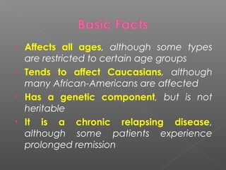
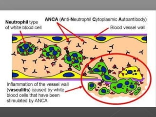
Dr. Angelo Smith M.D
WHPL
• Affects all ages, although some types
are restricted to certain age groups
• Tends to affect Caucasians, although
many African-Americans are affected
• Has a genetic component, but is not
heritable
• It is a chronic relapsing disease,
although some patients experience
prolonged remission
Inflammatory destruction of
blood vessels
•Infiltration of vessel wall with
inflammatory cells
– Leukocytoclasis
– Elastic membrane
disruption
•Fibrinoid necrosis of the
vessel wall
•Ischemia, occlusion,
thrombosis
•Aneurysm formation
•Rupture, hemorrhage
 A clinicopathologic
process characterized
by inflammatory
destruction of blood
vessels that results in
occlusion or
destruction of the
vessel and ischemia of
the tissues supplied by
that vessel.
 “Systemic vasculitides”
Definition
1. Unexplained signs and symptoms
2. Multisystem involvement
3. Unexplained elevated ESR/CRP
4. Skin lesions (palpable purpura)
5. Ischemic vascular changes (Raynaud’s,
gangrene, livedo, claudication)
6. Glomerulonephritis
7. Mononeuritis multiplex
8. Intestinal angina
9. Inflammatory ocular disease
10. Arthralgia's/arthritis, myalgia's
11. Sudden visual loss/headache
1. Fatigue
2. Weakness
3. Fever
4. Abdominal pain
5. Proteinuria , hematuria , casts
6. Nerve problems (numbness , weakness)
7. Skin rash
 Based on
› Size of Vessels involved
› Site of involvement
› Characteristic Features
• Large-vessel vasculitis
– Giant cell arteritis, Takayasu’s arteritis
– Behcet’s disease, Cogan’s syndrome
• Medium-vessel vasculitis
– Polyarteritis nodosa
– Buerger’s disease, Central nervous system
vasculitis, Kawasaki’s disease, Rheumatoid
vasculitis
• Small-vessel vasculitis
– Wegener’s Granulomatosis , microscopic
polyangiitis, Churg-Strauss (ANCA
associated)
– Cryoglobulinemic vasculitis, Henoch-
Schönlein purpura (Non ANCA associated)
 Can occur exclusively but often seen
with PMR
 Rare: 15/100,000
 Age >50
 Cause unknown
 Involves the medium/large blood
vessels of the head and neck
including the blood vessels that supply
the optic nerve
 Unknown trigger causes inflammatory
response with the release of IL-1 and
IL-6.
 This leads to systemic symptoms and
the infiltration of inflammatory cells
into the adventitia of the temporal
and other involved arteries
 Typical histologic pattern: Giant Cells
 Temporal Artery Biopsy is the gold
standard
 Elevated ESR and CRP, usually levels
higher than in PMR
 Anemia
 Elevated LFTs not uncommon
 High dose Steroids (60 mg/day) is the
only drug that works
 Slow taper over time usually 1-2 years.
Some patients require low dose (<10
mg/day) chronically
 Blindness
 Scalp Necrosis
 Lingual Infarction
 Aortic Dissection/Aneurysm
 Complications from high dose steroids:
osteoporosis, cataracts, elevated blood
sugars, wt. gain etc.
70 %
Pathergy- An erythematous papule larger than 2 mm
at the prick site 48 hours after the application of a 20-
to 22-gauge sterile needle, which obliquely penetrated
avascular skin to a depth of 5 mm as read by a
physician at 48 hours
 Medium vessel vasculitis
 Can be caused by Hep B
 5/million cases
 Peak incidence 50’s & 60’s, slightly
more common in males
 In Hep B assoc cases immune complexes
play significant role
 In non Hep B cases, the pathophysiology
is less understood
• Systemic: fever, fatigue, wt loss
• Abdominal pain due to mesenteric
angina/ischemia
• Mononeuritis multiplex
• Myalgias/arthalgias/mild arthritis
• Renal: uremia, Hypertension
• Skin: livedo reticularis, palpable purpura,
fingertip ulceration, subcutaneous nodules
• Testicular pain or tenderness
Livedo
reticularis
 Chronic renal failure
 Bowel perforation
 Stroke/cerebral
hemorrhage due to HTN
 Foot/wrist drop
 Elevation of acute phase reactants (ESR,
CRP etc)
 Absence of ANCA
 Elevated transaminases, decreased
albumin
 +/- Hep B
 Urine: proteinuria and hematuria without
casts
 Mesenteric and/or renal angiography is
the test of choice
 Biopsies seldom done
STRING OF PEARLS
 High dose steroids and
Cyclophosphamide
 Methotrexate or Azathioprine is used as
steroid sparing agents later once the
disease is controlled
 Treatment for Hep B with antivirals.
Sometimes plasma exchange is used to
remove immune complexes
Necrotizing Granuloma
Hypereosinophilia
•Sinusitis
•Subglottic stenosis
•Pulmonary nodules
•Orbital pseudotumor
•Asthma
•Pulmonary infiltrates
•Myocarditis
•Pulmonary capillaritis
•Glomerulonephritis
•Sensory neuropathy
•Mononeuritis multiplex
 Potentially fatal vasculitis involving
small vessels
 Rare: 3-14/million, more common in
whites, any age but rare in children
 Pathology shows necrotizing
granulomas usually in upper airways,
lungs and kidneys
SADDLE NOSE
• Variable, multisystem involvement
 Presence of c-ANCA (cytoplasmic staining
pattern antineutrophil cytoplasmic
antibodies + clinical picture is often enough
to make the diagnosis. It is + 80-90% of
generalized WG.
 Tissue biopsy of lung or kidney
 Elevated CRP and ESR
 Anemia, leukocytosis, & thrombocytosis
 Elevated Cr
 Active urine sediment with red cell casts,
hematuria and proteinuria
 Prior to immunosuppression therapies,
WG was uniformly fatal. Now survival
rates almost 90% with aggressive
treatment.
 High dose steroids and
Cyclophosphamide are cornerstone of
therapy. Methotrexate or Azathioprine
sometimes used as steroid sparing
agents.
 No specific diagnostic laboratory markers exist
 The plasma coagulation factor XIII is reduced
in about 50% of patients
 Urinalysis reveals hematuria. Proteinuria may
also be found
 CBC can show leukocytosis with eosinophilia
and a left shift. Thrombocytosis is present in
67% of cases
 Serum IgA levels are increased in about 50% of
patients during the acute phase of illness
 The antistreptolysin O (ASO) titer is elevated in
30% of cases
• Remission induction:
– Cyclophosphamide 2mg/kg po qd x 3-6 months
[or 15 mg/kg IV q 2 wk x3 then q 3 weeks x 6-12 months]
– Prednisone 1mg/kg po qd x 1 month, then taper
– [Bactrim, Calcium, Vitamin D]
• Remission maintenance (minimum 2 years)
– Methotrexate 20-25 mg po q week + folate
– Azathioprine 2mg/kg po qd
– Mycophenolate mofetil 1.5 g po BID
– Leflunomide 20-30 mg po BID
• Large-vessel vasculitis
– MRI/MRA chest/abdomen/pelvis every 6-12
months
• Medium-vessel vasculitis
– Mesenteric angiogram to assess disease
activity
– EMG/NCV to monitor nerve damage
– Wound care for cutaneous ulcers
• Small-vessel vasculitis
– Chest CT every 6-12 months
– Blood and urine tests every 1-4 weeks
• Large-vessel vasculitis
– Blindness, Stroke
– Claudication: “Angina” of the arms
• Medium-vessel vasculitis
– Foot drop: inability to lift a foot
– Wrist drop: inability to lift a hand
– Cutaneous ulcerations
• Small-vessel vasculitis
– Oxygen dependence
– Renal insufficiency/failure

Vasculitis

  • 1.
  • 2.
    • Affects allages, although some types are restricted to certain age groups • Tends to affect Caucasians, although many African-Americans are affected • Has a genetic component, but is not heritable • It is a chronic relapsing disease, although some patients experience prolonged remission
  • 3.
    Inflammatory destruction of bloodvessels •Infiltration of vessel wall with inflammatory cells – Leukocytoclasis – Elastic membrane disruption •Fibrinoid necrosis of the vessel wall •Ischemia, occlusion, thrombosis •Aneurysm formation •Rupture, hemorrhage  A clinicopathologic process characterized by inflammatory destruction of blood vessels that results in occlusion or destruction of the vessel and ischemia of the tissues supplied by that vessel.  “Systemic vasculitides” Definition
  • 5.
    1. Unexplained signsand symptoms 2. Multisystem involvement 3. Unexplained elevated ESR/CRP 4. Skin lesions (palpable purpura) 5. Ischemic vascular changes (Raynaud’s, gangrene, livedo, claudication) 6. Glomerulonephritis 7. Mononeuritis multiplex 8. Intestinal angina 9. Inflammatory ocular disease 10. Arthralgia's/arthritis, myalgia's 11. Sudden visual loss/headache
  • 7.
    1. Fatigue 2. Weakness 3.Fever 4. Abdominal pain 5. Proteinuria , hematuria , casts 6. Nerve problems (numbness , weakness) 7. Skin rash
  • 9.
     Based on ›Size of Vessels involved › Site of involvement › Characteristic Features
  • 12.
    • Large-vessel vasculitis –Giant cell arteritis, Takayasu’s arteritis – Behcet’s disease, Cogan’s syndrome • Medium-vessel vasculitis – Polyarteritis nodosa – Buerger’s disease, Central nervous system vasculitis, Kawasaki’s disease, Rheumatoid vasculitis • Small-vessel vasculitis – Wegener’s Granulomatosis , microscopic polyangiitis, Churg-Strauss (ANCA associated) – Cryoglobulinemic vasculitis, Henoch- Schönlein purpura (Non ANCA associated)
  • 16.
     Can occurexclusively but often seen with PMR  Rare: 15/100,000  Age >50  Cause unknown  Involves the medium/large blood vessels of the head and neck including the blood vessels that supply the optic nerve
  • 17.
     Unknown triggercauses inflammatory response with the release of IL-1 and IL-6.  This leads to systemic symptoms and the infiltration of inflammatory cells into the adventitia of the temporal and other involved arteries  Typical histologic pattern: Giant Cells
  • 20.
     Temporal ArteryBiopsy is the gold standard  Elevated ESR and CRP, usually levels higher than in PMR  Anemia  Elevated LFTs not uncommon
  • 21.
     High doseSteroids (60 mg/day) is the only drug that works  Slow taper over time usually 1-2 years. Some patients require low dose (<10 mg/day) chronically
  • 22.
     Blindness  ScalpNecrosis  Lingual Infarction  Aortic Dissection/Aneurysm  Complications from high dose steroids: osteoporosis, cataracts, elevated blood sugars, wt. gain etc.
  • 26.
  • 29.
    Pathergy- An erythematouspapule larger than 2 mm at the prick site 48 hours after the application of a 20- to 22-gauge sterile needle, which obliquely penetrated avascular skin to a depth of 5 mm as read by a physician at 48 hours
  • 30.
     Medium vesselvasculitis  Can be caused by Hep B  5/million cases  Peak incidence 50’s & 60’s, slightly more common in males
  • 31.
     In HepB assoc cases immune complexes play significant role  In non Hep B cases, the pathophysiology is less understood
  • 32.
    • Systemic: fever,fatigue, wt loss • Abdominal pain due to mesenteric angina/ischemia • Mononeuritis multiplex • Myalgias/arthalgias/mild arthritis • Renal: uremia, Hypertension • Skin: livedo reticularis, palpable purpura, fingertip ulceration, subcutaneous nodules • Testicular pain or tenderness
  • 34.
  • 35.
     Chronic renalfailure  Bowel perforation  Stroke/cerebral hemorrhage due to HTN  Foot/wrist drop
  • 36.
     Elevation ofacute phase reactants (ESR, CRP etc)  Absence of ANCA  Elevated transaminases, decreased albumin  +/- Hep B  Urine: proteinuria and hematuria without casts
  • 37.
     Mesenteric and/orrenal angiography is the test of choice  Biopsies seldom done STRING OF PEARLS
  • 38.
     High dosesteroids and Cyclophosphamide  Methotrexate or Azathioprine is used as steroid sparing agents later once the disease is controlled  Treatment for Hep B with antivirals. Sometimes plasma exchange is used to remove immune complexes
  • 45.
    Necrotizing Granuloma Hypereosinophilia •Sinusitis •Subglottic stenosis •Pulmonarynodules •Orbital pseudotumor •Asthma •Pulmonary infiltrates •Myocarditis •Pulmonary capillaritis •Glomerulonephritis •Sensory neuropathy •Mononeuritis multiplex
  • 46.
     Potentially fatalvasculitis involving small vessels  Rare: 3-14/million, more common in whites, any age but rare in children  Pathology shows necrotizing granulomas usually in upper airways, lungs and kidneys
  • 47.
  • 48.
  • 51.
     Presence ofc-ANCA (cytoplasmic staining pattern antineutrophil cytoplasmic antibodies + clinical picture is often enough to make the diagnosis. It is + 80-90% of generalized WG.  Tissue biopsy of lung or kidney  Elevated CRP and ESR  Anemia, leukocytosis, & thrombocytosis  Elevated Cr  Active urine sediment with red cell casts, hematuria and proteinuria
  • 52.
     Prior toimmunosuppression therapies, WG was uniformly fatal. Now survival rates almost 90% with aggressive treatment.  High dose steroids and Cyclophosphamide are cornerstone of therapy. Methotrexate or Azathioprine sometimes used as steroid sparing agents.
  • 58.
     No specificdiagnostic laboratory markers exist  The plasma coagulation factor XIII is reduced in about 50% of patients  Urinalysis reveals hematuria. Proteinuria may also be found  CBC can show leukocytosis with eosinophilia and a left shift. Thrombocytosis is present in 67% of cases  Serum IgA levels are increased in about 50% of patients during the acute phase of illness  The antistreptolysin O (ASO) titer is elevated in 30% of cases
  • 59.
    • Remission induction: –Cyclophosphamide 2mg/kg po qd x 3-6 months [or 15 mg/kg IV q 2 wk x3 then q 3 weeks x 6-12 months] – Prednisone 1mg/kg po qd x 1 month, then taper – [Bactrim, Calcium, Vitamin D] • Remission maintenance (minimum 2 years) – Methotrexate 20-25 mg po q week + folate – Azathioprine 2mg/kg po qd – Mycophenolate mofetil 1.5 g po BID – Leflunomide 20-30 mg po BID
  • 63.
    • Large-vessel vasculitis –MRI/MRA chest/abdomen/pelvis every 6-12 months • Medium-vessel vasculitis – Mesenteric angiogram to assess disease activity – EMG/NCV to monitor nerve damage – Wound care for cutaneous ulcers • Small-vessel vasculitis – Chest CT every 6-12 months – Blood and urine tests every 1-4 weeks
  • 65.
    • Large-vessel vasculitis –Blindness, Stroke – Claudication: “Angina” of the arms • Medium-vessel vasculitis – Foot drop: inability to lift a foot – Wrist drop: inability to lift a hand – Cutaneous ulcerations • Small-vessel vasculitis – Oxygen dependence – Renal insufficiency/failure