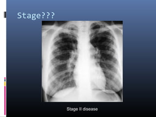
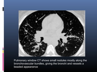
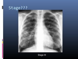
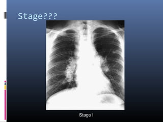
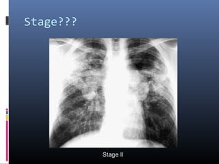
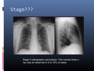
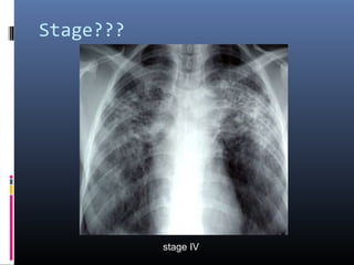
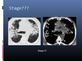
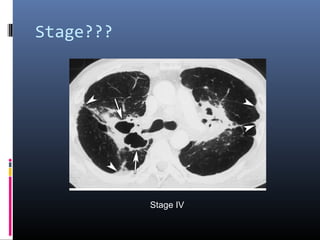
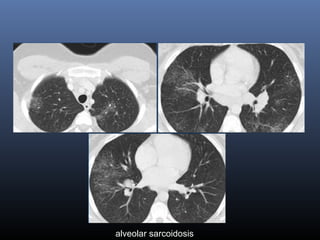
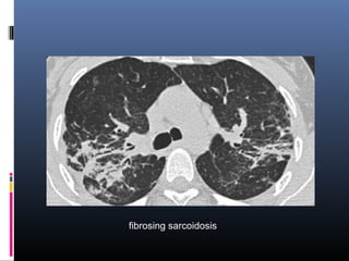
 Sarcoidosis is a systemic disorder of unknown 
origin. 
 It is characterized by non-caseating 
granulomas in multiple organs, that may 
resolve spontaneously or progress to fibrosis. 
 Pulmonary manifestations are present in 90% 
of patients.
 Systemic symptoms such as fatigue, night 
sweats and weight loss are common. 
 Löfgren's syndrome, an acute presentation of 
sarcoidosis, consists of arthritis, erythema 
nodosum, bilateral hilar adenopathy.
 Two third of patients have a remission within 
ten years. 
 One third have continuing disease leading to 
clinically significant organ impairment. 
 Less than 5% of patients die from sarcoidosis 
usually as a result of pulmonary fibrosis.
Radiographic staging of 
Sarcoidosis 
 Stage O is a normal chest radiograph 
 Stage I BHL without pulmonary infiltrates 
 Stage II BHL with pulmonary infiltrates 
 Stage III Pulmonary infiltrates without BHL 
 Stage IV Extensive pulmonary fibrosis
A PA view of the chest demonstrates right paratracheal (red 
arrow) and bilateral hilar (white arrows) lymphadenopathy
The CT scan confirms bilateral hilar lymphadenopathy (arrows. The lung 
parenchyma was normal. Differential diagnosis included primary and 
secondary neoplasms and granulomatous disease
Stage??? 
Stage I disease
Stage??? 
Stage II disease
Pulmonary window CT shows small nodules mostly along the 
bronchovascular bundles, giving the bronchi and vessels a 
beaded appearance
Stage??? 
Stage III
Stage??? 
Stage I
Stage??? 
Stage II
Stage??? 
Stage 0 radiographic sarcoidosis. This normal chest x-ray 
may be observed in 5 to 15% of cases
Stage??? 
stage IV
Stage??? 
Stage IV
Stage??? 
Stage IV
Conventional chest radiographs 
 Over 90% of patients with sarcoidosis manifest 
abnormalities on chest radiographs 
 Commonest feature-BHL (50-80%) 
 Concomitant enlargement of right paratracheal 
lymph nodes is common 
 Pulmonary parenchymal infiltrates (25 to 50%)
 Parenchymal infiltrates are often symmetrical 
and bilateral, with a predilection for the 
central (rather than peripheral) regions and 
upper lobes (particularly posterior and apical 
segments) 
 Reticular, reticulonodular, or focal alveolar 
opacities are most characteristic
 Destruction of lung parenchyma may lead to 
-architectural distortion 
-hilar retraction 
-upper-lobe volume loss 
-broad and coarse septal bands 
-honeycomb change 
-large bullae
 With advanced stage III or IV sarcoidosis, 
enlarged pulmonary arteries (attributable to 
secondary pulmonary arterial hypertension) 
and bronchiectasis may be observed.
Reticular opacities
Reticulonodular opacities
Focal alveolar opacities
Posteroanterior chest radiograph demonstrates extensive 
ground-glass, miliary infiltrates throughout both lung fields
Posteroanterior chest radiograph reveals multiple dense alveolar 
nodular. Bilateral hilar lymphadenopathy is also present
Unusual chest radiographic 
features 
The prevalence of atypical features is higher in 
sarcoid patients presenting after the age of 50 
 Pleural effusion 
 Pleural thickening 
 Pneumothorax 
 Cavitation 
 Bronchostenosis 
 Vascular involvement (pulmonary vessels) 
 Unilateral hilar lymphadenopathy
Computed tomographic scanning 
in Sarcoidosis 
 INDICATIONS 
(a) atypical clinical and/or chest radiographic 
findings. 
(b) detection of complications of the lung 
disease. 
(c) a normal chest radiograph but a clinical 
suspicion for sarcoidosis.
 CT scan provide improved anatomic lung 
detail and are more sensitive than plain chest 
radiographs in delineating parenchymal, 
mediastinal, and hilar structures. 
 CT scan may detect enlarged lymph nodes or 
parenchymal infiltrates that are below the 
resolution of conventional chest radiographs
 Enlarged lymph nodes are often observed in 
paratracheal, pretracheal, para-aortic, internal 
mammary, subcarinal, or axillary regions, 
which are not appreciated on chest 
radiographs. 
 Calcified hilar or mediastinal lymph nodes may 
be observed in patients with longstanding 
sarcoidosis
 Calcified lymph nodes may also be observed 
in tuberculosis, silicosis, and other chronic 
granulomatous disorders. 
 The lymph nodes in sarcoidosis are larger 
than those in tuberculosis, more often focal, 
and less likely to be completely calcified
 Lymph node calcification is bilateral in most 
of patients with sarcoidosis, but usually 
unilateral in most of patients with 
tuberculosis
para-aortic lymph node enlargement
Bilateral hilar lymphadenopathy and marked widening of 
the carina due to enlarged subcarinal lymph nodes (arrow)
HRCT findings in 
sarcoidosis 
 Common findings: 
 Small nodules in a perilymphatic distribution (i.e. 
along subpleural surface and fissures, along 
interlobular septa and the peribronchovascular 
bundle). 
 Upper and middle zone predominance. 
 Lymphadenopathy in left hilus, right hilus and 
paratracheal .Often with calcifications
 Uncommon findings: 
 Conglomerate masses in a perihilar location. 
 Larger nodules (> 1cm in diameter, in < 20%) 
 Grouped nodules or coalescent nodlues 
surrounded by multiple satellite nodules (Galaxi 
sign) 
 Nodules so small and dense that they appear as 
ground glass or even as consolidations (alveolar 
sarcoidosis)
small nodules along bronchovascular bundles (yellow 
arrow) and along fissures (red arrows)
small nodules in a perilymphatic distribution along 
bronchovascular bundles and along fissures (yellow arrows)
conglomerate masses of fibrous tissue
Sarcoidosis with fibrosis in the upper lobes
alveolar sarcoidosis
fibrosing sarcoidosis
Differential diagnosis of 
sarcoidosis 
 Lymphadenopathy: 
Primary TB: asymmetrical adenopathy 
Histoplasmosis 
Lymphoma 
Small cell lung cancer with nodal metastases 
 Nodular pattern: 
Silicosis / Pneumoconiosis: predominantly 
centrilobular and subpleural nodules. 
Miliary TB: random nodules
 Fibrotic pattern: 
Usual Interstitial Pneumonia (UIP): basal and 
peripheral fibrosis, honeycombing. 
Chronic Hypersensitivity Pneumonitis: mid zone 
fibrosis with mosaic pattern. 
Tuberculosis (more unilateral).
Pulmonary sarcoidosis

Pulmonary sarcoidosis

  • 2.
     Sarcoidosis isa systemic disorder of unknown origin.  It is characterized by non-caseating granulomas in multiple organs, that may resolve spontaneously or progress to fibrosis.  Pulmonary manifestations are present in 90% of patients.
  • 3.
     Systemic symptomssuch as fatigue, night sweats and weight loss are common.  Löfgren's syndrome, an acute presentation of sarcoidosis, consists of arthritis, erythema nodosum, bilateral hilar adenopathy.
  • 4.
     Two thirdof patients have a remission within ten years.  One third have continuing disease leading to clinically significant organ impairment.  Less than 5% of patients die from sarcoidosis usually as a result of pulmonary fibrosis.
  • 5.
    Radiographic staging of Sarcoidosis  Stage O is a normal chest radiograph  Stage I BHL without pulmonary infiltrates  Stage II BHL with pulmonary infiltrates  Stage III Pulmonary infiltrates without BHL  Stage IV Extensive pulmonary fibrosis
  • 6.
    A PA viewof the chest demonstrates right paratracheal (red arrow) and bilateral hilar (white arrows) lymphadenopathy
  • 7.
    The CT scanconfirms bilateral hilar lymphadenopathy (arrows. The lung parenchyma was normal. Differential diagnosis included primary and secondary neoplasms and granulomatous disease
  • 8.
  • 9.
  • 10.
    Pulmonary window CTshows small nodules mostly along the bronchovascular bundles, giving the bronchi and vessels a beaded appearance
  • 11.
  • 12.
  • 13.
  • 14.
    Stage??? Stage 0radiographic sarcoidosis. This normal chest x-ray may be observed in 5 to 15% of cases
  • 15.
  • 16.
  • 17.
  • 18.
    Conventional chest radiographs  Over 90% of patients with sarcoidosis manifest abnormalities on chest radiographs  Commonest feature-BHL (50-80%)  Concomitant enlargement of right paratracheal lymph nodes is common  Pulmonary parenchymal infiltrates (25 to 50%)
  • 19.
     Parenchymal infiltratesare often symmetrical and bilateral, with a predilection for the central (rather than peripheral) regions and upper lobes (particularly posterior and apical segments)  Reticular, reticulonodular, or focal alveolar opacities are most characteristic
  • 20.
     Destruction oflung parenchyma may lead to -architectural distortion -hilar retraction -upper-lobe volume loss -broad and coarse septal bands -honeycomb change -large bullae
  • 21.
     With advancedstage III or IV sarcoidosis, enlarged pulmonary arteries (attributable to secondary pulmonary arterial hypertension) and bronchiectasis may be observed.
  • 22.
  • 23.
  • 24.
  • 25.
    Posteroanterior chest radiographdemonstrates extensive ground-glass, miliary infiltrates throughout both lung fields
  • 26.
    Posteroanterior chest radiographreveals multiple dense alveolar nodular. Bilateral hilar lymphadenopathy is also present
  • 27.
    Unusual chest radiographic features The prevalence of atypical features is higher in sarcoid patients presenting after the age of 50  Pleural effusion  Pleural thickening  Pneumothorax  Cavitation  Bronchostenosis  Vascular involvement (pulmonary vessels)  Unilateral hilar lymphadenopathy
  • 28.
    Computed tomographic scanning in Sarcoidosis  INDICATIONS (a) atypical clinical and/or chest radiographic findings. (b) detection of complications of the lung disease. (c) a normal chest radiograph but a clinical suspicion for sarcoidosis.
  • 29.
     CT scanprovide improved anatomic lung detail and are more sensitive than plain chest radiographs in delineating parenchymal, mediastinal, and hilar structures.  CT scan may detect enlarged lymph nodes or parenchymal infiltrates that are below the resolution of conventional chest radiographs
  • 30.
     Enlarged lymphnodes are often observed in paratracheal, pretracheal, para-aortic, internal mammary, subcarinal, or axillary regions, which are not appreciated on chest radiographs.  Calcified hilar or mediastinal lymph nodes may be observed in patients with longstanding sarcoidosis
  • 31.
     Calcified lymphnodes may also be observed in tuberculosis, silicosis, and other chronic granulomatous disorders.  The lymph nodes in sarcoidosis are larger than those in tuberculosis, more often focal, and less likely to be completely calcified
  • 32.
     Lymph nodecalcification is bilateral in most of patients with sarcoidosis, but usually unilateral in most of patients with tuberculosis
  • 33.
  • 34.
    Bilateral hilar lymphadenopathyand marked widening of the carina due to enlarged subcarinal lymph nodes (arrow)
  • 35.
    HRCT findings in sarcoidosis  Common findings:  Small nodules in a perilymphatic distribution (i.e. along subpleural surface and fissures, along interlobular septa and the peribronchovascular bundle).  Upper and middle zone predominance.  Lymphadenopathy in left hilus, right hilus and paratracheal .Often with calcifications
  • 36.
     Uncommon findings:  Conglomerate masses in a perihilar location.  Larger nodules (> 1cm in diameter, in < 20%)  Grouped nodules or coalescent nodlues surrounded by multiple satellite nodules (Galaxi sign)  Nodules so small and dense that they appear as ground glass or even as consolidations (alveolar sarcoidosis)
  • 37.
    small nodules alongbronchovascular bundles (yellow arrow) and along fissures (red arrows)
  • 38.
    small nodules ina perilymphatic distribution along bronchovascular bundles and along fissures (yellow arrows)
  • 39.
    conglomerate masses offibrous tissue
  • 40.
    Sarcoidosis with fibrosisin the upper lobes
  • 41.
  • 42.
  • 43.
    Differential diagnosis of sarcoidosis  Lymphadenopathy: Primary TB: asymmetrical adenopathy Histoplasmosis Lymphoma Small cell lung cancer with nodal metastases  Nodular pattern: Silicosis / Pneumoconiosis: predominantly centrilobular and subpleural nodules. Miliary TB: random nodules
  • 44.
     Fibrotic pattern: Usual Interstitial Pneumonia (UIP): basal and peripheral fibrosis, honeycombing. Chronic Hypersensitivity Pneumonitis: mid zone fibrosis with mosaic pattern. Tuberculosis (more unilateral).

Editor's Notes

  • #9 Sarcoidosis, thoracic. Stage I disease. Standard posteroanterior chest radiograph in a 28-year-old man shows extensive bilateral hilar and mediastinal lymph node enlargement not associated with a pulmonary abnormality
  • #10 Sarcoidosis, thoracic. Stage II disease. Chest radiograph in a 36-year-old woman shows mediastinal lymph node enlargement and bilateral pulmonary opacities.
  • #11 Sarcoidosis, thoracic. Pulmonary window CT image in the same patient as in the previous image shows small nodules mostly along the bronchovascular bundles, giving the bronchi and vessels a beaded appearance. This distribution along the bronchovascular bundles accounts for the fact that transbronchial biopsy is usually successful for obtaining tissue for diagnosis.
  • #12 Sarcoidosis, thoracic.Stage III Posteroanterior (PA) chest radiograph in a 38-year-old man shows stage III disease associated with a tumefactive type of lung parenchymal involvement: opacities in the right and left mid lung zones that mimic neoplasm.
  • #13 Stage I sarcoidosis. Posteroanterior chest radiograph demonstrates bilateral hilar and right paratracheal lymphadenopathy. Lung fields are clear.
  • #14 Stage II sarcoidosis. Posteroanterior chest radiograph demonstrates massive bilateral hilar lymphadenopathy as well as bilateral parenchymal infiltrates. Note the predilection for mid and upper lung zones.
  • #15 Stage 0 radiographic sarcoidosis. This normal chest x-ray may be observed in 5 to 15% of cases
  • #16 typical chest film of long standing sarcoidosis (stage IV) with fibrosis in the upper zones and volume loss of the upper lobes resulting in hilar elevation. Fibrosis results in obliteration of pulmonary vessels, which can lead to pulmonary hypertension.
  • #17 Stage IV pulmonary sarcoidosis in a 48-year-old man. (a) Chest CT scan (lung window) demonstrates traction bronchiectasis (arrowheads) and fibrotic lesions with extensive calcification, findings that indicate stage IV disease. (b) Chest CT scan (mediastinal window) demonstrates calcification in the fibrotic lesions, mediastinal adenopathy, and irregularly thickened pleura (arrowheads).
  • #18 Stage IV pulmonary sarcoidosis in a 60-year-old man. Chest CT scan demonstrates extensive fibrotic change and cavitary lesions with a central distribution (arrows) that distort the lung parenchyma. Irregular thickening of the pleura (arrowheads) and overinflation of the peripheral lung parenchyma are also seen.
  • #20 Diffuse miliary or ground-glass patterns are exceptionally rare Confluent alveolar opacities with consolidation or multiple well-circumscribed pulmonary nodules are less common features of sarcoidosis
  • #23 Reticular opacities
  • #24 Reticulonodular opacities
  • #25 Focal alveolar opacities
  • #26 Sarcoidosis. Posteroanterior chest radiograph demonstrates extensive ground-glass, miliary infiltrates throughout both lung fields in a 22-year-old black female with a 4-month history of dyspnea. Transbronchial lung biopsies demonstrated confluent, noncaseating granuloma. Special stains and cultures for acid-fast bacilli and fungi were negative. Corticosteroids (prednisone 40 mg/day) led to dramatic improvement.
  • #27 (A) Nodular sarcoidosis. Posteroanterior chest radiograph reveals multiple dense alveolar nodular infiltrates in a 36-year-old woman. Bilateral hilar lymphadenopathy is also present. Transbronchial lung biopsies demonstrated confluent noncaseating granulomas consistent with sarcoidosis. The infiltrates resolved following institution of corticosteroid therapy.
  • #34 Chest computed tomographic (CT) scan from a patient with stage I sarcoidosis at the level of the carina shows extensive para-aortic lymph node enlargement (arrows).
  • #35 Chest CT scan from the same patient following intravenous contrast. Bilateral hilar lymphadenopathy and marked widening of the carina due to enlarged subcarinal lymph nodes (arrow) are present
  • #38 a typical presentation of sarcoidosis with hilar lymphadenopathy and small nodules along bronchovascular bundles (yellow arrow) and along fissures (red arrows).
  • #39 typical presentation of sarcoidosis with mediastinal lymphadenopathy and small nodules in a perilymphatic distribution along bronchovascular bundles and along fissures (yellow arrows).Always look for small nodules along the fissures, because this is a very specific and typical sign of sarcoidosis.
  • #40 Sarcoidosis with conglomerate masses of fibrous tissue
  • #41 Sarcoidosis with fibrosis in the upper lobes. Typical HRCT findings. stage IV sarcoidosis.; Notice the distribution of the conglomerate masses of fibrosis in the posterior part of the lungs. In addition there are multiple small well-defined nodules. Some of these nodules have the typical sub pleural distribution.
  • #42 a case of alveolar sarcoidosis. In this case the appearance resembles a ground glass attenuation, but with a close look you may appreciate that the increased attenuation is the result of many tiny grouped nodules. Also notice the hilar lymphadenopathy.
  • #43 a case of fibrosing sarcoidosis, showing fibrosis, traction bronchiectases and crowding of the involved bronchi, predominantly in the perihilar region and upper lobes. Nodular abnormalities are absent, but the appearance and the location of the fibrosis are very suggestive of the diagnosis of sarcoidosis.