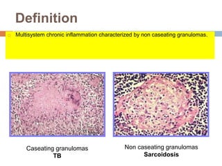
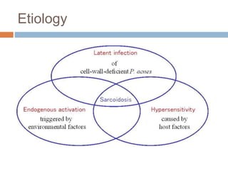
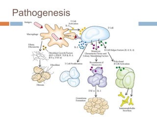
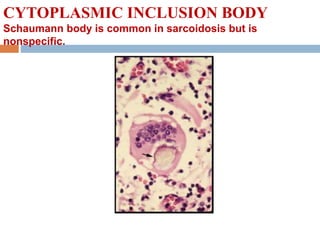
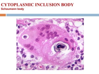
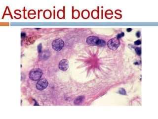
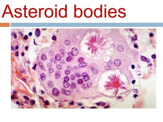
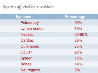
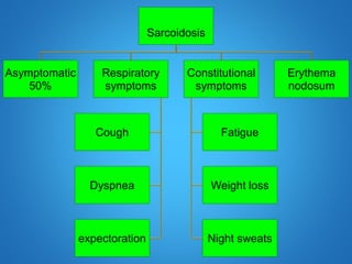
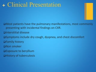
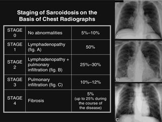
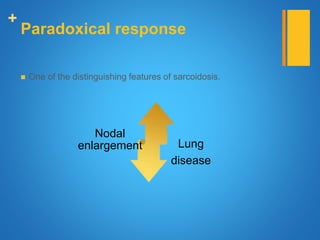
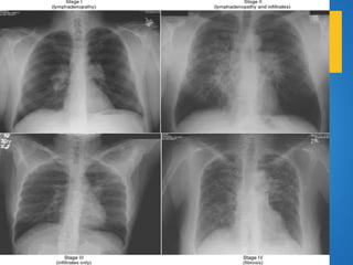
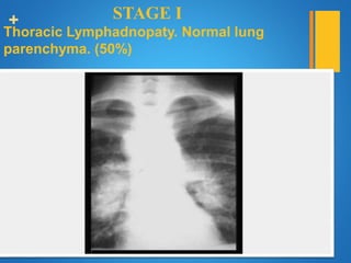
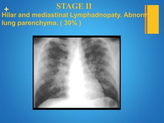
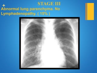
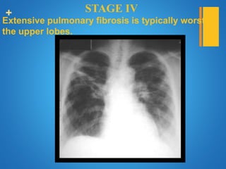
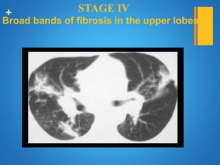
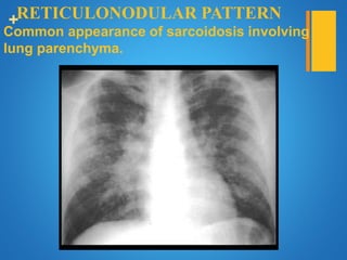
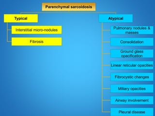
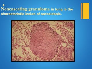
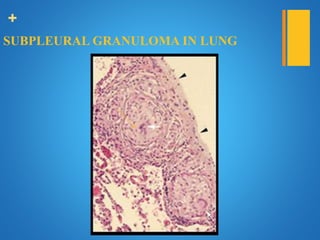
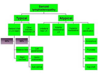
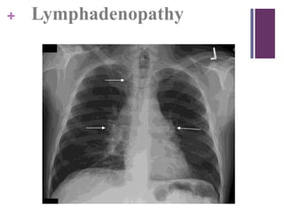
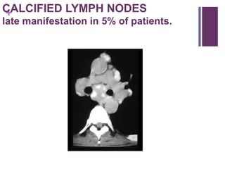
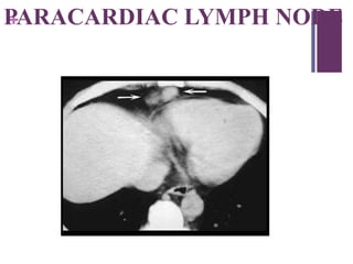
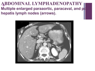
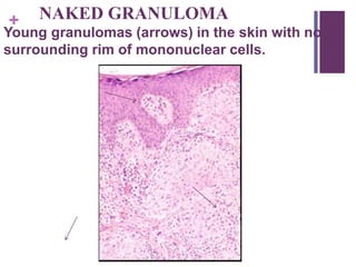
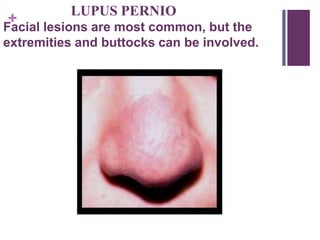
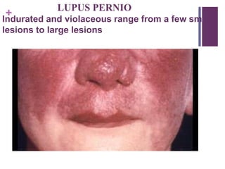
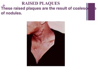
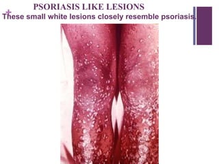
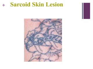
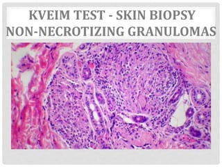
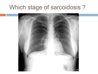
SARCOIDOSIS
Thomas Kurian
Mortimer’s Malady
Sarcoid
Definition
 Multisystem chronic inflammation characterized by non caseating granulomas.
Caseating granulomas
TB
Non caseating granulomas
Sarcoidosis
Epidemiology
 It occurs mainly in 3rd or 7th decade of life
 F>M
 Affects siblings of first- or second- degree relatives in
15% of patients with sarcoidosis.
Etiology
Etiology and Pathogenesis
 Unknown
 disordered immune regulation in genetically
predisposed individual
Etiology and Pathogenesis
Genetic Factor
 HLA
Environment factor
 Mycobacteria
 Propionobacterium acnes
 Rickettsia species
 World Trade Center
 Wood product exposure
Pathogenesis
Morphology
 classic well formed noncaseating granuloma, each
composed of an aggregates of tightly clustered
epithelioid cell
 langhans or foreign body giant cell
 central necrosis is unusual
 with chronicity the granulomas may become
enclosed within fibrous rims or may be eventually be
replaced by hyaline fibrous scars.
Morphology
 Laminated concentration composed of calcium and
proteins known as schaumann bodies and stellate
inclusion as asteroid bodies enclosed with in giant
cells are formed in approximately 60% of granulomas.
 Characteristic
LANGHANS' GIANT CELL
Langhans' giant cell in center of granuloma is surrounded
by epithelioid cells
Epitheliod cells produce collagenase, elastase, lysozyme and ACE
ADVANCED COLLAGENOUS FIBROSIS
Elongated fibroblasts with extensive collagenous tissue,
Giant cells
CYTOPLASMIC INCLUSION BODY
Schaumann body is common in sarcoidosis but is
nonspecific.
CYTOPLASMIC INCLUSION BODY
Schaumann body
Asteroid bodies
Asteroid bodies
HISTOLOGY IS NOT
SPECIFIC
Leprosy , tertiary syphilis, brucellosis, primary
biliary cirrhosis, Berylliosis
Differential Diagnosis of Noncaseating Granulo
TB
Fungal infections
Lymphoma
Epithelioid tumors of the breast
Lung cancer
Systems affected by sarcoidosis
Systems Percentage
Pulmonary 90%
Lymph nodes 70%
Hepatic 50-80%
Cardiac 30%
Cuteneous 25%
Ocular 20%
Spleen 18%
Bones 14%
Neurogenic 5%
+
PULM0NARY
SARCOIDOSIS
Sarcoidosis
Asymptomatic
50%
Respiratory
symptoms
Cough
Dyspnea
expectoration
Constitutional
symptoms
Fatigue
Weight loss
Night sweats
Erythema
nodosum
+ Clinical Presentation
Most patients have the pulmonary manifestations, most commonly
presenting with incidental findings on CXR.
Interstitial disease
Symptoms include dry cough, dyspnea, and chest discomfort
Family history
Non smoker
Exposure to beryllium
History of tuberculosis
+
+
Paradoxical response
 One of the distinguishing features of sarcoidosis.
Nodal
enlargement Lung
disease
+ ADENOPATHY AT TIME OF DIAGNOSIS
Marked enlarged hilar and mediastinal lymph nodes.
+ ADENOPATHY DECREASED 2 YRS LATER
Lymph nodes are smaller and there is parenchymal lung disease.
+
Calcified hilar & mediastinal LN
+
Hilar enlargement
Unilateral hilar enlargement Bilateral hilar enlargement
•Sarcoiosis
•Silicosis
Infection •TB •TB
Neoplasm •Bronchogenic carcinoma.
•Metastasis (lung, head & neck,
thyroid or testis).
•Lymphoma
•Metastases
Vascular •Post-stenotic dilatation (left side).
•Pulmonary artery aneurysm.
•Pulmonary hypertension.
•Pulmonary artery aneurysm.
+
LN enlargement vs pulmonary vascular
enlargement
LobulatedSmooth
+
+ STAGE I
Thoracic Lymphadnopaty. Normal lung
parenchyma. (50%)
+ STAGE II
Hilar and mediastinal Lymphadnopaty. Abnormal
lung parenchyma. ( 30% )
+ STAGE III
Abnormal lung parenchyma. No
Lymphadenopathy. ( 15% )
+ STAGE IV
Extensive pulmonary fibrosis is typically worst in
the upper lobes.
+ STAGE IV
Broad bands of fibrosis in the upper lobes
+RETICULONODULAR PATTERN
Common appearance of sarcoidosis involving the
lung parenchyma.
Parenchymal sarcoidosis
Typical
Interstitial micro-nodules
Fibrosis
Atypical
Pulmonary nodules &
masses
Consolidation
Ground glass
opacification
Linear reticular opacities
Fibrocystic changes
Miliary opacities
Airway involvement
Pleural disease
+
Typical parenchymal sarcoidosis
Interstitial micronodules
 Coalescent groups of microscopic granulomas.
 Well defined, with irregular margins.
 2-4 mm.
+
Interstitium
Central
Peri-broncho-
vascular
Peripheral
Interlobular Subpleural
+Upper lobe predominance
+
Bilateral perihiar predominance
+ Nodular thickening of the
peri-broncho-vascular interstitium
+ Nodular thickening of interlobular septa,
fissures
(.-.-.-.-.-.-)
+
Sub-pleural nodules
(pseudo-plaques)
+
Typical parenchymal sarcoidosis
Fibrosis
 Reticular opacities.
 Traction bronchiectasis.
 Architectural distortion.
+ MILIARY SARCOIDOSIS
CT shows well defined lung nodules less than 5mm in diameter. This
pattern is rare
+ ALVEOLAR SARCOIDOSIS
Multiple lung masses are an unusual form of
Sarcoidosis, resembles lung metastases.
+ ALVEOLAR SARCOIDOSIS
Computed tomography shows a mass which has air
containing bronchi within it.
+ CAVITARY SARCOIDOSIS
Rare pattern of multiple cavitary sarcoid lung lesions. Note
Lymphadnopaty.
+ ACINAR PATTERN
Poorly defined nodular opacities are the size of
pulmonary acini (6mm).
+ PNEUMONIC APPEARANCE
Confluent acinar opacities look similar to
pneumonic consolidation.
+
Lymph nodes with rim (eggshell) calcification are rare in
sarcoidosis but common in silicosis.
+
Sarcoid Galaxy sign
+Pleural disease
Rare < 1%
 Pleural effusion.
 Hemothorax.
 Chylothorax (due to involvement of the mediastinal LN or
thoracic duct).
+
Air way involvement
 May occur at any level
 Bronchial wall thickening
 Luminal abnormalities
 Obstruction of lobular or
segmental bronchi
 Mosaic perfusion
 Air-trapping on expiration
+
Noncaseating granuloma in lung is the
characteristic lesion of sarcoidosis.
+
SUBPLEURAL GRANULOMA IN LUNG
+PROGNOSIS OF PULMONARY
SARCOIDOSIS
Prognosis of
pulmonary sarcoidosis
3/4 1/3 1/5
Complete resolution
of
hilar
lymphadenopathy
Complete resolution
of
parenchymal
disease
Irreversible
pulmonary
fibrosis
+
Lymph nodes
sarcoidosis
Sarcoid
lymphadenopathy
Typical
Garland triad
(1,2,3 sign)
Bilateral hilar
Right
tracheal
95%
Other
tracheo-
bronchial LN
Left
paratracheal
Aorto-
pulmonary
Sub-carinal
50%
Atypical
Unilateral
hilar LN
Anterior
mediastinal
LN
Posterior
mediastinal
LN
calcification
Amorphous
Punctate
Popcorn
Egg shell
+
Sarcoidosis of Lymph nodes
Lymphadenopathy
nodes are characteristically enlarged discrete
and sometimes calcified
Tonsil may affected in about quarter to one
third of the cases
+ Lymphadenopathy
Typical
1.Bilateral hilar & right paratracheal LN,
2.Middle mediastinal LN occur in 50% of cases.
3.Left paratracheal, aorto-pulmonary & subcarinal
LN.
1-2-3 sign present in 95% of cases. This is called
Garland triad
+ Lymphadenopathy
+CALCIFIED LYMPH NODES
late manifestation in 5% of patients.
+PARACARDIAC LYMPH NODE
+ABDOMINAL LYMPHADENOPATHY
Multiple enlarged paraaortic, paracaval, and porta
hepatis lymph nodes (arrows).
+
Cuteneous
sarcoidosis
+ Skin
 33% have skin lesions
 Cutaneous anergy is common.
+ NAKED GRANULOMA
Young granulomas (arrows) in the skin with no
surrounding rim of mononuclear cells.
+
Erythema Nodosum
+Erythema nodosum
+ Skin
 Lupus pernio- indurated blue purple swollen
shiny lesions on nose, cheeks, lips, ears and
fingers.
 Papules, nodules, and plaques
 Psoriatic like lesions
 Lesions in scars and tattoos
+
LUPUS PERNIO
Facial lesions are most common, but the
extremities and buttocks can be involved.
+
LUPUS PERNIO
Indurated and violaceous range from a few small
lesions to large lesions
+
RAISED PLAQUES
These raised plaques are the result of coalescence
of nodules.
+
PSORIASIS LIKE LESIONS
These small white lesions closely resemble psoriasis.
+ Sarcoid Skin Lesion
Lofgren syndrome
Acute sarcoidosis characterized by triad
Lofgren
syndrome
Bilateral
hilar
lymphadenopathy
Arthritis
Erythema
nodosum
Heerfordt’s syndrome
Acute sarcoidosis characterized by uveoparotitis
Heerfordt’s
syndrome
Uveitis Parotitis Facial palsy
+
Heerfordt’s syndrome
+
OCULAR
sarcoidosis
+ Eyes
25% have eye lesions
Blurred vision, pain, photophobia and dry eyes
Chronic uveitis leads to glaucoma, cataracts and
blindness
Keratoconjunctivitis sicca
Papilledema
+ CONJUNCTIVITIS
+ PAPILLEDEMA
Often associated with 7th nerve facial palsy.
+Sarcoid Choreoretinitis
+ Sarcoid Uveitis
+
liver
sarcoidosis
+ Liver
33% have hepatomegaly or biochemical evidence
of disease
Symptoms usually absent
Cholestasis, fibrosis, cirrhosis, portal
hypertension, and the Budd-Chiari syndrome
have been seen
+SPLEEN & LIVER GRANULOMAS
The small low attenuation lesions in the liver and
spleen in sarcoidosis.
+
EARLY COLLAGEN FORMATION
Extracellular collagen is being produced by
fibroblasts
+
Musculoskeletal
SARCOIDOSIS
+ Musculoskeletal
 Acute polyarthritis with fever is common
 Arthritis is self limited
 Chronic destructive bone disease with deformity is
rare
+
PUNCHED OUT LYTIC LESIONS
Focal osteolytic lesions in the fingers are most common
abnormality.
+LACY TRABECULAR PATTERN
Osteolysis has left a lacy trabecular pattern in this
phalanx
+ SCLEROTIC LESION
Rare and often in the axial skeleton.
+ NASAL BONE LESION
Nasal sarcoidosis can lead to osteolysis of the
nasal bone.
+
Nervous System
SARCOIDOSIS
+ Nervous System
Cranial nerves, and peripheral nerves can be involved
7th nerve facial palsy is most common
Acute, transient, and can be unilateral or bilateral
HEREFORDT'S SYNDROME; facial palsy accompanied by
fever, uveitis, and enlargement of the parotid gland
+
T1-W POST GADOLINIUM MR IMAGE
Post contrast image of high signal intensity temporal lobe sarcoid lesion
+ Nervous System
 Optic nerve dysfunction
 Papilledema
 Palate dysfunction
 Hearing abnormalities
 Paresthesias
 Meningeal granulomas
 Encephalopathy
+
KIDNEY
SARCOIDOSIS
+ Kidney
Granulomatous interstitial nephritis produces renal failure
Develops over a period of weeks to months
Rapid response to steroid therapy
Kidney stones (nephrolithiasis) and nephrocalcinosis are
very unusual secondary to hypercalcemia and
hypercalciuria
+
NEPHROCALCINOSIS
There are multiple calcifications of the kidneys.
Enlarged retroperitoneal lymph nodes
+ Kidney
Increased calcium absorption in the gut
Related to high levels of circulating 1,25-dihydroxy
vitamin D produced by mononuclear phagocytes in
granulomas
+ GASTRIC SARCOID
Granuloma involves the gastric antrum leading to
irregular nonspecific narrowing.
+
COLONIC SARCOID
Irregular narrowing of the rectosigmoid has the
appearance of inflammatory disease or malignancy.
DIAGNOSIS
LAB ABNORMALITIES
 Lymphocytopenia
 Mild eosinophilia
 Increased E.S.R
 Hyperglobulinemia
 Serum Calcium, urinary calcium
LAB ABNORMALITIES
Elevated level of angiotensin converting enzyme
Gallium 67 lung scan showing a pattern of diffused
uptake.
Bronchiole alveolar lavage shows increased
lymphocytes, CD4/CD8 levels
LUNG FUNCTION TEST
• Decreased lung volumes
• Decrease in DLCO
• Airflow obstruction
RADIOLOGY
DIAGNOSIS
Identify noncaseating granulomas
Variety of infections
Transbronchial biopsies positive in 65-95%, even if no
lung parenchymal abnormalities imaged.
Tissue from mediastinoscopy
Scalene node biopsy
DIAGNOSIS
• Difficult to differentiate from chronic infections, fungal
diseases, T.B. and lymphoma.
• Based on combined clinical, radiologic and histologic
findings.
• Laboratory tests seldom important
THE KVEIM-SILTZBACH TEST
• If positive, is considered to be diagnostic of sarcoidosis
• Test material
• Positive test
• No corticosteroids
• Why does this reaction occur?
THE KVEIM-SILTZBACH TEST
• Difficulties
Test not always positive
FDA
KVEIM TEST - SKIN BIOPSY
NON-NECROTIZING GRANULOMAS
Do all patients require treatment?
Prednisolone 20 mg
When is it considered refractory?
Treatment
 When ?
 Chloroquine , hydroxychloroquine
 Methotrexate
 Azathioprine
 Leflunomide
 Mycophenolate
 Infliximab
Corticosteroid sparing drugs
 Pulmonary Hypertension
 Fatigue
 Aspergilloma
 Lung transplantation
Management of
Complications
 Good
 50% have some permanent organ dysfunction
 In 15-20% remains active or recurs intermittently.
Prognosis
QUESTIONS
Which stage of sarcoidosis ?
Identify the syndrome
+

Sarcoidosis