This document provides an overview of sarcoidosis, including:
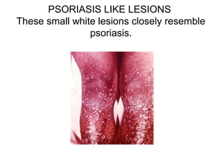
- It is a multisystem granulomatous disorder of unknown cause that commonly affects the lungs, skin and eyes.
- Risk factors include genetics and environmental exposures, and it has the highest rates in the United States and Sweden.
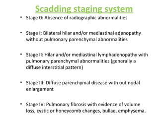
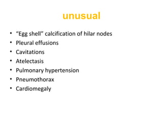
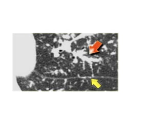
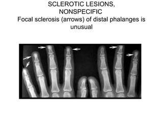
- Clinical presentation varies from asymptomatic to involvement of multiple organ systems. Lung involvement is most common and is staged based on chest x-ray findings.
- Diagnosis involves ruling out other causes and may include biopsy showing non-caseating granulomas. Treatment involves corticosteroids and prognosis is generally good with many experiencing remission.
5. DEFINITION
• Sarcoidosis is a multisystem non-caseous
granulomatous disorder of unknown aetiology
characterized by depression of cutaneous
delayed type hypersenstivity and heightened
Th1 immune response in affected organs.
• Most commonly affecting young adults
• Presenting most frequently with bilateral hilar
lymphadenopathy, pulmonary infiltration and
skin or eye lesions.
6. HISTORICAL PERSPECTIVE
• Jonathan Hutchinson 1877- Mortimer’s malady
• 1889- Besnier - “lupus pernio.”
• 1899- Caesar Boeck- “sarcoid” (multiple benign sarcoids of the
skin.)
• 1909- heerfordt- uveoparotid fever
• Jorgen Schaumann - 1914 “lymphogranuloma benigne”
• 1940+ universal acceptance of word sarcoidosis.
7. EPIDEMIOLOGY
• occurs worldwide
• highest incidence - United States and Sweden.
• lower - Asian
• Prevalence - 10 and 40 cases per 100,000
• mortality - 1 to 5 percent
8. • Environmental exposures - winter and
early spring months
• Geographic variation and time–space
clustering
• Occupational association- health care
professionals,firefighters, military
personnel.
9. • Age : 20-40 yrs
• Sex : female ; bimodal
• 16 times more common in blacks
• genetic influence
monozygotic : dizygotic twins (13: 1)
10. Aetiology
• Unknown
• exposure to pine pollen or beryllium
• infection with Mycobacterium, viruses and
fungi,
• protoplast or L form of the tubercle bacillus
may be a cause of sarcoidosis
11. ACCESS STUDY
• - A Case Control Etiologic Study of
Sarcoidosis ;706 subjects
-Absence of occupational and environmental
association
-Weak assoc.-insecticide;mold;musty odour
-No association-berylium;wood ;rural; smoking
-No single dominant factor
-Gene environmental factors
13. • Borrelia
• chlamydiae
• Rickettsiae
• Viruses : EBV,CMV,herpes 6
• High titres of these viruses may be reflect
generalised B cell activation in sarcoidosis.
16. GENETICS
• STRONG EVIDENCE
• ACCESS STUDY
• siblings are higher risk than parents
• More of mother-child than father-child
associations.
17. ROLE OF HLA
• HLAB 8 – most consistently associated.
• HLA-B1,B8, DR3- also associated with
abnormal immune responsiveness.
• DR1, DR 4 – protective.
• HLA-B8, DR3, Cw7 associated with good
prognosis in caucasians.
• Other alleles are associated with chronic
disease, favorable outcomes.
18.
19. Non-HLA genes:
• TNF genes.(polymorphisms associated
with 1.5 fold increased risk)
• CC chemokine receptors.
• ACE complement receptor 1.
• German study – Chromosome 6
• SAGA study – Chromosome 5
20. Non-HLA Candidate Genes Evaluated in Sarcoidosis
Vascular endothelial growth factor þ813 CT and TT genotypes associated
with protection
Angiotensin converting enzyme (ACE) Moderate association between II
genotype
C-C Chemokine receptor 2 Associated with protection/Lo ¨ fgren‘s
syndrome
Heat shock protein 70 like HSP(þ2437)CC associated with
susceptibility/Lo ¨ fgren’s syndrome
Vitamin D receptor (VDR BsmI allele associated with sarcoidosis
Cystic fibrosis transmembrane
regulator
(CFTR)
R75Q increases risk
IL1a,IL1 8 The IL-1 a-889 1.1 ; Genotype -607CA
increased risk over AA
Interferon gamma IFNA17 polymorphism (551T!G) and
IFNA10 [60A]- IFNA17 [551G]
haplotype increase risk
23. • IL-12 contributes to proliferation of activated T
cells in early disease.
• Increased levels of IL-12 and IL-18 stimulate
IFN-gamma production.
• TGF-beta inhibits IL-12 and IFN-gamma-
downregulation of granulomatous
inflammation in sarcoidosis.
• Elevated levels of IL-6 and IL-8 in BAL in active
sarcoidosis.
24. Histopathology
• the pathologic hallmark is presence of non caseating
epitheloid cell granuloma.
• active sarcoidosis has nodular collections of large closely
packed, pale-staining histiocytes - epithelioid cells with
giant cells.
• Necrosis doesn’t occur.
• Electron microscopy showed that epitheloid cells tend
to be central and macrophages peripheral.
• As lesion ages, reticulin fibres between epitheloid cells
ramify and converted to collagen.
25. • The cytoplasm of giant cells have inclusion bodies
1) Schaumann bodies- round or oval and vary in size from
that of a leucocyte to about 100µm in diameter.The larger
of these bodies ( conchoid bodies) seem to be formed of
basophilic concentric lamellae that appear to contain
calcium and iron
2) Doubly refractile crystalline inclusion bodies are 1–20 µm
in diameter and may take up stains for calcium and iron.
3) Asteroid bodies consist of a central mass 2.3–3µm in
diameter with radiating straight or centred spinous
projections, the whole being 5–25µm in diameter
4) Hamazaki wesenberg bodies.
26.
27.
28. DIFFERENCES BETWEEN GRANULOMA OF TB and
SARCOIDOSIS
TUBERCULOSIS SARCOIDOSIS
EPITHELOID CELLS PRESENT PRESENT
NECROSIS COMMON ABSENT
CONFLUENT GRANULOMA USUAL DISCRETE
GIANT CELLS MULTIPLE FEW
RETICULIN IN GRANULOMA LOST PRESERVED
AFB MAY BE PRESENT ABSENT
29. • Histological features of sarcoid lesion are not
specific . Can also be seen in
tuberculosis
leprosy
tertiary syphilis
brucellosis
primary biliary cirrhosis
hypogammaglobulinemia
Brucellosis
fungal infections.
30.
31.
32. CLINICAL PRESENTATION
Acute Sarcoidosis
• Lofgren's syndrome - acute erythema nodosum with
bilateral hilar lymphadenopathy, fever, and
polyarthritis, non granulomatous uveitis
• Symptoms are typically abrupt in onset and have a
transient course.
• A vesicular or maculopapular rash, acute iritis,
conjunctivitis, and Bell's palsy may also be features of
acute disease
33. Chronic Sarcoidosis
• more gradual, insidious onset, is more likely to
develop chronic disease.
• Other risk factors for chronic disease include the
presence of lupus pernio, which is a persistent,
disfiguring, violaceous rash over the nose, cheeks,
and ears, and the presence of multiorgan
involvement at the time of diagnosis.
• Chronic eye involvement includes chronic uveitis,
cataracts, glaucoma, or keratoconjunctivitis sicca,
which can be confused with Sjogren's syndrome
34.
35. Major Clinical Manifestations
Organ System
(percent clinical
disease)
Major clinical features
Pulmonary (>90%) Restrictive and/or obstructive disease
fibrocystic disease ;bronchiectasis
Upper respiratory
tract and oral cavity
(5-10%)
Hoarseness
Laryngeal or tracheal obstruction
Nasal congestion
Sinusitis
Ocular (20-30%) Anterior and posterior uveitis
Chorioretinitis ,Conjunctivitis
Optic neuritis
38. Renal (<5%) Renal calculi
Nephrocalcinosis
Renal failure
Genitourinary
(<5%)
Ovarian or uterine mass
Dysmenorrhea
Testicular mass
Epididymitis
Psychosocial
manifestations (30-
60%)
Depression
39. Lungs
• First site involved
• Begins with alveolitis involving small bronchi and small
blood vessels
• Alveolitis either clears up spontaneously or leads to
granuloma and fibrosis.
40. • Around 50% patients asymptomatic.
• Dry cough and dyspnoea
• Chest pain-rare
• Wheezing secondary to endobronchial disease,
extrinsic compression by lymphadenopathy or
bronchial distrortion secondary to fibrosis.
• Productive cough- traction bronchiectasis
• Hemoptysis- bronchiectasis or aspergilloma
41.
42.
43. Radiographic features
• Chest radiograph abnormal in 90% of sarcoidosis
patients.
• Bilateral hilar lymphadenopathy in 50-85%cases.
• Lymph nodes big and sharply defined with clear line
of transluscency between mediastinum and lymph
nodes- POTATO NODES.
• Unilateral lymphadenopathy- rare
• Pulmonary infiltrates in 25-60% cases
44. Scadding staging system
• Stage 0: Absence of radiographic abnormalities
• Stage I: Bilateral hilar and/or mediastinal adenopathy
without pulmonary parenchymal abnormalities
• Stage II: Hilar and/or mediastinal lymphadenopathy with
pulmonary parenchymal abnormalities (generally a
diffuse interstitial pattern)
• Stage III: Diffuse parenchymal disease with out nodal
enlargement
• Stage IV: Pulmonary fibrosis with evidence of volume
loss, cystic or honeycomb changes, bullae, emphysema.
45. CXR :
Radiographic staging of intrathoracic sarcoidosis
Staging also helps in prognosis.
stage Hilar
adenopathy
Parenchymal
lesions
% at onset
% with
resolution
0 No No <10 NA
1 Yes No 50 65
2 Yes Yes 30 20-50
3 No Yes 10-15 <20
4 No fibrosis 10-15 <20
46.
47. Sarcoidosis stage I: left and right hilar and
paratracheal adenopathy (1-2-3 sign)
55. ADENOPATHY AT TIME OF DIAGNOSIS
Marked enlarged hilar and mediastinal lymph
nodes.
56. Radiographic abnormalities on chest x ray.
1) disseminated miliary lesions
2) disseminated nodular lesions
3) linear type of infiltration extending fan-wise from the hilum
4) diffuse and confluent patchy shadows;
5) diffuse fibrosis
6) diffuse fibrosis with cavitation
7) diffuse ground-glass shadowing
8) changes similar to chronic tuberculosis as regards location
and distribution
9) bilateral confluent massive opacities resembling areas of
pneumonia
10) atelectasis.
58. • HRCT findings in Sarcoidosis.
• Common findings:
– Small nodules in a perilymphatic distribution (i.e.
along subpleural surface and fissures, along
interlobular septa and the peribronchovascular
bundle).
– Upper and middle zone predominance.
– Lymphadenopathy in left hilus, right hilus and
paratracheal (1-2-3 sign). Often with calcifications.
•
62. • Uncommon findings:
– Conglomerate masses in a perihilar location.
– Larger nodules (> 1cm in diameter, in < 20%)
– Grouped nodules or coalescent nodules
surrounded by multiple satellite nodules
(sarcoid galaxy sign)
– Nodules so small and dense that they appear
as ground glass or even as consolidations
(alveolar sarcoidosis)
– Reverse halo sign
65. Gallium 67 scan
• Gallium 67 concentrated in metabolically and mitotically
active tissues.,
• Intravenous injection of 11X107
Bq of gallium 67 citrate,
simultaneous anterior and posterior scans performed 3 days
later.
• Close correlations between gallium uptake and total number
of lymphocytes recovered in BAL.
• Not specific for sarcoidosis.
• Expensive and radiation exposure.
66. the parotid, salivary, andlacrimal gland region (panda sign) is
pathognomic for sarcoidosis.
67.
68. Gallium scan showing
increased uptake in
the lacrimal and
parotid glands and
pulmonary regions in
a patient with active
sarcoidosis
68
69. Other intrathoracic manifestations
• Pleural invovlment in 5-10% cases.( effusions and
pleural thickening)
• Aspergilloma
• Bronchiectasis
• Necrotising sarcoid angitis
• Superior venacaval syndrome
• Mediastinal lymph node calcification
• Mediastinal fibrosis
• Vanishing lung syndrome(giant bulla)
70. Functional abnormalties
• Seen in 20%-40% in patients with normal chest x ray
and 50%-70% when x ray is visibly abnormal.
• Abnormalities in vital capacity, diffusion capacity,
PaO2 at rest, PaO2 at exercise and lung compliance.
• Usually restrictive defect noted on PFT but
obstructive defect in endobronchial involvement
leading to airflow obstruction, also from airway
distortion secondary to lung fibrosis.
72. Upper respiratory tract
• Uncommon but disabling.
• Nasal mucosa- crusting,obstruction, discharge. The
mucosa erythematous and granular with polypoid
hypertrophy.
• Laryngeal and pharyngeal mucosa- hoarseness,
cough,dysphagia,dypsnoea
• Patients with sarcoidosis of upper respiratory tract
have 50% chance of developing lupus pernio
• Nasal septal and palatal perforations in untreated
cases.
• d/d tuberculosis, wegeners granulomatosis, leprosy
73. • Krespi staging system
• Stage I : mild nasal disease without paranasal sinus
disease. - saline nasal spray, nasal irrigation, and topical
nasal steroids.
• Stage II - moderate disease, with involvement of both
nasal and paranasal sinuses; it is typically treated with
both stage I therapy and intralesional steroids.
• Stage III - severe, often irreversible, nasal and sinus
disease that usually requires the therapeutic
interventions of stages I and II, as well as systemic
therapy.
74. Lymphatic system
• hilar and mediastinal lymph nodes (>90% of
patients);
• peripheral lymphadenopathy (5%–30%).
• cervical, axillary, epitrochlear, and inguinal
regions.
• nontender, mobile
• Of superficial nodes, right scalene group most
common
75. Eyes
• upto 25% of sarcoidosis patients and uveitis most
common manifestation.
• Anterior uveitis - acutely, with pain, photophobia,
lacrimation, and redness, self limiting
• Posterior uveitis is typically gradual in onset and is
more likely to result in visual morbidity, chronic form
of disease.
• Chronic uveitis leads to glaucoma, cataracts and
blindness ,Keratoconjunctivitis sicca
Papilledema
• 10% of patients with sarcoid-associated uveitis
develop blindness in at least one eye
80. Skin
• Most common manifestation- erythema nodosum.
• Erythema nodosum typically presents as raised, tender, red
nodules, 1 to 2 cm in diameter, on the anterior surface of the
lower legs.
• lupus pernio is a rare lesion,most severe dermatological
manifesation - purplish plaques typically found over the
nose, cheeks, lips, and ears. Associated with poor prognosis
and severe pulmonary disease.
• skin abnormalities include red-brown to orange macules and
papules, keloids, and hyper- or hypopigmentation.
81. • LOFGREN'S SYNDROME; acute triad of
erythema nodosum,polyartropathy, bilateral
hilar adenopathy and non granulomatous
uveitis
86. Liver
• 33% have hepatomegaly or biochemical evidence of
disease
• Symptoms usually absent
• Cholestasis, fibrosis, cirrhosis, portal hypertension,
and the Budd-Chiari syndrome have been seen
87. Musculoskeletal
• Bone involvement most commonly affects terminal
phalanges of hands and feet and proximal bones in
severe cases.
• Three types of bony lesions:
lytic
permeative
destructive
• Bone lesions not affected by corticosteroids.
88. PUNCHED OUT LYTIC LESIONS
Focal osteolytic lesions in the fingers are most
common abnormality.
90. • Subcutaneous swellings also noted along with
digit abnormalities but they respond to
corticosteroid therapy.
• Skeletal granulomas commonly affecting
pectoral,shoulder, arm and calf muscles.
91. Nervous System
• Peripheral neuropathy or mononeuritis multiplex
• Cranial nerve palsy- 7th nerve facial palsy is most
common (sarcoidosis is most common cause of bilateral
facial nerve palsy)
Acute, transient, and can be unilateral or bilateral
HEERFORDT'S SYNDROME: facial palsy
accompanied by fever, uveitis, and enlargement of the
parotid gland
• Lymphocytic meningitis
• Meningoencephalitis
• SOLs
• Epilepsy
92. • Hematopoietic system: splenomegaly
common
• Genitourinary system: nephrocalcinosis due
to unexplained increase in senstivity to
vitamin D leading to increased absorption of
calcium from gut.
• Rarely glomerulonephritis and sarcoidosis of
epididymis.
93. cardiac
• extensive pulmonary fibrosis leading to cor
pulmonale.
• Involvement of myocardium leading to
dysrhythmias,conduction disorders, heart failure and
sudden death.
• PAH may occur due to parenchymal fibrosis
distorting the pulmonary vasculature, granulomatous
inflammation of pulmonary vasculature,hypoxic
pulm arterial vasoconstriction and from elevated left
ventricular diastolic pressure of cardiac involvement.
94. ATS criteria diagnosis of
pulmonary sarcoidosis
(1) presence of a consistent clinical and radiographic
picture
(2) demonstration of noncaseating granulomas on
biopsy
(3) exclusion of other conditions that can produce
granulomatous inflammation
95. Recommended Test for all patients :
Complete blood count with differential count
Chest radiograph
PFT-spirometry, diffusion capacity, lung volumes
Ophthalmologic examination
Complete metabolic profile
ECG
PPD skin test. (<10mm reaction to 5TU has 100% sensitivity but not
specific. About 2/3rd
of patients with active disease fail to react to
100TU and 1/4th
to 100TU and less than 1/10th
to 10TU.)
Serum & Urine Calcium
96. Organ Specific Test :
Cardiac-
ECG
Holter monitoring
Thalium or sestamibi myocardial scan
Cardiac MRI
Cardiac PET.
Neurologic :
Brain or spine MRI with gadolinium enhancement
CSF examination
EMG or nerve conduction studies.
97. URT :
Flow-volume loop
ENT evaluation.
X-Ray PNS
CT Scan PNS
Endocrine :
Pituitary function test
Thyroid profile.
Biopsy :
Tissue biopsy is essential.
Biopsy almost always +ve if skin, lymphnodes, conjunctiva involved,
accessible sites.
98. Cont
Transbronchial lung biopsy is usually performed because high yield
and relative safe.
The diagnostic yield of TBLB is ranges 40-90% if atleast 4 biopsies
are taken.
Higher yield if pulmonary infiltrates an CXR or CT scan shows.
TBLB in advanced fibrocystic sarcoidosis has a low yield.
TBNA has diagnostic sensitivity of 100%
Combining EBUS with TBNA increases the sensitivity.
99. BAL Findings :
Increased lymphocytes.
Raised CD4: CD8 ratio >3.5 to 4.0 supports the diagnosis of sarcoidosis.
When FOB is non diagnostic –
Mediastionscopy
VATS
Open lung biopsy establish the diagnosis at >90% diagnostic yield.
For organs that are difficult to biopsy image techniques such as gallium
67 scan or more recently 18F – fluoro deoxyglucose position emmison
tomography (FDG-PET) may help.
F-methyltyrosine-PET uses amino acids that is preferentially taken up
and expressed on tumor cells and is negative in sarcoidosis.
100. Tissues from mediastinoscopy +ve in 95%.
Scalene lymph node biopsy positive in 80%
101. Serum ACE levels (SACE levels) :
• Libermann first to report raised ACE in sarcoidosis
• Produced by epitheloid cells.
• Elevated in 30 to 80% of patients.
• Elevated levels are seen in infections ,graulomatous disease,
lymphoma, hepatitis, DM, thyroid disease. Thus SACE is not
recommended as a diagnostic test.
• Neither sensitive nor specific to be used as a diagnostic tool.
• Provides good monitor of disease activity.
102. Kveim-stiltzbach test :
Intradermal injection of homogenised tissue of organs
involved with sarcoidosis causes delayed cutaneous
reaction in 4 – 6 weeks.
Method of testing
• The test is performed by injecting intradermally 0.1- 0.2
ml of suspension of human sarcoid tissue , usually
obtained from cervical gland.
• Rarely , splenectomy for splenomegaly due to sarcoidosis
allows the preparation of large quantities of test
substance.
103. • Within 2-3 wks positive test shows purplish red
nodule at the site of injection.
• Biopsy at 4- 6 wks reveals sarcoid tissue on
histological examination.
• Value of the test : false positive reactions are rare,
only 1 – 2% .
• Positive test can be regarded as a virtual proof of
active sarcoidosis.
• Positive results are obtained in 75% of patients with
clinical evidence of disease.
105. Assessment of disease activity
• Best is by clinical assessment by worsening or
persistence of symptoms, with skin lesions and
changes in chest radiograph and PFTs.
• Serum ACE levels
• Others:
blood( lysozyme, neopterin,soluble IL-2 receptor)
BAL fluid(high lymphocytes, CD4/CD8 ratio, TNF-
alpha, collagenase etc.,)
106. Differential Diagnosis
S.NO
Cause Granulomatous Disease Confirmatory Data
1) Infectious agents
a) Mycobacteria
Tuberculosis, atypical
mycobacterial infection
PPD; sputum or BAL culture
b) Fungi
Histoplasmosis,
coccidioidomycosis
Serologic testing (histo); sputum
or biopsy stains, serologies, skin
tests (cocci)
c) Spirochetes Syphilis RPR
d) Parasites
Leishmaniasis,
toxoplasmosis
Tissue biopsy, culture, leishmanin
test (leishmaniasis); serologies;
BAL, lung biopsy cultures
(toxoplasmosis)
2) Neoplasms
Carcinoma, sarcomas,
malignant nasal granuloma
Full sectioning and
histopathologic examination of
lymph nodes
107. s.no
Cause Granulomatous Disease Confirmatory Data
HP
Farmers’ lung, bird breeder's
lung, bagassosis
history exposures; biopsies
; precipitins
Metals
Chronic beryllium disease,
zirconium or aluminum
granulomas
history of exposure; blood
and/or BAL beryllium
lymphocyte proliferation
test
Silicates
Silicosis with granulomatous
inflammation
history of exposure;
radiographic findings
silicosis; biopsy
microanalysis for dust
Vasculitic
granulomato
ses and
autoimmune
disorders
Wegener's granulomatosis,
Churg-Strauss syndrome,
lymphomatoid granulomatosis,
bronchocentric granulomatosis,
SLE; PAN , primary biliary
cirrhosis, juvenile rheumatoid
arthritis
Pathologic review for
features of vasculitis;
autoantibody studies,
including c-ANCA, p-ANCA,
ANA, anti-dsDNA,
rheumatoid factor
Other
conditions
Chronic granulomatous disease
(children), Whipple's disease
108.
109.
110. Diagnostic criteria for diagnosis of
tuberculous sarcoidosis
Age: 25-45 years
Gender: No gender predisposition
History of previous tuberculosis infection which was adequately
treated with ATT with adequate drug combinations, dosages and
duration.
H/O close contact with pt’s having tuberculosis infection or family
H/O tubeculosis or association with type II DM.
Onset: Asymtomatic or with mild fever, anorexia and loss of weight.
111. Contd
Important symtoms: Chronic cough, brethlessness on exertion.
Important signs: Involvement of multiple nodes, Eg- Scalene or
cervical group of lymph nodes, at Multiple locations.
B/L bibasilar end inspiratory velcrow crackles.
112. • The Marshall Protocol is a medical treatment
While other treatments for chronic disease
use palliative medications in an effort to cover
up symptoms, the Marshall Protocol is a
curative treatment, which strives to address
the root cause of the disease process.
113. TREATMENT- WHEN TO TREAT??
• Pulmonary disease:
stage 1 disease, Asymptomatic with or without erythema
nodosum without extrapulm disease- NO TREATMENT
Stage 2 disease without symptoms and mild functional lung
impairment- followed up without treatment
Stage 2 disease with symptoms- treated
Stage 2, asymptomatic with severe lung impairment- treated.
Stage 3 disease with or without symptoms-treated
Stage 4 disease- respond poorly or not at all to corticosteroids
or immunosuppressive therapy.
114. • Extrapulmonary disease:
Ocular, cardiac, neurological, upper airway
involvement- treated with higher doses.(40-
60mg/day)
Glandular, splenic, parotid, cutaneous lesions treated
with modest doses.(20-30mg/day)
Hepatic no treatment required but regular followup
required.
115. Choice of drug
• Corticosteroids drug of choice. Act by gene
transcription leading to inactivation of nuclear
factor-kappa B which inhibits synthesis of most
known cytokines.
• Prednisolone 40mg daily maximum dose
recommended for acute pulmonary sarcoidosis
which is tapered after 3-6months to maintainance
dose of 5-10mg/day in patients responding to
therapy with close monitoring for relapse and
treatment continued for 12months.
• Insufficient data to recommend inhalational
corticosteroids in pulmonary disease.
116. • Major complication of corticosteroids therapy is
osteoporosis for which calcium and vitamin D
considered after ruling out hypercalcemia and
hypercalciuria.
• Use of bisphosphonates recommended in patients
with >5mg prednisolone for >3 months.
• Others: hypertension, diabetes, increased
susceptibility to infection, mood changes, skin
thinning, cataracts, weight gain etc.,
117. Alternative drugs
• CYTOTOXIC DRUGS:
Rationale use of cytotoxic drugs with low doses of
corticosteroids enhances efficacy and reduces
toxicity.
Methotrexate preferred 2nd
line drug. 10-15mg once a
week given often in combination with steroid
therapy.
Others: azathioprine, chlorambucil,
cyclophosphamide.
118. • ANTI MALARIAL DRUGS:
Good response in upper airway, sarcoid related
hypercalcemia and neurosarcoid.
Chloroquine and hydroxychloroquine
Hydroxychloroquine 200-400mg daily.
Side effects of irreversible retinopathy and blindness.
(more with chloroquine)
Rare side effects- agranulocytosis and myopathy.
119. • TNF APLHA ANTAGONISTS:
Reports clinical improvement in refractory
neurosarcoidosis, cardiac sarcoid, upper airway
sarcoid and skin lesions.
Drugs: infliximab and adalimumab
Infliximab- 3-5mg/kg intravenous infusion on weeks
0 and 2 with repeat dose every 4-8 weeks thereafter.
Concern is reactivation of latent tuberculosis and
other opportunistic infections. And also lymphomas,
new onset and worsening of congestive cardiac
failure and demyelinating diseases.
120. • Other rare drugs include:
leflunomide, mycophenolate,colchicine,
pentoxyfylline, cyclosporin A.
121. Treat how long??
• Duration individualised based on symptoms, organ
involved and response.
• Mostly for 1 year.
• Some from 6 months to even 10years.
• If needed for longer periods, alternate day regimen to
minimise side effects.
• Recently showed that in acute exacerbations of
pulmonary sarcoidosis a 20mg prednsiolone for 21days
improved spirometry back to baseline and improved
clinical symptoms.
122. Treatment of complications
• Sarcoidosis associated fatigue- dexmethylphenidate
hydrochloride
• Bronchiectasis- antibiotic therapy, postural drainage,
anti inflammatory therap
• Extensive lung fibrosis- long term oxygen therapy
• Sarcoid cardiomyopathy- decongestive therapy
• Refractory neurosarcoidosis- whole brain irradiation
• Bronchostenosis following sarcoidosis- bronchoscopic
balloon dilatation with topical mitomycin C
• Pulmonary HTN- sildenafil and bosentan. Candidates for
lung transplantation.
123. PROGNOSIS AND MORTALITY
• Prognosis related to severity of disease.
• Mortality rates of 1-6%
• Fibrosis in lung and forced vital capacity of less than
1.5litres are predictive of death due to respiratory
failure caused by sarcoidosis.
• In survival analysis, sarcoidosis has better prognosis
at 5 years (91.6% survival) compared to other
diffuse interstitial lung diseases.
Access- a case controlled etiological study of sarcoidosis.
Fibrosis can form, causing the lung to stiffen and making breathing even more difficult.
Acute uveitis (anterior or posterior) presents with discomfort, photophobia, blurred vision, and a red eye.
When the parotid gland is involved the mouth is dry also.
Lesions resolve spontaneously in weeks.
The arthritis ranges from mild with no physical findings to severe with swelling and tenderness.
Arthritis is self limited and usually lasts for weeks to months.