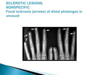
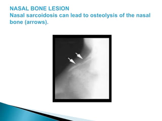
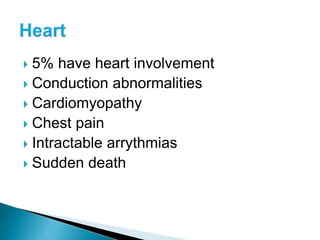
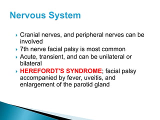
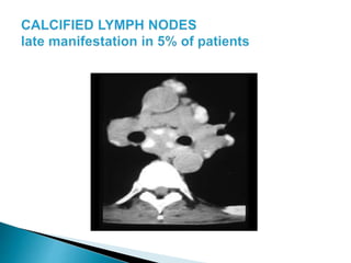
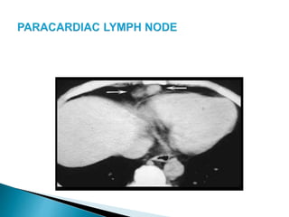
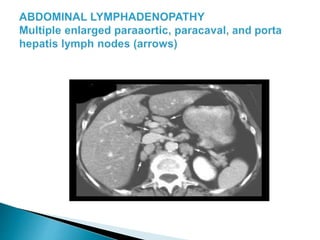
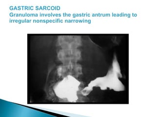
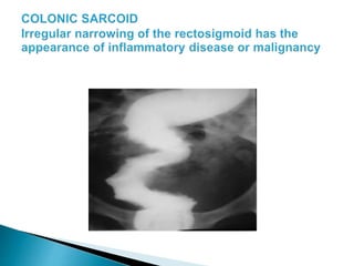
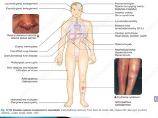
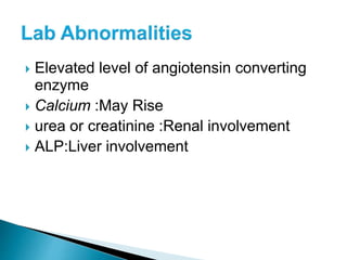
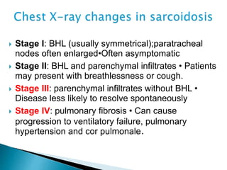
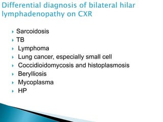
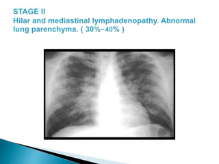
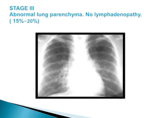
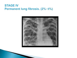
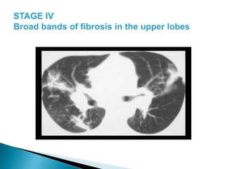
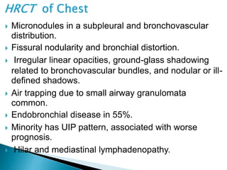
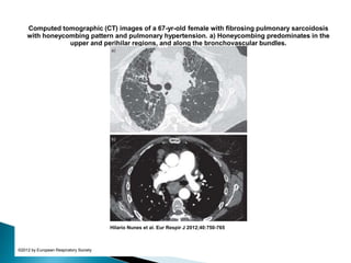
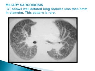
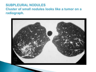
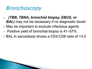
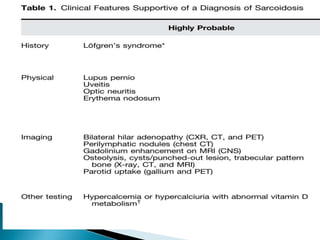
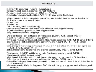
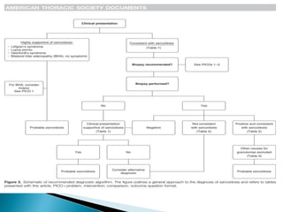
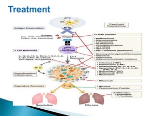
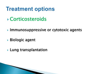
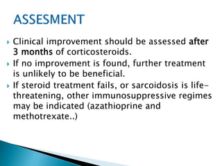
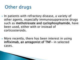
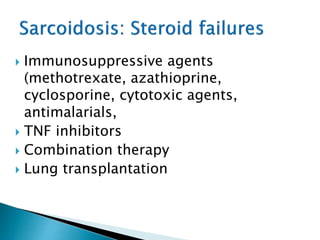
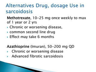
PRESENTED BY
DR.MD.SHARIF AHSAN
MD RESIDENT(PULMONOLOGY) PHASE-B
 Chronic multisystem disorder
 Unknown cause
 Most affected organ is the lung
 Skin, eyes and lymph nodes are
frequently involved
 Acute or sub acute and self limiting
 Waxing and waning over years
 A defect in the immune system
 An unidentified toxic substance
 An unknown environmental cause
 An inherited or genetic cause
 A viral or bacterial infection
 In all races and both sexes
 Risk greatest in a young black woman(20-40 years)
 The condition is more frequently described in
colder parts of northern Europe(Scandinavian,
German, Irish, or Puerto Rican origin)
 It also appears to be more common and more
severe in those from a West Indian or Asian
background
 Eskimos, Arabs and Chinese are rarely affected
 5/100,000 whites in the US have
sarcoidosis
 40/100,000 blacks
 Black women gets sarcoidosis twice than
black men
 White women equal white men
 Accumulation of mononuclear inflammatory
cells and T helper lymphocytes
 Formation of granulomas, aggregates of
macrophages, epithelioid cells and
multinucleated giant cells
 Giant cells in the central part of the granuloma
 The central epithelioid and giant cells are
surrounded by a rim of lymphocytes, mostly T-
helper cells
 T-cell lymphocytes are increased in areas of
active granulomas
 T-helper cells to T-suppressor cells ratio is
increased
 Exaggerated T-cell activity indicates an
altered immune response
 Hyper globulinemia
 Mass affect of granulomas damages the
tissues
 Asymptomatic: abnormal routine chest X-ray
(~30%) or abnormal liver function tests
 Respiratory and constitutional symptoms (20–
30%)
 Erythema nodosum and arthralgia (20–30%)
 Ocular symptoms (5–10%)
 Skin sarcoid (including lupus pernio) (5%)
 Superficial lymphadenopathy (5%)
 Other (1%), e.g. hypercalcaemia, diabetes
insipidus, cranial nerve palsies, cardiac
arrhythmias, nephrocalcinosis
 Symptoms reflect the specific organs involved
by the granulomas
 First site involved
 Begins with alveolitis involving small bronchi
and small blood vessels
 Alveolitis either clears up spontaneously or
leads to granuloma
 Fibrosis
 25% have eye lesions
 Blurred vision, pain, photophobia and dry eyes
 Chronic uveitis leads to glaucoma, cataracts and
blindness
 Keratoconjunctivitis sicca
 Papilledema
 33% have skin lesions
 Cutaneous anergy is common.
 LOFGREN'S SYNDROME; acute triad
of erythema nodosum, joint pains, and
bilateral hilar adenopathy
 Lupus pernio- indurated blue purple swollen
shiny lesions on nose, cheeks, lips, ears and
fingers.
 Papules, nodules, and plaques
 Psoriatic like lesions
 Lesions in scars and tattoos
 33% have hepatomegaly or biochemical
evidence of disease
 Symptoms usually absent
 Cholestasis, fibrosis, cirrhosis, portal
hypertension, and the Budd-Chiari syndrome
have been seen
 Acute polyarthritis with fever is common
 Arthritis is self limited
 Chronic destructive bone disease with deformity
is rare
 Polymyositis and chronic myopathy
 Muscle disease is rare
 5% have heart involvement
 Conduction abnormalities
 Cardiomyopathy
 Chest pain
 Intractable arrythmias
 Sudden death
 Cranial nerves, and peripheral nerves can be
involved
 7th nerve facial palsy is most common
 Acute, transient, and can be unilateral or
bilateral
 HEREFORDT'S SYNDROME; facial palsy
accompanied by fever, uveitis, and
enlargement of the parotid gland
 Optic nerve dysfunction
 Papilledema
 Palate dysfunction
 Hearing abnormalities
 Paresthesias
 Meningeal granulomas
 Encephalopathy
 Granulomatous interstitial nephritis produces
renal failure
 Develops over a period of weeks to months
 Rapid response to steroid therapy
 Kidney stones (nephrolithiasis) and
nephrocalcinosis are very unusual secondary
to hypercalcemia and hypercalciuria
 Increased calcium absorption in the gut
 Related to high levels of circulating 1,25-
dihydroxy vitamin D produced by mononuclear
phagocytes in granulomas
 Lymphadenopathy
 Intrathoracic nodes enlarged in 75-90% patients
including hilar nodes and paratracheal nodes.
 Peripheral lymphadenopathy
Differential diagnosis of granuloma on
lung biopsy
• Fungal infections:
• Cryptococcosis
• Aspergillosis
• Coccidioidomycosis
• Blastomycosis
• Aspiration of foreign
material
• Primary biliary cirrhosis
• Sarcoid-like reaction to
malignancy.
• Sarcoidosis
• TB
• HP
• Wegener’s
granulomatosis
• Drug reactions
• NTM
 Lymphocytopenia
 Mild eosinphilia
 Increased E.S.R
 Hyperglobulenemia
 Elevated level of angiotensin converting
enzyme
 Calcium :May Rise
 urea or creatinine :Renal involvement
 ALP:Liver involvement
 Stage I: BHL (usually symmetrical);paratracheal
nodes often enlarged•Often asymptomatic
 Stage II: BHL and parenchymal infiltrates • Patients
may present with breathlessness or cough.
 Stage III: parenchymal infiltrates without BHL •
Disease less likely to resolve spontaneously
 Stage IV: pulmonary fibrosis • Can cause
progression to ventilatory failure, pulmonary
hypertension and cor pulmonale.
 Sarcoidosis
 TB
 Lymphoma
 Lung cancer, especially small cell
 Coccidioidomycosis and histoplasmosis
 Berylliosis
 Mycoplasma
 HP
 Micronodules in a subpleural and bronchovascular
distribution.
 Fissural nodularity and bronchial distortion.
 Irregular linear opacities, ground-glass shadowing
related to bronchovascular bundles, and nodular or ill-
defined shadows.
 Air trapping due to small airway granulomata
common.
 Endobronchial disease in 55%.
 Minority has UIP pattern, associated with worse
prognosis.
 Hilar and mediastinal lymphadenopathy.
Computed tomographic image of pulmonary sarcoidosis with bilateral hilar lymphadenopathy
and micronodules with a perilymphatic distribution including spreading along the fissures.
Hilario Nunes et al. Eur Respir J 2012;40:750-765
©2012 by European Respiratory Society
a) Chest radiograph and b) computed tomographic image of “alveolar” sarcoidosis associated
with profuse micronodular involvement and cavitary lesions on the left.
Hilario Nunes et al. Eur Respir J 2012;40:750-765
©2012 by European Respiratory Society
Computed tomographic images of a 35-yr-old female with pulmonary sarcoidosis and extensive
ground-glass opacification.
Hilario Nunes et al. Eur Respir J 2012;40:750-765
©2012 by European Respiratory Society
Computed tomographic images of a) the “sarcoid galaxy” sign (irregular nodule resulting of the
confluence of numerous micronodules), b) the “sarcoid cluster” sign (clusters of micronodules
without confluence) and c) the “reversed halo” sign (ring of microno...
Hilario Nunes et al. Eur Respir J 2012;40:750-765
©2012 by European Respiratory Society
Computed tomographic (CT) images of a 67-yr-old female with fibrosing pulmonary sarcoidosis
with honeycombing pattern and pulmonary hypertension. a) Honeycombing predominates in the
upper and perihilar regions, and along the bronchovascular bundles.
Hilario Nunes et al. Eur Respir J 2012;40:750-765
©2012 by European Respiratory Society
 (TBB, TBNA, bronchial biopsy, EBUS, or
BAL) may not be necessary if no diagnostic doubt
 May be important to exclude infectious agents
 Positive yield of bronchial biopsy is 41–57%.
 BAL in sarcoidosis shows a CD4:CD8 ratio of >3.5
 Central or paratracheal nodes or open lung biopsy:
90% positive yield
 May be necessary to exclude lymphoma
 Surgical biopsy is not usually necessary, but, if
other procedures have not yielded a definitive
diagnosis, it may be required
 Lymph node ± lung (usually via VATS) can be
biopsied
skin, liver, etc., if indicated, as these may be easier to
biopsy in order to make a diagnosis
 ECG
 TTE
 MRI of HEART
• May be positive in areas of disease activity.
• Not reliable for studying brain or heart.
 Rarely used now, as non-specific and
expensive. Areas of active inflammation are
positive, with a classic ‘panda pattern
 Typically grade 0 in sarcoidosis.
 Positive Mantoux or Heaf test make sarcoidosis a
less likely diagnosis although does not necessarily
make TB more likely.
 No longer performed clinically, due to risks of
transmissible diseases.
 It involved injecting homogenized splenic tissue from
a patient with sarcoidosis to see if a granulomatous
reaction occurred
 PFT :Pulmonary sarcoidosis gives a restrictive
defect with decreased TLC and VC. TLCO
provides the most sensitive measurement of
change, although many use a properly
performed VC as an alternative. Likely to
improve with steroids. Airflow obstruction may
also occur
 CXR may improve with time or treatment
 HRCT can help with determining burden of
active disease
 Difficult to differentiate from chronic
infections, fungal diseases, T.B. and
lymphoma.
 Based on combined clinical, radiologic and
histologic findings.
 Exclusion of other defferentials
 Laboratory tests seldom important
 Good
 50% have some permanent organ dysfunction
 In 15-20% remains active or recurs intermittently.
 The overall mortality is low (1–5%) and usually
reflects cardiac involvement or pulmonary
fibrosis
 Age over 40
 Afro-Caribbean ethnicity
 Persistent symptoms for more than 6 months
 The involvement of more than three organs
 Lupus pernio
 A stage III/IV chest X-ray
 In patients with severe disease
 No known cure
 Corticosteroids, primary treatment
for inflammation and granuloma
formation.
 Corticosteroids
 Immunosuppressive or cytotoxic agents
 Biologic agent
 Lung transplantation
Does Sarcoidosis mandate
treatment?
 The initial treatment decision confronting the
clinician is whether or not to institute therapy for
the patient with sarcoidosis.
 Many patients do not require treatment,
especially when the disease is causing neither
significant symptoms nor significant functional
organ involvement..
 Because the disease may undergo
spontaneous remission and there are
significant potential toxicities from therapy.
 For patients with asymptomatic disease and
stage I disease there is no evidence to
support the need for corticosteroids.
 Many of these patients resolve spontaneously
and therefore can be monitored closely for
signs of progression
 which resolves spontaneously without
treatment. However, a minority of patients with
chronic sarcoidosis develop progressive
fibrosis.
 Systemic corticosteroids have been the
mainstay of therapy for decades, and is still
widely used today
 Symptomatic or progressive stages II and III
disease ( Increasing symptoms, deteriorating
PFTs, and worsening CXR infiltrates)
 Cardiac sarcoidosis
 Neurosarcoidosis
 Sight-threatening ocular sarcoidosis
 Hypercalcaemia
 Lupus pernio
 Splenic, hepatic, or renal sarcoidosis
 The drug of choice
 The initial dose is usually 20 to 40 mg per
day, although the dose may be higher in
cases of ocular or neurological involvement.
 The patient should be monitored closely for
8 to 12 weeks after the initiation of steroids.
 If there are signs of clinical improvement, the
steroids can be tapered over the next 6 to 12
months , as shorter courses of therapy are
associated with relapse
 in Sarcoidosis Individualize prednisolone
dose: Initial: 40 mg/day
 For CNS or Cardiac, 1 mg/kg/d and taper to
40 mg q.o.d < 3 months
 In steroid responders we decrease gradually
to 5 -10 mg/d or alternatively
 Relapses often occur when treatment is stopped
and may require the reintroduction of steroids,
or the increase of steroid dose.
 Duration and dose of steroids is dictated by site
and response to treatment
 Prescribe gastric and bone protection with
steroids when necessary
 Avoid futile steroid treatment for end-
stage disease, such as honeycomb lungs
 No effect on established fibrosis or
destroyed lung architecture
 Uveitis may be treated by topical
steroids, and
 skin manifestations may be amenable to
steroid creams or steroid injections.
 Inhaled steroids have been tried for
pulmonary disease
 Clinical improvement should be assessed after
3 months of corticosteroids.
 If no improvement is found, further treatment
is unlikely to be beneficial.
 If steroid treatment fails, or sarcoidosis is life-
threatening, other immunosuppressive regimes
may be indicated (azathioprine and
methotrexate..)
 In patients with refractory disease, a variety of
other agents, especially immunosuppressive drugs
such as methotrexate and cyclophosphamide, have
been used, either with or instead of
corticosteroids.
 More recently, there has been interest in using
infliximab, an antagonist of TNF- in selected
cases.
 Immunosuppressive agents
(methotrexate, azathioprine,
cyclosporine, cytotoxic agents,
antimalarials,
 TNF inhibitors
 Combination therapy
 Lung transplantation
Methotrexate, 10-25 mg once weekly to max
of 1 year or 2 yrs
 Chronic or worsening disease,
 common second line drug
 Effect may take 6 months
Azathioprine (Imuran), 50-200 mg QD
 Chronic or worsening disease
 Advanced fibrotic sarcoidosis
 Cyclophosphamide (Cytoxan) 50-150 mg QD or
500-2,000 mg q 2wk IV Refractory cases only
 Hydroxychloroquine (Plaquenil), 200-400 mg QD
Cutaneous manifestations, hypercalcemia, chronic
pulmonary fibrotic disease Corneal deposits,
retinopathy. Ophtho exam prior to treatment and
q 6 month
 Biologic agents that has changed the
landscape of treatment of refractory disease
 Infliximab is the biologic agent with the most
comprehensive data
 More agents, such as canakinumab,
roflumilast, and the nicotine patch, are
currently being investigated by ongoing
clinical trials.
 Rare indication for lung transplant
 Consider if patient has end-stage lung
disease, rapidly progressive disease despite
treatment, or if they are oxygen dependent
 Should be considered when all alternative
treatments for sarcoidosis have been
exhausted
 This should include, at a minimum, a
reasonable trial of corticosteroids and at least
one alternative agent
 In the cases of pulmonary sarcoidosis,
transplantation should be considered if the
disease is not only severe but also
progressive
 Sarcoidosis is a systemic disease that can affect
any organ, with the lungs and intrathoracic
lymph nodes being the most frequently
affected sites.
 The diagnosis of sarcoidosis relies on the
presence of noncaseating granuloma on
histopathologic examination, compatible
clinical presentation, and exclusion of other
causes of granulomatous inflammation.
 Treatment is generally reserved for patients
with disabling symptoms or with progressive
organ damage/dysfunction because
spontaneous remission is frequent.
 Glucocorticoids are the first-line therapy,
whereas glucocorticoid-sparing agents and
biologic agents are used in
refractory/recurrent cases.
Sarcoidosis
Sarcoidosis

Sarcoidosis

  • 2.
    PRESENTED BY DR.MD.SHARIF AHSAN MDRESIDENT(PULMONOLOGY) PHASE-B
  • 3.
     Chronic multisystemdisorder  Unknown cause  Most affected organ is the lung  Skin, eyes and lymph nodes are frequently involved  Acute or sub acute and self limiting  Waxing and waning over years
  • 4.
     A defectin the immune system  An unidentified toxic substance  An unknown environmental cause  An inherited or genetic cause  A viral or bacterial infection
  • 5.
     In allraces and both sexes  Risk greatest in a young black woman(20-40 years)  The condition is more frequently described in colder parts of northern Europe(Scandinavian, German, Irish, or Puerto Rican origin)  It also appears to be more common and more severe in those from a West Indian or Asian background  Eskimos, Arabs and Chinese are rarely affected
  • 6.
     5/100,000 whitesin the US have sarcoidosis  40/100,000 blacks  Black women gets sarcoidosis twice than black men  White women equal white men
  • 7.
     Accumulation ofmononuclear inflammatory cells and T helper lymphocytes  Formation of granulomas, aggregates of macrophages, epithelioid cells and multinucleated giant cells
  • 11.
     Giant cellsin the central part of the granuloma  The central epithelioid and giant cells are surrounded by a rim of lymphocytes, mostly T- helper cells  T-cell lymphocytes are increased in areas of active granulomas
  • 13.
     T-helper cellsto T-suppressor cells ratio is increased  Exaggerated T-cell activity indicates an altered immune response  Hyper globulinemia  Mass affect of granulomas damages the tissues
  • 14.
     Asymptomatic: abnormalroutine chest X-ray (~30%) or abnormal liver function tests  Respiratory and constitutional symptoms (20– 30%)  Erythema nodosum and arthralgia (20–30%)  Ocular symptoms (5–10%)
  • 15.
     Skin sarcoid(including lupus pernio) (5%)  Superficial lymphadenopathy (5%)  Other (1%), e.g. hypercalcaemia, diabetes insipidus, cranial nerve palsies, cardiac arrhythmias, nephrocalcinosis  Symptoms reflect the specific organs involved by the granulomas
  • 16.
     First siteinvolved  Begins with alveolitis involving small bronchi and small blood vessels  Alveolitis either clears up spontaneously or leads to granuloma  Fibrosis
  • 21.
     25% haveeye lesions  Blurred vision, pain, photophobia and dry eyes  Chronic uveitis leads to glaucoma, cataracts and blindness  Keratoconjunctivitis sicca  Papilledema
  • 25.
     33% haveskin lesions  Cutaneous anergy is common.  LOFGREN'S SYNDROME; acute triad of erythema nodosum, joint pains, and bilateral hilar adenopathy
  • 27.
     Lupus pernio-indurated blue purple swollen shiny lesions on nose, cheeks, lips, ears and fingers.  Papules, nodules, and plaques  Psoriatic like lesions  Lesions in scars and tattoos
  • 33.
     33% havehepatomegaly or biochemical evidence of disease  Symptoms usually absent  Cholestasis, fibrosis, cirrhosis, portal hypertension, and the Budd-Chiari syndrome have been seen
  • 35.
     Acute polyarthritiswith fever is common  Arthritis is self limited  Chronic destructive bone disease with deformity is rare  Polymyositis and chronic myopathy  Muscle disease is rare
  • 41.
     5% haveheart involvement  Conduction abnormalities  Cardiomyopathy  Chest pain  Intractable arrythmias  Sudden death
  • 42.
     Cranial nerves,and peripheral nerves can be involved  7th nerve facial palsy is most common  Acute, transient, and can be unilateral or bilateral  HEREFORDT'S SYNDROME; facial palsy accompanied by fever, uveitis, and enlargement of the parotid gland
  • 45.
     Optic nervedysfunction  Papilledema  Palate dysfunction  Hearing abnormalities  Paresthesias  Meningeal granulomas  Encephalopathy
  • 46.
     Granulomatous interstitialnephritis produces renal failure  Develops over a period of weeks to months  Rapid response to steroid therapy  Kidney stones (nephrolithiasis) and nephrocalcinosis are very unusual secondary to hypercalcemia and hypercalciuria
  • 48.
     Increased calciumabsorption in the gut  Related to high levels of circulating 1,25- dihydroxy vitamin D produced by mononuclear phagocytes in granulomas
  • 49.
     Lymphadenopathy  Intrathoracicnodes enlarged in 75-90% patients including hilar nodes and paratracheal nodes.  Peripheral lymphadenopathy
  • 57.
    Differential diagnosis ofgranuloma on lung biopsy • Fungal infections: • Cryptococcosis • Aspergillosis • Coccidioidomycosis • Blastomycosis • Aspiration of foreign material • Primary biliary cirrhosis • Sarcoid-like reaction to malignancy. • Sarcoidosis • TB • HP • Wegener’s granulomatosis • Drug reactions • NTM
  • 58.
     Lymphocytopenia  Mildeosinphilia  Increased E.S.R  Hyperglobulenemia
  • 59.
     Elevated levelof angiotensin converting enzyme  Calcium :May Rise  urea or creatinine :Renal involvement  ALP:Liver involvement
  • 60.
     Stage I:BHL (usually symmetrical);paratracheal nodes often enlarged•Often asymptomatic  Stage II: BHL and parenchymal infiltrates • Patients may present with breathlessness or cough.  Stage III: parenchymal infiltrates without BHL • Disease less likely to resolve spontaneously  Stage IV: pulmonary fibrosis • Can cause progression to ventilatory failure, pulmonary hypertension and cor pulmonale.
  • 62.
     Sarcoidosis  TB Lymphoma  Lung cancer, especially small cell  Coccidioidomycosis and histoplasmosis  Berylliosis  Mycoplasma  HP
  • 67.
     Micronodules ina subpleural and bronchovascular distribution.  Fissural nodularity and bronchial distortion.  Irregular linear opacities, ground-glass shadowing related to bronchovascular bundles, and nodular or ill- defined shadows.  Air trapping due to small airway granulomata common.  Endobronchial disease in 55%.  Minority has UIP pattern, associated with worse prognosis.  Hilar and mediastinal lymphadenopathy.
  • 68.
    Computed tomographic imageof pulmonary sarcoidosis with bilateral hilar lymphadenopathy and micronodules with a perilymphatic distribution including spreading along the fissures. Hilario Nunes et al. Eur Respir J 2012;40:750-765 ©2012 by European Respiratory Society
  • 69.
    a) Chest radiographand b) computed tomographic image of “alveolar” sarcoidosis associated with profuse micronodular involvement and cavitary lesions on the left. Hilario Nunes et al. Eur Respir J 2012;40:750-765 ©2012 by European Respiratory Society
  • 70.
    Computed tomographic imagesof a 35-yr-old female with pulmonary sarcoidosis and extensive ground-glass opacification. Hilario Nunes et al. Eur Respir J 2012;40:750-765 ©2012 by European Respiratory Society
  • 71.
    Computed tomographic imagesof a) the “sarcoid galaxy” sign (irregular nodule resulting of the confluence of numerous micronodules), b) the “sarcoid cluster” sign (clusters of micronodules without confluence) and c) the “reversed halo” sign (ring of microno... Hilario Nunes et al. Eur Respir J 2012;40:750-765 ©2012 by European Respiratory Society
  • 72.
    Computed tomographic (CT)images of a 67-yr-old female with fibrosing pulmonary sarcoidosis with honeycombing pattern and pulmonary hypertension. a) Honeycombing predominates in the upper and perihilar regions, and along the bronchovascular bundles. Hilario Nunes et al. Eur Respir J 2012;40:750-765 ©2012 by European Respiratory Society
  • 75.
     (TBB, TBNA,bronchial biopsy, EBUS, or BAL) may not be necessary if no diagnostic doubt  May be important to exclude infectious agents  Positive yield of bronchial biopsy is 41–57%.  BAL in sarcoidosis shows a CD4:CD8 ratio of >3.5
  • 76.
     Central orparatracheal nodes or open lung biopsy: 90% positive yield  May be necessary to exclude lymphoma  Surgical biopsy is not usually necessary, but, if other procedures have not yielded a definitive diagnosis, it may be required  Lymph node ± lung (usually via VATS) can be biopsied
  • 77.
    skin, liver, etc.,if indicated, as these may be easier to biopsy in order to make a diagnosis
  • 78.
     ECG  TTE MRI of HEART
  • 79.
    • May bepositive in areas of disease activity. • Not reliable for studying brain or heart.
  • 80.
     Rarely usednow, as non-specific and expensive. Areas of active inflammation are positive, with a classic ‘panda pattern
  • 81.
     Typically grade0 in sarcoidosis.  Positive Mantoux or Heaf test make sarcoidosis a less likely diagnosis although does not necessarily make TB more likely.
  • 82.
     No longerperformed clinically, due to risks of transmissible diseases.  It involved injecting homogenized splenic tissue from a patient with sarcoidosis to see if a granulomatous reaction occurred
  • 83.
     PFT :Pulmonarysarcoidosis gives a restrictive defect with decreased TLC and VC. TLCO provides the most sensitive measurement of change, although many use a properly performed VC as an alternative. Likely to improve with steroids. Airflow obstruction may also occur  CXR may improve with time or treatment  HRCT can help with determining burden of active disease
  • 84.
     Difficult todifferentiate from chronic infections, fungal diseases, T.B. and lymphoma.  Based on combined clinical, radiologic and histologic findings.  Exclusion of other defferentials  Laboratory tests seldom important
  • 88.
     Good  50%have some permanent organ dysfunction  In 15-20% remains active or recurs intermittently.  The overall mortality is low (1–5%) and usually reflects cardiac involvement or pulmonary fibrosis
  • 89.
     Age over40  Afro-Caribbean ethnicity  Persistent symptoms for more than 6 months  The involvement of more than three organs  Lupus pernio  A stage III/IV chest X-ray  In patients with severe disease
  • 90.
     No knowncure  Corticosteroids, primary treatment for inflammation and granuloma formation.
  • 92.
     Corticosteroids  Immunosuppressiveor cytotoxic agents  Biologic agent  Lung transplantation
  • 93.
  • 94.
     The initialtreatment decision confronting the clinician is whether or not to institute therapy for the patient with sarcoidosis.  Many patients do not require treatment, especially when the disease is causing neither significant symptoms nor significant functional organ involvement..
  • 95.
     Because thedisease may undergo spontaneous remission and there are significant potential toxicities from therapy.  For patients with asymptomatic disease and stage I disease there is no evidence to support the need for corticosteroids.  Many of these patients resolve spontaneously and therefore can be monitored closely for signs of progression
  • 96.
     which resolvesspontaneously without treatment. However, a minority of patients with chronic sarcoidosis develop progressive fibrosis.  Systemic corticosteroids have been the mainstay of therapy for decades, and is still widely used today
  • 97.
     Symptomatic orprogressive stages II and III disease ( Increasing symptoms, deteriorating PFTs, and worsening CXR infiltrates)  Cardiac sarcoidosis  Neurosarcoidosis  Sight-threatening ocular sarcoidosis  Hypercalcaemia  Lupus pernio  Splenic, hepatic, or renal sarcoidosis
  • 99.
     The drugof choice  The initial dose is usually 20 to 40 mg per day, although the dose may be higher in cases of ocular or neurological involvement.  The patient should be monitored closely for 8 to 12 weeks after the initiation of steroids.  If there are signs of clinical improvement, the steroids can be tapered over the next 6 to 12 months , as shorter courses of therapy are associated with relapse
  • 100.
     in SarcoidosisIndividualize prednisolone dose: Initial: 40 mg/day  For CNS or Cardiac, 1 mg/kg/d and taper to 40 mg q.o.d < 3 months  In steroid responders we decrease gradually to 5 -10 mg/d or alternatively
  • 101.
     Relapses oftenoccur when treatment is stopped and may require the reintroduction of steroids, or the increase of steroid dose.  Duration and dose of steroids is dictated by site and response to treatment  Prescribe gastric and bone protection with steroids when necessary
  • 102.
     Avoid futilesteroid treatment for end- stage disease, such as honeycomb lungs  No effect on established fibrosis or destroyed lung architecture
  • 103.
     Uveitis maybe treated by topical steroids, and  skin manifestations may be amenable to steroid creams or steroid injections.  Inhaled steroids have been tried for pulmonary disease
  • 104.
     Clinical improvementshould be assessed after 3 months of corticosteroids.  If no improvement is found, further treatment is unlikely to be beneficial.  If steroid treatment fails, or sarcoidosis is life- threatening, other immunosuppressive regimes may be indicated (azathioprine and methotrexate..)
  • 105.
     In patientswith refractory disease, a variety of other agents, especially immunosuppressive drugs such as methotrexate and cyclophosphamide, have been used, either with or instead of corticosteroids.  More recently, there has been interest in using infliximab, an antagonist of TNF- in selected cases.
  • 106.
     Immunosuppressive agents (methotrexate,azathioprine, cyclosporine, cytotoxic agents, antimalarials,  TNF inhibitors  Combination therapy  Lung transplantation
  • 107.
    Methotrexate, 10-25 mgonce weekly to max of 1 year or 2 yrs  Chronic or worsening disease,  common second line drug  Effect may take 6 months Azathioprine (Imuran), 50-200 mg QD  Chronic or worsening disease  Advanced fibrotic sarcoidosis
  • 108.
     Cyclophosphamide (Cytoxan)50-150 mg QD or 500-2,000 mg q 2wk IV Refractory cases only  Hydroxychloroquine (Plaquenil), 200-400 mg QD Cutaneous manifestations, hypercalcemia, chronic pulmonary fibrotic disease Corneal deposits, retinopathy. Ophtho exam prior to treatment and q 6 month
  • 109.
     Biologic agentsthat has changed the landscape of treatment of refractory disease  Infliximab is the biologic agent with the most comprehensive data  More agents, such as canakinumab, roflumilast, and the nicotine patch, are currently being investigated by ongoing clinical trials.
  • 110.
     Rare indicationfor lung transplant  Consider if patient has end-stage lung disease, rapidly progressive disease despite treatment, or if they are oxygen dependent  Should be considered when all alternative treatments for sarcoidosis have been exhausted
  • 111.
     This shouldinclude, at a minimum, a reasonable trial of corticosteroids and at least one alternative agent  In the cases of pulmonary sarcoidosis, transplantation should be considered if the disease is not only severe but also progressive
  • 112.
     Sarcoidosis isa systemic disease that can affect any organ, with the lungs and intrathoracic lymph nodes being the most frequently affected sites.  The diagnosis of sarcoidosis relies on the presence of noncaseating granuloma on histopathologic examination, compatible clinical presentation, and exclusion of other causes of granulomatous inflammation.
  • 113.
     Treatment isgenerally reserved for patients with disabling symptoms or with progressive organ damage/dysfunction because spontaneous remission is frequent.  Glucocorticoids are the first-line therapy, whereas glucocorticoid-sparing agents and biologic agents are used in refractory/recurrent cases.