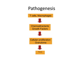
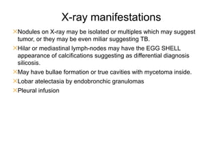
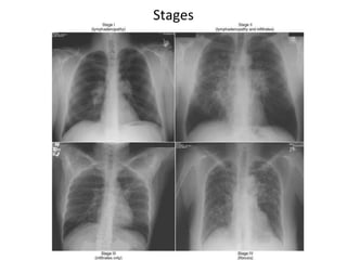
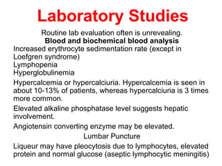
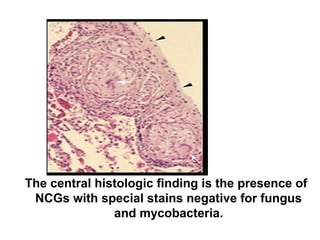
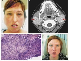
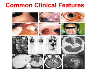
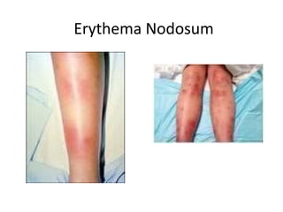
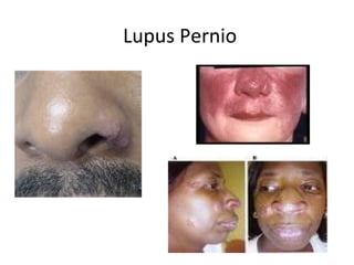
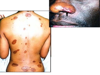
SlideShare a Scribd company logo
Kursk State Medical University
Department of Internal Disease N 1
Sarcoidosis
Student: Gustavo Duarte Viana
Group: 17
Course: 6th
year 2nd
semester
Sarcoidosis (or Bersier Boeck-
Schaumann disease)
• Chronic multisystemic inflammatory disorder of
unknown etiology that lead to non ceseous
granuloma formation, it most commonly affects
the lungs, but can also affect other organs mainly
the eyes, skin and bones.
• The name sarcoidosis is derived from a
misunderstanding, because doctor in the ninth
century thought that it was a benign form of
extreme aggressive neoplasm.
History of sarcoidosis
• In 1899, the pioneering Norwegian
dermatologist Caesar Boeck describe skin
nodules characterized by compact,
sharply defined foci of "epithelioid cells
with large pale nuclei and also a few giant
cells .
• Thinking this resembled sarcoma, he
called the condition "multiple benign
sarcoid of the skin.
Epidemiology
It is a worldwide disease, but there are some
prevalence in Scandinavian countries. But it may
affect all ethnic groups.
•It has a biphasic peak: Mostly in the ages around 20-
40, but it also appears around the 60.
•M-F ratio 2:1.
•Affects siblings of first- or second- degree relatives in
15% of patients with sarcoidosis.
•It is known that US black people have more severe
cases of pulmonary and skin sarcoidosis, but Japonese
have more cases of eyes sarcoidosis.
Etiology and Pathogenesis
• Cause is unknown, although both genetic and environmental
factors suspected.
• Theory that disease develops in genetically predetermined hosts
who are exposed to certain environmental agents that trigger an
exaggerated inflammatory immune response leading to
granuloma formation, specially because the organs most affects
(lungs, skin and eyes) are in direct exposure to the triggers.
• The trigger may be some insecticides, fungi, firefighting chemical,
wood-burning stoves, tree pollen, heavy metals, some dusts or
even bacteria such as Propionibacterium acnes (its DNA was
detected by PCR in the granulomas).
• Dust, miners or civil workers are more affected, there was a
break out of sarcoidosis in the workers helping in the 11-09-2001
• Hallmark is non caseaous granulomasn formation, composed
of a compact core of macrophages and around it a layer
oflymphocytes T CD+4 and lesser quantity lymphocytes B,
sometimes the macrophages can even fuse together forming
a multinucleated giant cell. There are a surrounding group of
macrophages forming the epithelioid histocytes.
• It is made in the porpose to surround and protect the
organism of a still unknown trigger.
• If the inflammatory process is persistent, it realease
chemiotaxis to fibroblasts which circumscribe the granuloma
and lead to formation of fibrotic tissue (scar) of granuloma
and surrounding tissue. It may lead to abnormalities in
normal architecture of the organ and further dysfunction.
T cells, Macrophages
Chemoattractants
Growth Factors
Cellular proliferation
Granuloma
Fibrosis
Pathogenesis
Curiosities
 In severe cases of AIDS, there is a very low account of T lymphocytes T CD4+, It lead to sarcoidosis
“spontaneous remission”, but when patient starts properly the antiretroviral therapy, sarcoidosis return
as the level of CD4+ raise up to normal level, it is one of the phenomenon of the IMMUNE
RECONSTITUTION SYNDROME.
 Kveim Siltzbach test: if positive, it is considered of diagnostic value, for rarely done due to difficulties in
the method and standardization of the method itself. It consists of injection a known homogenized
sarcoidosis tissue to the subcutaneous tissue of the patient, if after 4-6 weeks appear a papule, it is
considered positive. Mechanism is due to the Kveim Siltzbach reaction. It differs from a hypersensitivity
reaction because it only occurs after 4-6 weeks.
 It is possible to find markers of other disease, which my difficult at first the diagnosis, markers such as
Rheumatoid factor, antinuclear antibodies, anti T lymphocyte antibodies.
 As long as the disease persists untreated, more likely is to develop secondary organ disfunction.
Clinical Presentation
• One third have non-specific symptoms of fever, fatigue, weight
loss and malaise.
• It may be very difficult to diagnose sarcoidosis due to lack of
specific symptoms, in average it takes 1 year to diagnosis if
disease affects mostly the lungs and 6 months if affects mostly
the skin.
Forms: asymptomatic
Acute
Chronic
 Asymptomatic: 30-50% of patients and are diagnosed on routine CXR, usually with the classical SYMMETRICAL
BILATERAL HILAR ADENOPATHY with or without infiltrate.
 Acute: with evolution within few weeks with generalized symptoms as fever, severe asthenia and anorexia and
symptoms of pulmonary affection as dyspnea, cough and chest discomfort.
there are two acute syndromes distinguished separately in acute sarcoidosis
Loefgren syndrome, it is mostly frequent in Scandinavian women, from inland and Puerto Rico.
1- Uveitis
2- Erythema Nodosum
3- X-ray finding of symmetrical bilateral hilar adenopathy or parabrachial.
4- acute peripheral arthritis or poliarthralgia.
Heerfordt Walderstrom syndrome or AKA eveoparotid fever
1- Fever
2- parotid enlargement
3- anterior uveitis
4- facial paralysis.
 Chronic: insidious form lasting for months, strongly linked with respiratory symptoms and in lesser stand with
generalized symptoms, it lead to permanent irreversible organ changes, in specially lungs.
Specific organ involvement
✕ Lungs: more than 90% of patients with sarcoidosis will evolve to abnormalities shown in the X-ray, in very early stages
where interstitial pneumonia is present before formation of the granulomas. The infiltrate is usually in the upper lobes (in
contrary to other non infectious pneumopathy).
One third of the patients shows restrictive pneumopathy in pulmonary function test and one third of the patients show
criteria of obstructive pneumopathy.
There is the classical symmetrical bilateral hilar adenopathy which is very suggestive, but not pathognomonic because it
may be also present in fungic infections and neoplasia like bronchogenic carcinoma.
The main symptoms are dyspnea, dry cough and retrosternal dysconfort, others symptoms are seldom seen as pleural
involvement, pleural infusion, hemoptesis.
✕ Upper air ways: 20% of patients has involvement of the nasal mucosa leading to nasal obstruction, but all naso-oral
mucosa may be involved leading to high dyspnea, wheezing and hoarseness (Laryngeal sarcoidosis).
✕ Lymph-nodes: intrathoracic are enlarged in more than 90% of the patients with the classic symmetrical bilateral hilar
adenopathy, other lymph- nodes may be also involved as cervical chain, axillary and inguinal and they are painless,
mobile and non ulcerative and usually not perceived by the patient.
✕ Skin: about one third of patients which
• Erythema nodosum: they are erythematous painful nodules in the anterior surface of the legs, they
represent a subcutaneous vasculitis.
• Maculopapulous eruptions: they are due to granulomas itself and may be up to 3cm with a plateau
surface and of wax appearance, they may be found all over the body.
• Dischromias: usually painless and of a violate tin.
• Subcutanous nodules: in trunk and extrimities
• Lupus pernio: it forms complex of shining cyanotic harden lessions around the nose, libs, checks and
ears, it may erode to bones and cartilages around.
• It may develop over surgical scars and tattoo.
✕ Eyes: 25% of patients
Anterior uveitis: photophobia, epiphora and blurred vision.
Sicca keroconjutivitis
Blindness
✕ Bone marrow: 40% of patients, but lead only to a mild lymphopenia, very rare hematological
alterations.
✕ Spleen: 10%, but rarely lead to hiper-splenism syndrome.
✕ Liver: 50% reveals granulomas during autopsy, but rarely lead to hepatogram abnormalities, usually
only alkaline phosphatase is increased.
✕ Pancreas and intestine are rare affects, only granulomas at autopsy.
✕ Kidney: rare formation of granuloma, but sarcoidosis indirectly may lead to renal morbidity due to
increasing of active D vitamin (Granuloma has 25-hydroxivitamin D), which leads to hyperkalemia and
therefor nephrocalcinosis and repeated nephrolithiasis. All patients with sarcoidosis must routinely
check urine level of calcium and in blood analysis.
✕ Central and peripheral nervous system or neurosarcoidosis: A) cranial nerve paralysis B)
migraines C) ataxia D) cognitive dysfunction E) Asthenia F) seizures G) mononeuropathy or even
polyneuropathies of peripheral nerves leading to sensorial loss or paresthesy H) Incidious lymphocytic
meningitis (sarcoidosis meningitis) I) Diabetes insipidus due to granulomatous lesion to hypothalamus.
✕ Endocrine glands: 10% involving the parotid gland (Heerfordt syndrome) and rarely suprarenal cortex
insufficiency.
✕ Heart: 25% reveal granulomas in cardiac tissue, but only 5% will have symptoms, except Japoneses
where one forth of them will have heart sarcoidosis affection.
it may lead to: cardiac blocks, arrhythmias, angina, aneurisms, pericardial effusion, chronic cardiac
insufficiency. Cor pumonale is rare, but has association to pulmonary fibrosis in advanced sarcoidosis.
✕ Bones: affects mostly the phalanges, metacarpus and metatarsus leading to deformities in fingers and
nails. May lead to osteoporosis and cist formation
✕ Joints: arthritis or polyarthritis of big joints as ankles, knees, wrists and elbow. Patients with gout may
have worsening of the clinical picture because the granuloma itself produces purines.
✕ Muscles: acute myositis and chronic myopathy.
✕ Breats
✕ Tests
✕ Ovaries
✕ Stomach
✕ Others
Common Clinical Features
Erythema Nodosum
Lupus Pernio
X-ray manifestations
✕Nodules on X-ray may be isolated or multiples which may suggest
tumor, or they may be even miliar suggesting TB.
✕Hilar or mediastinal lymph-nodes may have the EGG SHELL
appearance of calcifications suggesting as differential diagnosis
silicosis.
✕May have bullae formation or true cavities with mycetoma inside.
✕Lobar atelectasia by endobronchic granulomas
✕Pleural infusion
4 Stages of Pulmonary Sarcoidosis
I Bilateral hilar lymphadenopathy
and paratracheal adenopathy
55-90%
remission
II Mediastinal adenopathy with
pulmonary parenchymal
involvements
40-70%
III Pulmonary parenchymal without
adenopathy
10-20%
IV Pulmonary fibrosis with
honeycombing
0-5%
Stages
Laboratory Studies
Routine lab evaluation often is unrevealing.
Blood and biochemical blood analysis
Increased erythrocyte sedimentation rate (except in
Loefgren syndrome)
Lymphopenia
Hyperglobulinemia
Hypercalcemia or hypercalciuria. Hypercalcemia is seen in
about 10-13% of patients, whereas hypercalciuria is 3 times
more common.
Elevated alkaline phosphatase level suggests hepatic
involvement.
Angiotensin converting enzyme may be elevated.
Lumbar Puncture
Liqueur may have pleocytosis due to lymphocytes, elevated
protein and normal glucose (aseptic lymphocytic meningitis)
Biopsy specimen
• A biopsy specimen should be obtained from the
involved organ that is most easily accessed, such
as the skin, peripheral LN. (not in erythema
nodosum)
• Eyes, lacrimal glands and gums only if they are
affected.
• If diagnosis requires pulmonary tissue, trans-
bronchial biopsy by means of bronchoscopy has a
diagnostic yield of at least 85% when multiple lung
segments are sampled. It may also be obtained by
mediastinoscopy or thoracothomy.
The central histologic finding is the presence of
NCGs with special stains negative for fungus
and mycobacteria.
Diagnosis
Consists of 3 main points
•Clinical manifestations
•Chest X-ray showing typical picture
• biopsy showing non caseating granuloma
Differential diagnosis
•TB
•Fungal infections
•Lymphoma
•Epithelioid tumors of the breast
•Lung cancer
Treatment
60% has remission spontaneously
10-20% has remission by corticosteroids
The main goal is to prevent fibrosis (ocular and
pulmonary)
•Observation
•Initiating corticosteroid therapy when
appropriate
•Monitoring response to therapy
•Discontinuing corticosteroids when clinically or
physiologically indicated.
Treatment by X-stages
•Stage 1 – only observation, if worsening of clinical picture we should start
steroids.
•Stage 2 – Asymptomatic – observation for 6 months, if no improvement we should
treat.
Treatment only if symptomatic or Asymptomatic with elevated blood
ACE, alteration in relation CD4+CD8+ or enhanced absorption of gallium 67.
•Stage 3 – Treat
•Stage 4 – treat if there is still absorption of Gallium 67, if there is not absorption
that means that the fibrosis is already formed and disease is not active anymore,
so corticosteroids would be useless
If there is still absorption of Gallium 67, administer corticoids
Other organs sarcoidosis or extrapulmonary
We should always treat with corticoisteroid:
Vital organs as eyes, nervous system, heart, liver, kidney, spleen.
Affection of upper air ways, parotidis, thyroid, cutaneous, specially lupus pernio.
Severe forms of articular, bone or muscular sarcoidosis
Biochemical alterations such as hyperkalemia, elevated ACE.
Neurosarcoidosis: pulse metilprednisolone IV 500 mg every
second day or 1g every week for 4-6 weeks.
Ocular sarcoidosis: eyes drops for conjuctivitis or uveitis, but
if it is posterior uveitis systemic corticosteroids should be
administered.
Pulmonary sarcoidosis: inhaled
Upper air way sarcoidosis: inhaled
Cutaneous: with ointments, creams or local injections of
corticosteroids.
Treatment
• Topical therapy for cutaneous or ophthalmic
disease (eye drops).
• Systemic corticosteroids for patients with
unresponsive ophthalmic manifestations,
cardiac, neurologic and progressive pulmonary
involvement.
• Systemic therapy for patients with
hypercalcemia.
Treatment
• Prednisone, 20 to 60 mg/d in divided doses or
alternate-day dosing is used for organ
involvement that is not life threatening gradually
diminishing the dose until the matainence dose
of 5 – 10 mg daily or 10-20 alternate day.
• Higher dosage is used off-label for potentially life
threatening disease.
• High-dose inhaled corticosteroids may be useful
in patients with symptomatic pulmonary disease.
Treatment
• Clinical improvement should be assessed after 3
months of corticosteroids.
• If no improvement is found, further treatment is
unlikely to be beneficial.
• Long term adverse affects of therapy include
weight gain, mood swings, cataracts, GERD,
osteoporosis
Other drugs
they are not so useful as corticosteroids, but may help in association to it.
NSAIDS: if present erythema nodosum, polyarthritis or acute uveitis.
Hydroxichlorquine: chronic fibrotic lesions in lung or skin, 200mg in
alternate days for 9 months to avoid ocular toxicity and should be followed
by a ophthalmologist.
Metothrexate: Lupus pernio or chronic skin lesions, 10mg once per week
for 3 months.
Azathioprine (50mg 3x daily for 3 months), cyclophosphamide (50mg 1x
daily for 3 months) or chlorambucil (5mg 1x daily for 3 months) may be
also used to try to diminish corticosteroids intake.
Organ transplantation is a choice for whom have lost organs function
(pulmonary, liver or kidney), interesting fact is that after transplantation
there are formation of non caseating granuloma in the transplanted organ,
even in patients in immunosuppressive therapy.
Prognosis
Many patients do not require therapy, and their
conditions will spontaneously improve.
Markers for a poor prognosis include :
• Advanced CXR stage.
• Extrapulmonary disease (predominantly cardiac and
neurologic)
• Evidence of pulmonary hypertension.
• Multiple studies have demonstrated that the
most important marker for prognosis is the initial
CXR stage.
Remission
• 2/3 of patients with sarcoidosis generally have a
remission within a decade after diagnosis, with
few or no consequences; remission occurs for
more than half of patients within 3 years.
• Unfortunately, up to 1/3 of patients have
progressive disease, leading to clinically significant
organ impairment.
• A recurrence after 1 or more years of remission is
uncommon (affecting <5% of patients), but
recurrent disease may develop at any age and in
any organ.
Death
• Less than 5% of patients die from
sarcoidosis.
• death is usually the result of pulmonary
fibrosis with respiratory failure or of
cardiac or neurologic involvement.

More Related Content

What's hot

Systemic lupus erythematosus (SLE)
Systemic lupus erythematosus (SLE)Systemic lupus erythematosus (SLE)
Systemic lupus erythematosus (SLE)
Arwa H. Al-Onayzan
 
Rheumatic fever, RHD, and infective endocarditis
Rheumatic fever, RHD, and infective endocarditisRheumatic fever, RHD, and infective endocarditis
Rheumatic fever, RHD, and infective endocarditis
med zar
 
Sarcoidosis
SarcoidosisSarcoidosis
Sarcoidosis
Opeyemi Muyiwa
 
Scleroderma
SclerodermaScleroderma
Scleroderma
hodmedicine
 
Cardiac sarcoidosis
Cardiac sarcoidosisCardiac sarcoidosis
Cardiac sarcoidosis
Piyush Bhardwaj
 
6 cardiomyopathies
6 cardiomyopathies6 cardiomyopathies
6 cardiomyopathies
Engidaw Ambelu
 
Cardiac tumors
Cardiac tumorsCardiac tumors
Cardiac tumors
Malleswara rao Dangeti
 
Kearns–Sayre Syndrome: Symptoms, Causes, Diagnosis, and Treatment
Kearns–Sayre Syndrome: Symptoms, Causes, Diagnosis, and TreatmentKearns–Sayre Syndrome: Symptoms, Causes, Diagnosis, and Treatment
Kearns–Sayre Syndrome: Symptoms, Causes, Diagnosis, and Treatment
vipul srivastava
 
Pericarditis, Pericardial Effusion, & Cardiac Tamponade - BMH/Tele
Pericarditis, Pericardial Effusion, & Cardiac Tamponade - BMH/TelePericarditis, Pericardial Effusion, & Cardiac Tamponade - BMH/Tele
Pericarditis, Pericardial Effusion, & Cardiac Tamponade - BMH/TeleTeleClinEd
 
surgery.Congenital heart disease.(dr.aram)
surgery.Congenital heart disease.(dr.aram)surgery.Congenital heart disease.(dr.aram)
surgery.Congenital heart disease.(dr.aram)student
 
Marfan syndrome
Marfan syndromeMarfan syndrome
Marfan syndrome
Satyam Rajvanshi
 
Sarcoidosis
SarcoidosisSarcoidosis
Sarcoidosis
MilDoc
 
Dilated cardiomyopathy
Dilated cardiomyopathyDilated cardiomyopathy
Dilated cardiomyopathy
drucsamal
 
Sudden cardiac death
Sudden cardiac deathSudden cardiac death
Sudden cardiac death
Sunil Reddy D
 
Sarcoidosis
SarcoidosisSarcoidosis
Sarcoidosis
Thomas Kurian
 
Dermatomyositis
DermatomyositisDermatomyositis
Dermatomyositis
Omondi Larry
 
Cardiac Amyloidosis - Dr. Akif
Cardiac Amyloidosis - Dr. AkifCardiac Amyloidosis - Dr. Akif
Cardiac Amyloidosis - Dr. Akif
akifab93
 
Pericardial diseases
Pericardial  diseasesPericardial  diseases
Pericardial diseases
ikramdr01
 

What's hot (20)

Systemic lupus erythematosus (SLE)
Systemic lupus erythematosus (SLE)Systemic lupus erythematosus (SLE)
Systemic lupus erythematosus (SLE)
 
Rheumatic fever, RHD, and infective endocarditis
Rheumatic fever, RHD, and infective endocarditisRheumatic fever, RHD, and infective endocarditis
Rheumatic fever, RHD, and infective endocarditis
 
Sarcoidosis
SarcoidosisSarcoidosis
Sarcoidosis
 
Scleroderma
SclerodermaScleroderma
Scleroderma
 
Sarcoidosis
SarcoidosisSarcoidosis
Sarcoidosis
 
Cardiac sarcoidosis
Cardiac sarcoidosisCardiac sarcoidosis
Cardiac sarcoidosis
 
6 cardiomyopathies
6 cardiomyopathies6 cardiomyopathies
6 cardiomyopathies
 
Myocarditis
MyocarditisMyocarditis
Myocarditis
 
Cardiac tumors
Cardiac tumorsCardiac tumors
Cardiac tumors
 
Kearns–Sayre Syndrome: Symptoms, Causes, Diagnosis, and Treatment
Kearns–Sayre Syndrome: Symptoms, Causes, Diagnosis, and TreatmentKearns–Sayre Syndrome: Symptoms, Causes, Diagnosis, and Treatment
Kearns–Sayre Syndrome: Symptoms, Causes, Diagnosis, and Treatment
 
Pericarditis, Pericardial Effusion, & Cardiac Tamponade - BMH/Tele
Pericarditis, Pericardial Effusion, & Cardiac Tamponade - BMH/TelePericarditis, Pericardial Effusion, & Cardiac Tamponade - BMH/Tele
Pericarditis, Pericardial Effusion, & Cardiac Tamponade - BMH/Tele
 
surgery.Congenital heart disease.(dr.aram)
surgery.Congenital heart disease.(dr.aram)surgery.Congenital heart disease.(dr.aram)
surgery.Congenital heart disease.(dr.aram)
 
Marfan syndrome
Marfan syndromeMarfan syndrome
Marfan syndrome
 
Sarcoidosis
SarcoidosisSarcoidosis
Sarcoidosis
 
Dilated cardiomyopathy
Dilated cardiomyopathyDilated cardiomyopathy
Dilated cardiomyopathy
 
Sudden cardiac death
Sudden cardiac deathSudden cardiac death
Sudden cardiac death
 
Sarcoidosis
SarcoidosisSarcoidosis
Sarcoidosis
 
Dermatomyositis
DermatomyositisDermatomyositis
Dermatomyositis
 
Cardiac Amyloidosis - Dr. Akif
Cardiac Amyloidosis - Dr. AkifCardiac Amyloidosis - Dr. Akif
Cardiac Amyloidosis - Dr. Akif
 
Pericardial diseases
Pericardial  diseasesPericardial  diseases
Pericardial diseases
 

Viewers also liked

Granulomatous diseases in ENT
Granulomatous diseases in ENTGranulomatous diseases in ENT
Granulomatous diseases in ENT
sarita pandey
 
Lupus and the Lungs by Dr. Gilda Diaz-Fuentes
Lupus and the Lungs by Dr. Gilda Diaz-FuentesLupus and the Lungs by Dr. Gilda Diaz-Fuentes
Lupus and the Lungs by Dr. Gilda Diaz-FuentesLupusNY
 
Sarcoidosis
SarcoidosisSarcoidosis
Granulomatous conditions of larynx
Granulomatous conditions of larynxGranulomatous conditions of larynx
Granulomatous conditions of larynxVinay Bhat
 
Pulmonary sarcoidosis
Pulmonary sarcoidosisPulmonary sarcoidosis
Pulmonary sarcoidosis
airwave12
 
Approach to ct chest 578
Approach to ct chest  578Approach to ct chest  578
Approach to ct chest 578divitto1
 

Viewers also liked (9)

Granulomatous diseases in ENT
Granulomatous diseases in ENTGranulomatous diseases in ENT
Granulomatous diseases in ENT
 
Lupus and the Lungs by Dr. Gilda Diaz-Fuentes
Lupus and the Lungs by Dr. Gilda Diaz-FuentesLupus and the Lungs by Dr. Gilda Diaz-Fuentes
Lupus and the Lungs by Dr. Gilda Diaz-Fuentes
 
Sarcoidosis
SarcoidosisSarcoidosis
Sarcoidosis
 
Granulomatous conditions of larynx
Granulomatous conditions of larynxGranulomatous conditions of larynx
Granulomatous conditions of larynx
 
Pulmonary sarcoidosis
Pulmonary sarcoidosisPulmonary sarcoidosis
Pulmonary sarcoidosis
 
Neurosarcoidosis
NeurosarcoidosisNeurosarcoidosis
Neurosarcoidosis
 
Sarcoidosis
SarcoidosisSarcoidosis
Sarcoidosis
 
Chest xrays pneumonias
Chest xrays pneumoniasChest xrays pneumonias
Chest xrays pneumonias
 
Approach to ct chest 578
Approach to ct chest  578Approach to ct chest  578
Approach to ct chest 578
 

Similar to Sarcoidosis

Systemic Sclerosis 2017
Systemic Sclerosis 2017Systemic Sclerosis 2017
Systemic Sclerosis 2017
singlamanik
 
Papulosequamous disorder
Papulosequamous disorder Papulosequamous disorder
Papulosequamous disorder
ssuser9127b3
 
Sarcoidosis.ppt
Sarcoidosis.pptSarcoidosis.ppt
Sarcoidosis.ppt
ssuser227d6b
 
DIFFUSE LUNG DISEASES.pptx
DIFFUSE LUNG DISEASES.pptxDIFFUSE LUNG DISEASES.pptx
DIFFUSE LUNG DISEASES.pptx
farkhandashaheen4
 
Orbital Inflammation
Orbital InflammationOrbital Inflammation
Orbital Inflammation
DrArpita123
 
Sarcoidosis....pptxNNNNNNNNNNNNNNNNNNNNNNNNNNNNNNNNNNNNNNNNNN
Sarcoidosis....pptxNNNNNNNNNNNNNNNNNNNNNNNNNNNNNNNNNNNNNNNNNNSarcoidosis....pptxNNNNNNNNNNNNNNNNNNNNNNNNNNNNNNNNNNNNNNNNNN
Sarcoidosis....pptxNNNNNNNNNNNNNNNNNNNNNNNNNNNNNNNNNNNNNNNNNN
NiteshYadav723617
 
RHD and IE.pptx
RHD and IE.pptxRHD and IE.pptx
RHD and IE.pptx
HolaHumble
 
SARCOIDOSIS
SARCOIDOSISSARCOIDOSIS
uveitis Denis kamara.pptx
uveitis Denis kamara.pptxuveitis Denis kamara.pptx
uveitis Denis kamara.pptx
MURHEC
 
Sarcoidosis agreat mimic
Sarcoidosis agreat mimicSarcoidosis agreat mimic
Sarcoidosis agreat mimichythemhashim
 
Behçet Disease.pptx
Behçet Disease.pptxBehçet Disease.pptx
Behçet Disease.pptx
dr maria saeed
 
Neurocysticercosis
Neurocysticercosis Neurocysticercosis
Neurocysticercosis
Payoz Pandey
 
Systemic sclerosis-1.pptx
Systemic sclerosis-1.pptxSystemic sclerosis-1.pptx
Systemic sclerosis-1.pptx
AshikArefin1
 
Sarcoidosis
SarcoidosisSarcoidosis
Sarcoidosis
DR.SHARIF AHSAN
 
brucellosis.pptx
brucellosis.pptxbrucellosis.pptx
brucellosis.pptx
AhmadRbeeHefni
 
Neurocysticercosis
NeurocysticercosisNeurocysticercosis
Neurocysticercosis
PGIMER,DR.RML HOSPITAL
 
Superficial &Opportunistic infections.pptx
Superficial &Opportunistic infections.pptxSuperficial &Opportunistic infections.pptx
Superficial &Opportunistic infections.pptx
SOLOMONKIPSEREK
 
Cutaneous presentation of tumours
Cutaneous presentation of tumoursCutaneous presentation of tumours
Cutaneous presentation of tumoursmeducationdotnet
 
Sarcoidosis .pptx
Sarcoidosis .pptxSarcoidosis .pptx
Sarcoidosis .pptx
Ömer aslankan
 

Similar to Sarcoidosis (20)

Systemic Sclerosis 2017
Systemic Sclerosis 2017Systemic Sclerosis 2017
Systemic Sclerosis 2017
 
Papulosequamous disorder
Papulosequamous disorder Papulosequamous disorder
Papulosequamous disorder
 
Sarcoidosis.ppt
Sarcoidosis.pptSarcoidosis.ppt
Sarcoidosis.ppt
 
DIFFUSE LUNG DISEASES.pptx
DIFFUSE LUNG DISEASES.pptxDIFFUSE LUNG DISEASES.pptx
DIFFUSE LUNG DISEASES.pptx
 
Orbital Inflammation
Orbital InflammationOrbital Inflammation
Orbital Inflammation
 
Sarcoidosis....pptxNNNNNNNNNNNNNNNNNNNNNNNNNNNNNNNNNNNNNNNNNN
Sarcoidosis....pptxNNNNNNNNNNNNNNNNNNNNNNNNNNNNNNNNNNNNNNNNNNSarcoidosis....pptxNNNNNNNNNNNNNNNNNNNNNNNNNNNNNNNNNNNNNNNNNN
Sarcoidosis....pptxNNNNNNNNNNNNNNNNNNNNNNNNNNNNNNNNNNNNNNNNNN
 
RHD and IE.pptx
RHD and IE.pptxRHD and IE.pptx
RHD and IE.pptx
 
SARCOIDOSIS
SARCOIDOSISSARCOIDOSIS
SARCOIDOSIS
 
uveitis Denis kamara.pptx
uveitis Denis kamara.pptxuveitis Denis kamara.pptx
uveitis Denis kamara.pptx
 
Sarcoidosis agreat mimic
Sarcoidosis agreat mimicSarcoidosis agreat mimic
Sarcoidosis agreat mimic
 
Behçet Disease.pptx
Behçet Disease.pptxBehçet Disease.pptx
Behçet Disease.pptx
 
Neurocysticercosis
Neurocysticercosis Neurocysticercosis
Neurocysticercosis
 
Systemic sclerosis-1.pptx
Systemic sclerosis-1.pptxSystemic sclerosis-1.pptx
Systemic sclerosis-1.pptx
 
Sarcoidosis
SarcoidosisSarcoidosis
Sarcoidosis
 
brucellosis.pptx
brucellosis.pptxbrucellosis.pptx
brucellosis.pptx
 
Neurocysticercosis
NeurocysticercosisNeurocysticercosis
Neurocysticercosis
 
Child with lymphadenopathy
Child with lymphadenopathyChild with lymphadenopathy
Child with lymphadenopathy
 
Superficial &Opportunistic infections.pptx
Superficial &Opportunistic infections.pptxSuperficial &Opportunistic infections.pptx
Superficial &Opportunistic infections.pptx
 
Cutaneous presentation of tumours
Cutaneous presentation of tumoursCutaneous presentation of tumours
Cutaneous presentation of tumours
 
Sarcoidosis .pptx
Sarcoidosis .pptxSarcoidosis .pptx
Sarcoidosis .pptx
 

More from Gustavo Duarte Viana

Degenerative diseases in aging patients
Degenerative diseases in aging patientsDegenerative diseases in aging patients
Degenerative diseases in aging patientsGustavo Duarte Viana
 
Disseminated intravascular coagulation
Disseminated intravascular coagulationDisseminated intravascular coagulation
Disseminated intravascular coagulation
Gustavo Duarte Viana
 
Bystander effect
Bystander effectBystander effect
Bystander effect
Gustavo Duarte Viana
 
Inhibition of the central nervous system
Inhibition of the central nervous systemInhibition of the central nervous system
Inhibition of the central nervous system
Gustavo Duarte Viana
 
Details of the central nervous system
Details of the central nervous systemDetails of the central nervous system
Details of the central nervous system
Gustavo Duarte Viana
 

More from Gustavo Duarte Viana (8)

Panic disorder
Panic disorder Panic disorder
Panic disorder
 
Degenerative diseases in aging patients
Degenerative diseases in aging patientsDegenerative diseases in aging patients
Degenerative diseases in aging patients
 
Disseminated intravascular coagulation
Disseminated intravascular coagulationDisseminated intravascular coagulation
Disseminated intravascular coagulation
 
Bystander effect
Bystander effectBystander effect
Bystander effect
 
Regulation of blood circulation
Regulation of blood circulationRegulation of blood circulation
Regulation of blood circulation
 
Cerebrospinal fluid
Cerebrospinal fluidCerebrospinal fluid
Cerebrospinal fluid
 
Inhibition of the central nervous system
Inhibition of the central nervous systemInhibition of the central nervous system
Inhibition of the central nervous system
 
Details of the central nervous system
Details of the central nervous systemDetails of the central nervous system
Details of the central nervous system
 

Recently uploaded

Alcohol_Dr. Jeenal Mistry MD Pharmacology.pdf
Alcohol_Dr. Jeenal Mistry MD Pharmacology.pdfAlcohol_Dr. Jeenal Mistry MD Pharmacology.pdf
Alcohol_Dr. Jeenal Mistry MD Pharmacology.pdf
Dr Jeenal Mistry
 
Phone Us ❤85270-49040❤ #ℂall #gIRLS In Surat By Surat @ℂall @Girls Hotel With...
Phone Us ❤85270-49040❤ #ℂall #gIRLS In Surat By Surat @ℂall @Girls Hotel With...Phone Us ❤85270-49040❤ #ℂall #gIRLS In Surat By Surat @ℂall @Girls Hotel With...
Phone Us ❤85270-49040❤ #ℂall #gIRLS In Surat By Surat @ℂall @Girls Hotel With...
Savita Shen $i11
 
Hemodialysis: Chapter 3, Dialysis Water Unit - Dr.Gawad
Hemodialysis: Chapter 3, Dialysis Water Unit - Dr.GawadHemodialysis: Chapter 3, Dialysis Water Unit - Dr.Gawad
Hemodialysis: Chapter 3, Dialysis Water Unit - Dr.Gawad
NephroTube - Dr.Gawad
 
Lung Cancer: Artificial Intelligence, Synergetics, Complex System Analysis, S...
Lung Cancer: Artificial Intelligence, Synergetics, Complex System Analysis, S...Lung Cancer: Artificial Intelligence, Synergetics, Complex System Analysis, S...
Lung Cancer: Artificial Intelligence, Synergetics, Complex System Analysis, S...
Oleg Kshivets
 
BRACHYTHERAPY OVERVIEW AND APPLICATORS
BRACHYTHERAPY OVERVIEW  AND  APPLICATORSBRACHYTHERAPY OVERVIEW  AND  APPLICATORS
BRACHYTHERAPY OVERVIEW AND APPLICATORS
Krishan Murari
 
Tom Selleck Health: A Comprehensive Look at the Iconic Actor’s Wellness Journey
Tom Selleck Health: A Comprehensive Look at the Iconic Actor’s Wellness JourneyTom Selleck Health: A Comprehensive Look at the Iconic Actor’s Wellness Journey
Tom Selleck Health: A Comprehensive Look at the Iconic Actor’s Wellness Journey
greendigital
 
Antiulcer drugs Advance Pharmacology .pptx
Antiulcer drugs Advance Pharmacology .pptxAntiulcer drugs Advance Pharmacology .pptx
Antiulcer drugs Advance Pharmacology .pptx
Rohit chaurpagar
 
Cervical & Brachial Plexus By Dr. RIG.pptx
Cervical & Brachial Plexus By Dr. RIG.pptxCervical & Brachial Plexus By Dr. RIG.pptx
Cervical & Brachial Plexus By Dr. RIG.pptx
Dr. Rabia Inam Gandapore
 
Physiology of Chemical Sensation of smell.pdf
Physiology of Chemical Sensation of smell.pdfPhysiology of Chemical Sensation of smell.pdf
Physiology of Chemical Sensation of smell.pdf
MedicoseAcademics
 
ANATOMY AND PHYSIOLOGY OF URINARY SYSTEM.pptx
ANATOMY AND PHYSIOLOGY OF URINARY SYSTEM.pptxANATOMY AND PHYSIOLOGY OF URINARY SYSTEM.pptx
ANATOMY AND PHYSIOLOGY OF URINARY SYSTEM.pptx
Swetaba Besh
 
Report Back from SGO 2024: What’s the Latest in Cervical Cancer?
Report Back from SGO 2024: What’s the Latest in Cervical Cancer?Report Back from SGO 2024: What’s the Latest in Cervical Cancer?
Report Back from SGO 2024: What’s the Latest in Cervical Cancer?
bkling
 
Non-respiratory Functions of the Lungs.pdf
Non-respiratory Functions of the Lungs.pdfNon-respiratory Functions of the Lungs.pdf
Non-respiratory Functions of the Lungs.pdf
MedicoseAcademics
 
Surat @ℂall @Girls ꧁❤8527049040❤꧂@ℂall @Girls Service Vip Top Model Safe
Surat @ℂall @Girls ꧁❤8527049040❤꧂@ℂall @Girls Service Vip Top Model SafeSurat @ℂall @Girls ꧁❤8527049040❤꧂@ℂall @Girls Service Vip Top Model Safe
Surat @ℂall @Girls ꧁❤8527049040❤꧂@ℂall @Girls Service Vip Top Model Safe
Savita Shen $i11
 
ARTHROLOGY PPT NCISM SYLLABUS AYURVEDA STUDENTS
ARTHROLOGY PPT NCISM SYLLABUS AYURVEDA STUDENTSARTHROLOGY PPT NCISM SYLLABUS AYURVEDA STUDENTS
ARTHROLOGY PPT NCISM SYLLABUS AYURVEDA STUDENTS
Dr. Vinay Pareek
 
POST OPERATIVE OLIGURIA and its management
POST OPERATIVE OLIGURIA and its managementPOST OPERATIVE OLIGURIA and its management
POST OPERATIVE OLIGURIA and its management
touseefaziz1
 
Couples presenting to the infertility clinic- Do they really have infertility...
Couples presenting to the infertility clinic- Do they really have infertility...Couples presenting to the infertility clinic- Do they really have infertility...
Couples presenting to the infertility clinic- Do they really have infertility...
Sujoy Dasgupta
 
New Directions in Targeted Therapeutic Approaches for Older Adults With Mantl...
New Directions in Targeted Therapeutic Approaches for Older Adults With Mantl...New Directions in Targeted Therapeutic Approaches for Older Adults With Mantl...
New Directions in Targeted Therapeutic Approaches for Older Adults With Mantl...
i3 Health
 
Ophthalmology Clinical Tests for OSCE exam
Ophthalmology Clinical Tests for OSCE examOphthalmology Clinical Tests for OSCE exam
Ophthalmology Clinical Tests for OSCE exam
KafrELShiekh University
 
ARTIFICIAL INTELLIGENCE IN HEALTHCARE.pdf
ARTIFICIAL INTELLIGENCE IN  HEALTHCARE.pdfARTIFICIAL INTELLIGENCE IN  HEALTHCARE.pdf
ARTIFICIAL INTELLIGENCE IN HEALTHCARE.pdf
Anujkumaranit
 
Ozempic: Preoperative Management of Patients on GLP-1 Receptor Agonists
Ozempic: Preoperative Management of Patients on GLP-1 Receptor Agonists  Ozempic: Preoperative Management of Patients on GLP-1 Receptor Agonists
Ozempic: Preoperative Management of Patients on GLP-1 Receptor Agonists
Saeid Safari
 

Recently uploaded (20)

Alcohol_Dr. Jeenal Mistry MD Pharmacology.pdf
Alcohol_Dr. Jeenal Mistry MD Pharmacology.pdfAlcohol_Dr. Jeenal Mistry MD Pharmacology.pdf
Alcohol_Dr. Jeenal Mistry MD Pharmacology.pdf
 
Phone Us ❤85270-49040❤ #ℂall #gIRLS In Surat By Surat @ℂall @Girls Hotel With...
Phone Us ❤85270-49040❤ #ℂall #gIRLS In Surat By Surat @ℂall @Girls Hotel With...Phone Us ❤85270-49040❤ #ℂall #gIRLS In Surat By Surat @ℂall @Girls Hotel With...
Phone Us ❤85270-49040❤ #ℂall #gIRLS In Surat By Surat @ℂall @Girls Hotel With...
 
Hemodialysis: Chapter 3, Dialysis Water Unit - Dr.Gawad
Hemodialysis: Chapter 3, Dialysis Water Unit - Dr.GawadHemodialysis: Chapter 3, Dialysis Water Unit - Dr.Gawad
Hemodialysis: Chapter 3, Dialysis Water Unit - Dr.Gawad
 
Lung Cancer: Artificial Intelligence, Synergetics, Complex System Analysis, S...
Lung Cancer: Artificial Intelligence, Synergetics, Complex System Analysis, S...Lung Cancer: Artificial Intelligence, Synergetics, Complex System Analysis, S...
Lung Cancer: Artificial Intelligence, Synergetics, Complex System Analysis, S...
 
BRACHYTHERAPY OVERVIEW AND APPLICATORS
BRACHYTHERAPY OVERVIEW  AND  APPLICATORSBRACHYTHERAPY OVERVIEW  AND  APPLICATORS
BRACHYTHERAPY OVERVIEW AND APPLICATORS
 
Tom Selleck Health: A Comprehensive Look at the Iconic Actor’s Wellness Journey
Tom Selleck Health: A Comprehensive Look at the Iconic Actor’s Wellness JourneyTom Selleck Health: A Comprehensive Look at the Iconic Actor’s Wellness Journey
Tom Selleck Health: A Comprehensive Look at the Iconic Actor’s Wellness Journey
 
Antiulcer drugs Advance Pharmacology .pptx
Antiulcer drugs Advance Pharmacology .pptxAntiulcer drugs Advance Pharmacology .pptx
Antiulcer drugs Advance Pharmacology .pptx
 
Cervical & Brachial Plexus By Dr. RIG.pptx
Cervical & Brachial Plexus By Dr. RIG.pptxCervical & Brachial Plexus By Dr. RIG.pptx
Cervical & Brachial Plexus By Dr. RIG.pptx
 
Physiology of Chemical Sensation of smell.pdf
Physiology of Chemical Sensation of smell.pdfPhysiology of Chemical Sensation of smell.pdf
Physiology of Chemical Sensation of smell.pdf
 
ANATOMY AND PHYSIOLOGY OF URINARY SYSTEM.pptx
ANATOMY AND PHYSIOLOGY OF URINARY SYSTEM.pptxANATOMY AND PHYSIOLOGY OF URINARY SYSTEM.pptx
ANATOMY AND PHYSIOLOGY OF URINARY SYSTEM.pptx
 
Report Back from SGO 2024: What’s the Latest in Cervical Cancer?
Report Back from SGO 2024: What’s the Latest in Cervical Cancer?Report Back from SGO 2024: What’s the Latest in Cervical Cancer?
Report Back from SGO 2024: What’s the Latest in Cervical Cancer?
 
Non-respiratory Functions of the Lungs.pdf
Non-respiratory Functions of the Lungs.pdfNon-respiratory Functions of the Lungs.pdf
Non-respiratory Functions of the Lungs.pdf
 
Surat @ℂall @Girls ꧁❤8527049040❤꧂@ℂall @Girls Service Vip Top Model Safe
Surat @ℂall @Girls ꧁❤8527049040❤꧂@ℂall @Girls Service Vip Top Model SafeSurat @ℂall @Girls ꧁❤8527049040❤꧂@ℂall @Girls Service Vip Top Model Safe
Surat @ℂall @Girls ꧁❤8527049040❤꧂@ℂall @Girls Service Vip Top Model Safe
 
ARTHROLOGY PPT NCISM SYLLABUS AYURVEDA STUDENTS
ARTHROLOGY PPT NCISM SYLLABUS AYURVEDA STUDENTSARTHROLOGY PPT NCISM SYLLABUS AYURVEDA STUDENTS
ARTHROLOGY PPT NCISM SYLLABUS AYURVEDA STUDENTS
 
POST OPERATIVE OLIGURIA and its management
POST OPERATIVE OLIGURIA and its managementPOST OPERATIVE OLIGURIA and its management
POST OPERATIVE OLIGURIA and its management
 
Couples presenting to the infertility clinic- Do they really have infertility...
Couples presenting to the infertility clinic- Do they really have infertility...Couples presenting to the infertility clinic- Do they really have infertility...
Couples presenting to the infertility clinic- Do they really have infertility...
 
New Directions in Targeted Therapeutic Approaches for Older Adults With Mantl...
New Directions in Targeted Therapeutic Approaches for Older Adults With Mantl...New Directions in Targeted Therapeutic Approaches for Older Adults With Mantl...
New Directions in Targeted Therapeutic Approaches for Older Adults With Mantl...
 
Ophthalmology Clinical Tests for OSCE exam
Ophthalmology Clinical Tests for OSCE examOphthalmology Clinical Tests for OSCE exam
Ophthalmology Clinical Tests for OSCE exam
 
ARTIFICIAL INTELLIGENCE IN HEALTHCARE.pdf
ARTIFICIAL INTELLIGENCE IN  HEALTHCARE.pdfARTIFICIAL INTELLIGENCE IN  HEALTHCARE.pdf
ARTIFICIAL INTELLIGENCE IN HEALTHCARE.pdf
 
Ozempic: Preoperative Management of Patients on GLP-1 Receptor Agonists
Ozempic: Preoperative Management of Patients on GLP-1 Receptor Agonists  Ozempic: Preoperative Management of Patients on GLP-1 Receptor Agonists
Ozempic: Preoperative Management of Patients on GLP-1 Receptor Agonists
 

Sarcoidosis

  • 1. Kursk State Medical University Department of Internal Disease N 1 Sarcoidosis Student: Gustavo Duarte Viana Group: 17 Course: 6th year 2nd semester
  • 2. Sarcoidosis (or Bersier Boeck- Schaumann disease) • Chronic multisystemic inflammatory disorder of unknown etiology that lead to non ceseous granuloma formation, it most commonly affects the lungs, but can also affect other organs mainly the eyes, skin and bones. • The name sarcoidosis is derived from a misunderstanding, because doctor in the ninth century thought that it was a benign form of extreme aggressive neoplasm.
  • 3. History of sarcoidosis • In 1899, the pioneering Norwegian dermatologist Caesar Boeck describe skin nodules characterized by compact, sharply defined foci of "epithelioid cells with large pale nuclei and also a few giant cells . • Thinking this resembled sarcoma, he called the condition "multiple benign sarcoid of the skin.
  • 4. Epidemiology It is a worldwide disease, but there are some prevalence in Scandinavian countries. But it may affect all ethnic groups. •It has a biphasic peak: Mostly in the ages around 20- 40, but it also appears around the 60. •M-F ratio 2:1. •Affects siblings of first- or second- degree relatives in 15% of patients with sarcoidosis. •It is known that US black people have more severe cases of pulmonary and skin sarcoidosis, but Japonese have more cases of eyes sarcoidosis.
  • 5. Etiology and Pathogenesis • Cause is unknown, although both genetic and environmental factors suspected. • Theory that disease develops in genetically predetermined hosts who are exposed to certain environmental agents that trigger an exaggerated inflammatory immune response leading to granuloma formation, specially because the organs most affects (lungs, skin and eyes) are in direct exposure to the triggers. • The trigger may be some insecticides, fungi, firefighting chemical, wood-burning stoves, tree pollen, heavy metals, some dusts or even bacteria such as Propionibacterium acnes (its DNA was detected by PCR in the granulomas). • Dust, miners or civil workers are more affected, there was a break out of sarcoidosis in the workers helping in the 11-09-2001
  • 6. • Hallmark is non caseaous granulomasn formation, composed of a compact core of macrophages and around it a layer oflymphocytes T CD+4 and lesser quantity lymphocytes B, sometimes the macrophages can even fuse together forming a multinucleated giant cell. There are a surrounding group of macrophages forming the epithelioid histocytes. • It is made in the porpose to surround and protect the organism of a still unknown trigger. • If the inflammatory process is persistent, it realease chemiotaxis to fibroblasts which circumscribe the granuloma and lead to formation of fibrotic tissue (scar) of granuloma and surrounding tissue. It may lead to abnormalities in normal architecture of the organ and further dysfunction.
  • 7. T cells, Macrophages Chemoattractants Growth Factors Cellular proliferation Granuloma Fibrosis Pathogenesis
  • 8.
  • 9. Curiosities  In severe cases of AIDS, there is a very low account of T lymphocytes T CD4+, It lead to sarcoidosis “spontaneous remission”, but when patient starts properly the antiretroviral therapy, sarcoidosis return as the level of CD4+ raise up to normal level, it is one of the phenomenon of the IMMUNE RECONSTITUTION SYNDROME.  Kveim Siltzbach test: if positive, it is considered of diagnostic value, for rarely done due to difficulties in the method and standardization of the method itself. It consists of injection a known homogenized sarcoidosis tissue to the subcutaneous tissue of the patient, if after 4-6 weeks appear a papule, it is considered positive. Mechanism is due to the Kveim Siltzbach reaction. It differs from a hypersensitivity reaction because it only occurs after 4-6 weeks.  It is possible to find markers of other disease, which my difficult at first the diagnosis, markers such as Rheumatoid factor, antinuclear antibodies, anti T lymphocyte antibodies.  As long as the disease persists untreated, more likely is to develop secondary organ disfunction.
  • 10. Clinical Presentation • One third have non-specific symptoms of fever, fatigue, weight loss and malaise. • It may be very difficult to diagnose sarcoidosis due to lack of specific symptoms, in average it takes 1 year to diagnosis if disease affects mostly the lungs and 6 months if affects mostly the skin. Forms: asymptomatic Acute Chronic
  • 11.  Asymptomatic: 30-50% of patients and are diagnosed on routine CXR, usually with the classical SYMMETRICAL BILATERAL HILAR ADENOPATHY with or without infiltrate.  Acute: with evolution within few weeks with generalized symptoms as fever, severe asthenia and anorexia and symptoms of pulmonary affection as dyspnea, cough and chest discomfort. there are two acute syndromes distinguished separately in acute sarcoidosis Loefgren syndrome, it is mostly frequent in Scandinavian women, from inland and Puerto Rico. 1- Uveitis 2- Erythema Nodosum 3- X-ray finding of symmetrical bilateral hilar adenopathy or parabrachial. 4- acute peripheral arthritis or poliarthralgia. Heerfordt Walderstrom syndrome or AKA eveoparotid fever 1- Fever 2- parotid enlargement 3- anterior uveitis 4- facial paralysis.  Chronic: insidious form lasting for months, strongly linked with respiratory symptoms and in lesser stand with generalized symptoms, it lead to permanent irreversible organ changes, in specially lungs.
  • 12.
  • 13.
  • 14. Specific organ involvement ✕ Lungs: more than 90% of patients with sarcoidosis will evolve to abnormalities shown in the X-ray, in very early stages where interstitial pneumonia is present before formation of the granulomas. The infiltrate is usually in the upper lobes (in contrary to other non infectious pneumopathy). One third of the patients shows restrictive pneumopathy in pulmonary function test and one third of the patients show criteria of obstructive pneumopathy. There is the classical symmetrical bilateral hilar adenopathy which is very suggestive, but not pathognomonic because it may be also present in fungic infections and neoplasia like bronchogenic carcinoma. The main symptoms are dyspnea, dry cough and retrosternal dysconfort, others symptoms are seldom seen as pleural involvement, pleural infusion, hemoptesis. ✕ Upper air ways: 20% of patients has involvement of the nasal mucosa leading to nasal obstruction, but all naso-oral mucosa may be involved leading to high dyspnea, wheezing and hoarseness (Laryngeal sarcoidosis). ✕ Lymph-nodes: intrathoracic are enlarged in more than 90% of the patients with the classic symmetrical bilateral hilar adenopathy, other lymph- nodes may be also involved as cervical chain, axillary and inguinal and they are painless, mobile and non ulcerative and usually not perceived by the patient.
  • 15. ✕ Skin: about one third of patients which • Erythema nodosum: they are erythematous painful nodules in the anterior surface of the legs, they represent a subcutaneous vasculitis. • Maculopapulous eruptions: they are due to granulomas itself and may be up to 3cm with a plateau surface and of wax appearance, they may be found all over the body. • Dischromias: usually painless and of a violate tin. • Subcutanous nodules: in trunk and extrimities • Lupus pernio: it forms complex of shining cyanotic harden lessions around the nose, libs, checks and ears, it may erode to bones and cartilages around. • It may develop over surgical scars and tattoo.
  • 16.
  • 17.
  • 18. ✕ Eyes: 25% of patients Anterior uveitis: photophobia, epiphora and blurred vision. Sicca keroconjutivitis Blindness ✕ Bone marrow: 40% of patients, but lead only to a mild lymphopenia, very rare hematological alterations. ✕ Spleen: 10%, but rarely lead to hiper-splenism syndrome. ✕ Liver: 50% reveals granulomas during autopsy, but rarely lead to hepatogram abnormalities, usually only alkaline phosphatase is increased. ✕ Pancreas and intestine are rare affects, only granulomas at autopsy. ✕ Kidney: rare formation of granuloma, but sarcoidosis indirectly may lead to renal morbidity due to increasing of active D vitamin (Granuloma has 25-hydroxivitamin D), which leads to hyperkalemia and therefor nephrocalcinosis and repeated nephrolithiasis. All patients with sarcoidosis must routinely check urine level of calcium and in blood analysis. ✕ Central and peripheral nervous system or neurosarcoidosis: A) cranial nerve paralysis B) migraines C) ataxia D) cognitive dysfunction E) Asthenia F) seizures G) mononeuropathy or even polyneuropathies of peripheral nerves leading to sensorial loss or paresthesy H) Incidious lymphocytic meningitis (sarcoidosis meningitis) I) Diabetes insipidus due to granulomatous lesion to hypothalamus. ✕ Endocrine glands: 10% involving the parotid gland (Heerfordt syndrome) and rarely suprarenal cortex insufficiency.
  • 19.
  • 20.
  • 21.
  • 22. ✕ Heart: 25% reveal granulomas in cardiac tissue, but only 5% will have symptoms, except Japoneses where one forth of them will have heart sarcoidosis affection. it may lead to: cardiac blocks, arrhythmias, angina, aneurisms, pericardial effusion, chronic cardiac insufficiency. Cor pumonale is rare, but has association to pulmonary fibrosis in advanced sarcoidosis. ✕ Bones: affects mostly the phalanges, metacarpus and metatarsus leading to deformities in fingers and nails. May lead to osteoporosis and cist formation ✕ Joints: arthritis or polyarthritis of big joints as ankles, knees, wrists and elbow. Patients with gout may have worsening of the clinical picture because the granuloma itself produces purines. ✕ Muscles: acute myositis and chronic myopathy. ✕ Breats ✕ Tests ✕ Ovaries ✕ Stomach ✕ Others
  • 23.
  • 24.
  • 25.
  • 29.
  • 30. X-ray manifestations ✕Nodules on X-ray may be isolated or multiples which may suggest tumor, or they may be even miliar suggesting TB. ✕Hilar or mediastinal lymph-nodes may have the EGG SHELL appearance of calcifications suggesting as differential diagnosis silicosis. ✕May have bullae formation or true cavities with mycetoma inside. ✕Lobar atelectasia by endobronchic granulomas ✕Pleural infusion
  • 31. 4 Stages of Pulmonary Sarcoidosis I Bilateral hilar lymphadenopathy and paratracheal adenopathy 55-90% remission II Mediastinal adenopathy with pulmonary parenchymal involvements 40-70% III Pulmonary parenchymal without adenopathy 10-20% IV Pulmonary fibrosis with honeycombing 0-5%
  • 33. Laboratory Studies Routine lab evaluation often is unrevealing. Blood and biochemical blood analysis Increased erythrocyte sedimentation rate (except in Loefgren syndrome) Lymphopenia Hyperglobulinemia Hypercalcemia or hypercalciuria. Hypercalcemia is seen in about 10-13% of patients, whereas hypercalciuria is 3 times more common. Elevated alkaline phosphatase level suggests hepatic involvement. Angiotensin converting enzyme may be elevated. Lumbar Puncture Liqueur may have pleocytosis due to lymphocytes, elevated protein and normal glucose (aseptic lymphocytic meningitis)
  • 34. Biopsy specimen • A biopsy specimen should be obtained from the involved organ that is most easily accessed, such as the skin, peripheral LN. (not in erythema nodosum) • Eyes, lacrimal glands and gums only if they are affected. • If diagnosis requires pulmonary tissue, trans- bronchial biopsy by means of bronchoscopy has a diagnostic yield of at least 85% when multiple lung segments are sampled. It may also be obtained by mediastinoscopy or thoracothomy.
  • 35. The central histologic finding is the presence of NCGs with special stains negative for fungus and mycobacteria.
  • 36. Diagnosis Consists of 3 main points •Clinical manifestations •Chest X-ray showing typical picture • biopsy showing non caseating granuloma Differential diagnosis •TB •Fungal infections •Lymphoma •Epithelioid tumors of the breast •Lung cancer
  • 37. Treatment 60% has remission spontaneously 10-20% has remission by corticosteroids The main goal is to prevent fibrosis (ocular and pulmonary) •Observation •Initiating corticosteroid therapy when appropriate •Monitoring response to therapy •Discontinuing corticosteroids when clinically or physiologically indicated.
  • 38. Treatment by X-stages •Stage 1 – only observation, if worsening of clinical picture we should start steroids. •Stage 2 – Asymptomatic – observation for 6 months, if no improvement we should treat. Treatment only if symptomatic or Asymptomatic with elevated blood ACE, alteration in relation CD4+CD8+ or enhanced absorption of gallium 67. •Stage 3 – Treat •Stage 4 – treat if there is still absorption of Gallium 67, if there is not absorption that means that the fibrosis is already formed and disease is not active anymore, so corticosteroids would be useless If there is still absorption of Gallium 67, administer corticoids Other organs sarcoidosis or extrapulmonary We should always treat with corticoisteroid: Vital organs as eyes, nervous system, heart, liver, kidney, spleen. Affection of upper air ways, parotidis, thyroid, cutaneous, specially lupus pernio. Severe forms of articular, bone or muscular sarcoidosis Biochemical alterations such as hyperkalemia, elevated ACE.
  • 39. Neurosarcoidosis: pulse metilprednisolone IV 500 mg every second day or 1g every week for 4-6 weeks. Ocular sarcoidosis: eyes drops for conjuctivitis or uveitis, but if it is posterior uveitis systemic corticosteroids should be administered. Pulmonary sarcoidosis: inhaled Upper air way sarcoidosis: inhaled Cutaneous: with ointments, creams or local injections of corticosteroids.
  • 40. Treatment • Topical therapy for cutaneous or ophthalmic disease (eye drops). • Systemic corticosteroids for patients with unresponsive ophthalmic manifestations, cardiac, neurologic and progressive pulmonary involvement. • Systemic therapy for patients with hypercalcemia.
  • 41. Treatment • Prednisone, 20 to 60 mg/d in divided doses or alternate-day dosing is used for organ involvement that is not life threatening gradually diminishing the dose until the matainence dose of 5 – 10 mg daily or 10-20 alternate day. • Higher dosage is used off-label for potentially life threatening disease. • High-dose inhaled corticosteroids may be useful in patients with symptomatic pulmonary disease.
  • 42. Treatment • Clinical improvement should be assessed after 3 months of corticosteroids. • If no improvement is found, further treatment is unlikely to be beneficial. • Long term adverse affects of therapy include weight gain, mood swings, cataracts, GERD, osteoporosis
  • 43. Other drugs they are not so useful as corticosteroids, but may help in association to it. NSAIDS: if present erythema nodosum, polyarthritis or acute uveitis. Hydroxichlorquine: chronic fibrotic lesions in lung or skin, 200mg in alternate days for 9 months to avoid ocular toxicity and should be followed by a ophthalmologist. Metothrexate: Lupus pernio or chronic skin lesions, 10mg once per week for 3 months. Azathioprine (50mg 3x daily for 3 months), cyclophosphamide (50mg 1x daily for 3 months) or chlorambucil (5mg 1x daily for 3 months) may be also used to try to diminish corticosteroids intake. Organ transplantation is a choice for whom have lost organs function (pulmonary, liver or kidney), interesting fact is that after transplantation there are formation of non caseating granuloma in the transplanted organ, even in patients in immunosuppressive therapy.
  • 44. Prognosis Many patients do not require therapy, and their conditions will spontaneously improve. Markers for a poor prognosis include : • Advanced CXR stage. • Extrapulmonary disease (predominantly cardiac and neurologic) • Evidence of pulmonary hypertension. • Multiple studies have demonstrated that the most important marker for prognosis is the initial CXR stage.
  • 45. Remission • 2/3 of patients with sarcoidosis generally have a remission within a decade after diagnosis, with few or no consequences; remission occurs for more than half of patients within 3 years. • Unfortunately, up to 1/3 of patients have progressive disease, leading to clinically significant organ impairment. • A recurrence after 1 or more years of remission is uncommon (affecting <5% of patients), but recurrent disease may develop at any age and in any organ.
  • 46. Death • Less than 5% of patients die from sarcoidosis. • death is usually the result of pulmonary fibrosis with respiratory failure or of cardiac or neurologic involvement.