Sarcoidosis
Dr.Adil Al Sulami
KAUH
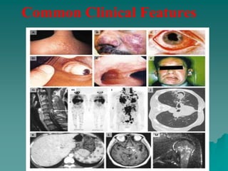
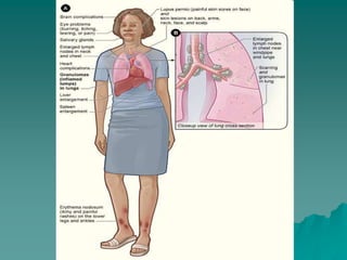
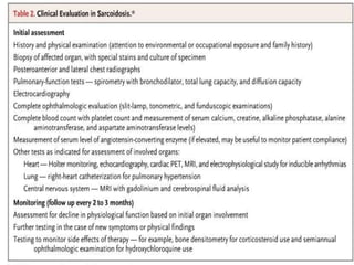
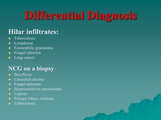
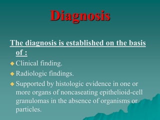
Sarcoidosis is a multisystem
inflammatory disease of unknown
etiology that predominantly affects
the lungs and intrathoracic lymph
nodes.
Sarcoidosis is manifested by
the presence of noncaseating
granulomas (NCGs) in
affected organ tissues.
The modern history of
sarcoidosis
 In 1899, the pioneering Norwegian
dermatologist Caesar Boeck describe skin
nodules characterized by compact, sharply
defined foci of "epithelioid cells with large pale
nuclei and also a few giant cells .
 Thinking this resembled sarcoma, he called the
condition "multiple benign sarcoid of the skin.
Epidemiology
 All racial .
 All ethnic groups.
 All ages (with the incidence peaking at 20 to 39 years).
 M-F ratio 2:1.
The incidence
 The highest annual incidence in northern
European countries 5 - 40 / 100,000.
 In Japan, the annual incidence 1 - 2 / 100,000.
 Among black Americans is roughly 3 times
that among white Americans (35.5 / 100,000,
as compared with 10.9 / 100,000.
Pathophysiology
T cells play a central role in the
development of sarcoidosis, as
they likely propagate an excessive
cellular immune reaction.
The cause of sarcoidosis is
unknown.
Efforts to identify a possible
infectious etiology have
been unsuccessful.
 Genetic and environmental factors seem to play
a role.
 As yet, no bacterial, fungal, or viral antigen has
been consistently isolated from the sarcoidosis
lesions.
 Sarcoidosis is neither a malignant nor an
autoimmune disease.
The following have been suggested as possible
candidates that might play a role in causing
sarcoidosis:
 Mycobacteria, such as Mycobacterium tuberculosis, and
atypical pathogens have been suggested.
 Fungi and viruses, particularly Mycoplasma, Chlamydia, and
Epstein-Barr virus, have been unconvincingly implicated.
Environmental Causes
 Some of the earliest studies of sarcoidosis reported
associations with exposures to irritants found in rural settings,
such as emissions from wood-burning stoves and tree pollen.
 More recently, associations with sarcoidosis and exposure to
inorganic particles ,insecticides ,and moldy environments
have been reported.
 Occupational studies have shown positive associations with
service in the U.S. Navy ,metalworking ,firefighting ,and the
handling of building supplies.
Genetic Features
 Familial sarcoidosis was first reported in 1923 in two
affected sisters .
 No formal twin study has been reported, but the
concordance appears to be higher in monozygotic
twins than in dizygotic twins .
 In A Case-Control Study, patients with sarcoidosis
stated 5 times as often as control subjects that they
had siblings or parents with sarcoidosis.
Common Clinical Features
 Presentation depends on the extent and severity of
the organ involved.
 Approximately 5% of cases are asymptomatic and
incidentally detected by CXR.
 Systemic symptoms occur in 45% of cases such as
:
 Fever.
 anorexia
 Fatigue.
 Night sweats .
 Weight loss .
 Pulmonary, dyspnea on exertion, cough, chest
pain, and hemoptysis (rare) occur in 50% of cases.
Löfgren's syndrome, an acute presentation
consisting of:
 Fever.
 Arthralgia.
 erythema nodosum.
 bilateral hilar adenopathy.
 occurs in 9 to 34% of patients .
Heerford's syndrome :
 Anterior Uveitis
 Fever
 Parotid enlargment
 Facial palsy
Physical finding
 Pulmonary findings.
 Dermatological manifestations.
 Ocular manifestations .
 Cardiac manifestations
 Neurologic manifestations (rare)
 Sarcoidal granulomas can involve any organ, but
in more than 90% of patients, clinical
sarcoidosis is manifested as intrathoracic LN
enlargement, pulmonary involvement, skin or
ocular signs and symptoms, or some
combination of these findings.
Organ Involvement
Pulmonary Involvement
 dyspnea, cough, vague chest discomfort, and wheezing.
 Chest radiographs in patients with sarcoidosis have been
classified into four stages:
– stage 1, bilateral hilar lymphadenopathy without infiltration.
– stage 2, bilateral hilar lymphadenopathy with infiltration.
– stage 3, infiltration alone.
– stage 4, fibrotic bands, bullae, hilar retraction, bronchiectasis,
and diaphragmatic tenting.
 These so-called stages represent radiographic patterns and do
not indicate disease chronicity or correlate with changes in
pulmonary function.
Stage 1
Stage II is BHL and infiltrates
Stage III is infiltrates alone
Cutaneous Involvement
Although not life-threatening, but can be
emotionally devastating.
 Erythema nodosum may occur.
 Lupus pernio is the most specific associated cutaneous lesion.
 Violaceous rash is often seen on the cheeks or nose.
 Osseous involvement may be present.
 Maculopapular plaques are possible.
 Lupus pernio is more common in women than in men and is associated
with chronic disease and extrapulmonary involvement.
 Erythema nodosum occurs in about 10% of patients with sarcoidosis and
usually lasts for about 3 weeks.
 Biopsy specimens of erythema nodosum lesions show nonspecific septal
panniculitis, which neither confirms nor negates the diagnosis of
sarcoidosis.
Liver and Spleen Involvement
 10% of all patients with sarcoidosis have elevated serum
aminotransferase and alkaline phosphatase levels.
 A cholestatic syndrome characterized by pruritus and jaundice,
hepatic failure, or portal hypertension can develop (liver
involvement is usually clinically silent).
 Detection of hepatic and splenic lesions on CT is described in
5% and 15% of patients.
 60% of patients with hepatic manifestations of sarcoidosis
have constitutional symptoms such as fever, night sweats,
anorexia, and weight loss.
 Portal hypertension with variceal bleeding, a hepatopulmonary
syndrome with refractory hypoxemia, and cirrhosis leading to
liver failure occur in only 1% of patients with sarcoidosis.
Neurologic Involvement
 CNS is involved in up to 25% of patients with sarcoidosis who undergo
autopsy, but only 10% of all patients with sarcoidosis present with
neurologic symptoms.
 The most common problems:
– cranial-nerve palsies.
– Headache.
– Ataxia.
– cognitive dysfunction.
– Weakness.
– seizures.
 CSF Analysis :
– nonspecific lymphocytic inflammation.
– measuring ACE levels .
– oligoclonal immunoglobulin bands in the CSF are elevated, making it difficult
to differentiate sarcoidosis from multiple sclerosis.
 Magnetic resonance imaging (MRI)
Ophthalmologic Complications
 The eye and adnexa are involved in 25 -80% of
patients with sarcoidosis,this necessitating routine
slit-lamp and funduscopic examination.
 Anterior or posterior granulomatous uveitis .
 Conjunctival lesions and scleral plaques may also be
noted.
 Ocular involvement may lead to blindness if
untreated.
 Anterior uveitis
(is the most common manifestation)
chronic anterior uveitis, with insidious symptoms
leading to glaucoma and vision loss, is more
common than acute anterior uveitis.
Cardiac manifestations
 Heart failure from cardiomyopathy rarely
occurs.
 Heart block and sudden death may occur.
 Approximately 25% of patients may have
NCGs at autopsy, but fewer than 5% have
clinical cardiac disease.
 Okada et al reported on cardiac infiltration
associated with a novel heterogenous mutation
(G481D in CARD15) in early-onset
sarcoidosis.
Differential Diagnosis
Hilar infiltrates:
 Tuberculosis.
 Lymphoma
 Eosinophilic granuloma
 Fungal infection
 Lung cancer
NCG on a biopsy :
 Berylliosis
 Catscratch disease
 Fungal infection
 Hypersensitivity pneumonitis
 Leprosy
 Primary biliary cirrhosis
 Tuberculosis.
Diagnosis
The diagnosis is established on the basis
of :
 Clinical finding.
 Radiologic findings.
 Supported by histologic evidence in one or
more organs of noncaseating epithelioid-cell
granulomas in the absence of organisms or
particles.
A diagnosis of sarcoidosis is reasonably certain
without biopsy in patients who present with
Löfgren's syndrome.
Laboratory Studies
 Routine lab evaluation often is unrevealing.
 Hypercalcemia or hypercalciuria may occur (NCGs
secrete 1,25 vitamin D).
 Hypercalcemia is seen in about 10-13% of patients,
whereas hypercalciuria is 3 times more common.
 An elevated alkaline phosphatase level suggests hepatic
involvement.
 Angiotensin converting enzyme (ACE) levels may be
elevated.
 NCGs secrete ACE, which may function as a cytokine.
 Serum ACE levels are elevated in 60% of patients at the
time of diagnosis.
 Levels may be increased in fluid from bronchoalveolar
lavage or in CSF.
 Sensitivity and specificity as a diagnostic test is limited (60
and 70%, respectively).
 There is no clear prognostic value.
 Serum ACE levels may decline in response to therapy.
 Decisions on treatment should not be based on the ACE
level alone.
Imaging Studies
 A chest radiograph is central to evaluation.
 Routine chest CT scan adds little.
 HRCT of the chest may be helpful.
Biopsy specimen
 A biopsy specimen should be obtained from
the involved organ that is most easily
accessed, such as the skin, peripheral LN,
lacrimal glands, or conjunctiva.
 If diagnosis requires pulmonary tissue,
transbronchial biopsy by means of
bronchoscopy has a diagnostic yield of at
least 85% when multiple lung segments are
sampled .
The central histologic finding is the presence of
NCGs with special stains negative for fungus and
mycobacteria.
 Sarcoidal granulomas have no unique histologic
features to differentiate them from other
granulomas.
 Special stains for acid-fast bacilli and fungi, as
well as cultures of such organisms, are essential.
 If the results of lung biopsy with bronchoscopy are
negative and other organs are not obviously
involved, biopsy of intrathoracic lymph nodes,
which are often enlarged in patients with
Treatment
Most patients (>75%) require only
symptomatic therapy NSAID.
 Approximately 10% of patients need
treatment for extrapulmonary disease.
15% of patients require treatment for
persistent pulmonary disease.
Corticosteroids are the mainstay
of therapy
 prednisone given daily and then tapered over a 6-month
course is adequate for pulmonary disease.
 Earlier recommendations suggested an initial dose of 1
mg/kg/d of prednisone; however, more recent expert
opinions endorse a lower dose (eg, 40 mg/d), which is
tapered to every other day long-term therapy over several
weeks.
 Most patients who require long-term steroids can be
treated using 10-15 mg of prednisone every other day.
 High-dose inhaled corticosteroids may be an option, but
conclusive data are lacking.
Data suggest that corticosteroid use may
be associated with increased relapse rates.
Occasionally, certain patients cannot
tolerate or do not respond to
corticosteroids.
Noncorticosteroid agents
Used more frequently.
Common indications :
 Steroid-resistant disease.
 Intolerable adverse effects.
 patient desire not to take corticosteroids.
 Methotrexate (MTX) has been a successful
alternative to prednisone and is a steroid-sparing
agent.
 Chloroquine and hydroxychloroquine are
antimalarial drugs with immunomodulating
properties, which have been used for cutaneous
lesions, hypercalcemia, neurological sarcoidosis, and
bone lesions.
 Chloroquine has also been shown to be efficacious
for the treatment and maintenance of chronic
 Cyclophosphamide has been rarely used with modest
success as a steroid-sparing treatment in patients with
refractory sarcoidosis.
 Azathioprine is another second-line therapy, which is best
used as a steroid-sparing agent rather than as a single-drug
treatment for sarcoidosis.
 Chlorambucil is an alkylating agent that may be beneficial
in patients with progressive disease unresponsive to
corticosteroids or when corticosteroids are contraindicated.
 Cyclosporine is a fungal cyclic polypeptide with
lymphocyte-suppressive properties that may be of limited
benefit in skin sarcoidosis or in progressive sarcoid resistant to
conventional therapy.
 Infliximab and thalidomide have been used for
refractory sarcoidosis, particularly for cutaneous
disease.
 Infliximab appears to be an effective treatment for
patients with systemic manifestations such as lupus
pernio, uveitis, hepatic sarcoidosis, and
neurosarcoidosis.
 Tetracyclines have shown promise for the
treatment of cutaneous sarcoidosis.
For pulmonary disease
 Asymptomatic PFT and/or CXR abnormalities are not an
indication for treatment.
 In patients with minimal symptoms, serial reevaluation is
prudent.
 Significant respiratory symptoms associated with PFT and
CXR abnormalities likely require therapy.
 For such patients, treatment is indicated if objective evidence
of recent deterioration in lung function exists.
 Corticosteroids can result in small improvements in the
functional vital capacity and in the radiographic appearance in
patients with more severe stage II and III disease.
One recent study demonstrated an approach
that may minimize the use of corticosteroids
without harming the patient.
This is accomplished by
 Withholding therapy unless the patient shows at least a 15%
decline in one spirometric measure associated with increasing
symptoms or,
 if asymptomatic, withholding therapy unless the patient shows
worsening PFTs and a change in CXR.
 For extrapulmonary sarcoidosis involving such
critical organs as the heart, liver, eyes, kidneys, or
central nervous system, corticosteroid therapy is
indicated.
 Topical corticosteroids are effective for ocular
disease.
 Inhaled corticosteroids are occasionally used, in
particular in patients with endobronchial disease.
NSAIDs are indicated for the treatment of
arthralgias and other rheumatic
complaints.
Patients with stage I sarcoidosis often
require only occasional treatment with
NSAIDs.
 Further Inpatient Care
– Monitor pulmonary function and CXR every 6-12 months.
– Assess for progression or resolution.
– Determine if previously uninvolved organs have become
affected.
 Further Outpatient Care
– Annual slit lamp eye examination and ECG are
recommended.
Follow-up
Many patients do not require therapy, and their
conditions will spontaneously improve.
Markers for a poor prognosis include :
 Advanced CXR stage.
 Extrapulmonary disease (predominantly cardiac and neurologic)
 Evidence of pulmonary hypertension.
 Multiple studies have demonstrated that the most
important marker for prognosis is the initial CXR
stage.
Prognosis
Remission
 2/3 of patients with sarcoidosis generally have a
remission within a decade after diagnosis, with few
or no consequences ;remission occurs for more than
half of patients within 3 years .
 Unfortunately, up to 1/3 of patients have progressive
disease, leading to clinically significant organ
impairment.
 A recurrence after 1 or more years of remission is
uncommon (affecting <5% of patients), but recurrent
disease may develop at any age and in any organ.
Death
 Less than 5% of patients die from
sarcoidosis.
 death is usually the result of pulmonary
fibrosis with respiratory failure or of
cardiac or neurologic involvement .
Sarcoidosis.ppt

Sarcoidosis.ppt

  • 1.
  • 2.
    Sarcoidosis is amultisystem inflammatory disease of unknown etiology that predominantly affects the lungs and intrathoracic lymph nodes.
  • 3.
    Sarcoidosis is manifestedby the presence of noncaseating granulomas (NCGs) in affected organ tissues.
  • 4.
    The modern historyof sarcoidosis  In 1899, the pioneering Norwegian dermatologist Caesar Boeck describe skin nodules characterized by compact, sharply defined foci of "epithelioid cells with large pale nuclei and also a few giant cells .  Thinking this resembled sarcoma, he called the condition "multiple benign sarcoid of the skin.
  • 5.
    Epidemiology  All racial.  All ethnic groups.  All ages (with the incidence peaking at 20 to 39 years).  M-F ratio 2:1.
  • 6.
    The incidence  Thehighest annual incidence in northern European countries 5 - 40 / 100,000.  In Japan, the annual incidence 1 - 2 / 100,000.  Among black Americans is roughly 3 times that among white Americans (35.5 / 100,000, as compared with 10.9 / 100,000.
  • 7.
    Pathophysiology T cells playa central role in the development of sarcoidosis, as they likely propagate an excessive cellular immune reaction.
  • 8.
    The cause ofsarcoidosis is unknown. Efforts to identify a possible infectious etiology have been unsuccessful.
  • 9.
     Genetic andenvironmental factors seem to play a role.  As yet, no bacterial, fungal, or viral antigen has been consistently isolated from the sarcoidosis lesions.  Sarcoidosis is neither a malignant nor an autoimmune disease.
  • 10.
    The following havebeen suggested as possible candidates that might play a role in causing sarcoidosis:  Mycobacteria, such as Mycobacterium tuberculosis, and atypical pathogens have been suggested.  Fungi and viruses, particularly Mycoplasma, Chlamydia, and Epstein-Barr virus, have been unconvincingly implicated.
  • 11.
    Environmental Causes  Someof the earliest studies of sarcoidosis reported associations with exposures to irritants found in rural settings, such as emissions from wood-burning stoves and tree pollen.  More recently, associations with sarcoidosis and exposure to inorganic particles ,insecticides ,and moldy environments have been reported.  Occupational studies have shown positive associations with service in the U.S. Navy ,metalworking ,firefighting ,and the handling of building supplies.
  • 12.
    Genetic Features  Familialsarcoidosis was first reported in 1923 in two affected sisters .  No formal twin study has been reported, but the concordance appears to be higher in monozygotic twins than in dizygotic twins .  In A Case-Control Study, patients with sarcoidosis stated 5 times as often as control subjects that they had siblings or parents with sarcoidosis.
  • 13.
  • 14.
     Presentation dependson the extent and severity of the organ involved.  Approximately 5% of cases are asymptomatic and incidentally detected by CXR.  Systemic symptoms occur in 45% of cases such as :  Fever.  anorexia  Fatigue.  Night sweats .  Weight loss .  Pulmonary, dyspnea on exertion, cough, chest pain, and hemoptysis (rare) occur in 50% of cases.
  • 15.
    Löfgren's syndrome, anacute presentation consisting of:  Fever.  Arthralgia.  erythema nodosum.  bilateral hilar adenopathy.  occurs in 9 to 34% of patients . Heerford's syndrome :  Anterior Uveitis  Fever  Parotid enlargment  Facial palsy
  • 16.
  • 17.
     Pulmonary findings. Dermatological manifestations.  Ocular manifestations .  Cardiac manifestations  Neurologic manifestations (rare)
  • 18.
     Sarcoidal granulomascan involve any organ, but in more than 90% of patients, clinical sarcoidosis is manifested as intrathoracic LN enlargement, pulmonary involvement, skin or ocular signs and symptoms, or some combination of these findings. Organ Involvement
  • 19.
    Pulmonary Involvement  dyspnea,cough, vague chest discomfort, and wheezing.  Chest radiographs in patients with sarcoidosis have been classified into four stages: – stage 1, bilateral hilar lymphadenopathy without infiltration. – stage 2, bilateral hilar lymphadenopathy with infiltration. – stage 3, infiltration alone. – stage 4, fibrotic bands, bullae, hilar retraction, bronchiectasis, and diaphragmatic tenting.  These so-called stages represent radiographic patterns and do not indicate disease chronicity or correlate with changes in pulmonary function.
  • 20.
  • 21.
    Stage II isBHL and infiltrates
  • 22.
    Stage III isinfiltrates alone
  • 23.
    Cutaneous Involvement Although notlife-threatening, but can be emotionally devastating.  Erythema nodosum may occur.  Lupus pernio is the most specific associated cutaneous lesion.  Violaceous rash is often seen on the cheeks or nose.  Osseous involvement may be present.  Maculopapular plaques are possible.  Lupus pernio is more common in women than in men and is associated with chronic disease and extrapulmonary involvement.  Erythema nodosum occurs in about 10% of patients with sarcoidosis and usually lasts for about 3 weeks.  Biopsy specimens of erythema nodosum lesions show nonspecific septal panniculitis, which neither confirms nor negates the diagnosis of sarcoidosis.
  • 26.
    Liver and SpleenInvolvement  10% of all patients with sarcoidosis have elevated serum aminotransferase and alkaline phosphatase levels.  A cholestatic syndrome characterized by pruritus and jaundice, hepatic failure, or portal hypertension can develop (liver involvement is usually clinically silent).  Detection of hepatic and splenic lesions on CT is described in 5% and 15% of patients.  60% of patients with hepatic manifestations of sarcoidosis have constitutional symptoms such as fever, night sweats, anorexia, and weight loss.  Portal hypertension with variceal bleeding, a hepatopulmonary syndrome with refractory hypoxemia, and cirrhosis leading to liver failure occur in only 1% of patients with sarcoidosis.
  • 27.
    Neurologic Involvement  CNSis involved in up to 25% of patients with sarcoidosis who undergo autopsy, but only 10% of all patients with sarcoidosis present with neurologic symptoms.  The most common problems: – cranial-nerve palsies. – Headache. – Ataxia. – cognitive dysfunction. – Weakness. – seizures.  CSF Analysis : – nonspecific lymphocytic inflammation. – measuring ACE levels . – oligoclonal immunoglobulin bands in the CSF are elevated, making it difficult to differentiate sarcoidosis from multiple sclerosis.  Magnetic resonance imaging (MRI)
  • 28.
    Ophthalmologic Complications  Theeye and adnexa are involved in 25 -80% of patients with sarcoidosis,this necessitating routine slit-lamp and funduscopic examination.  Anterior or posterior granulomatous uveitis .  Conjunctival lesions and scleral plaques may also be noted.  Ocular involvement may lead to blindness if untreated.  Anterior uveitis (is the most common manifestation) chronic anterior uveitis, with insidious symptoms leading to glaucoma and vision loss, is more common than acute anterior uveitis.
  • 29.
    Cardiac manifestations  Heartfailure from cardiomyopathy rarely occurs.  Heart block and sudden death may occur.  Approximately 25% of patients may have NCGs at autopsy, but fewer than 5% have clinical cardiac disease.  Okada et al reported on cardiac infiltration associated with a novel heterogenous mutation (G481D in CARD15) in early-onset sarcoidosis.
  • 32.
    Differential Diagnosis Hilar infiltrates: Tuberculosis.  Lymphoma  Eosinophilic granuloma  Fungal infection  Lung cancer NCG on a biopsy :  Berylliosis  Catscratch disease  Fungal infection  Hypersensitivity pneumonitis  Leprosy  Primary biliary cirrhosis  Tuberculosis.
  • 33.
    Diagnosis The diagnosis isestablished on the basis of :  Clinical finding.  Radiologic findings.  Supported by histologic evidence in one or more organs of noncaseating epithelioid-cell granulomas in the absence of organisms or particles.
  • 34.
    A diagnosis ofsarcoidosis is reasonably certain without biopsy in patients who present with Löfgren's syndrome.
  • 35.
    Laboratory Studies  Routinelab evaluation often is unrevealing.  Hypercalcemia or hypercalciuria may occur (NCGs secrete 1,25 vitamin D).  Hypercalcemia is seen in about 10-13% of patients, whereas hypercalciuria is 3 times more common.  An elevated alkaline phosphatase level suggests hepatic involvement.  Angiotensin converting enzyme (ACE) levels may be elevated.
  • 36.
     NCGs secreteACE, which may function as a cytokine.  Serum ACE levels are elevated in 60% of patients at the time of diagnosis.  Levels may be increased in fluid from bronchoalveolar lavage or in CSF.  Sensitivity and specificity as a diagnostic test is limited (60 and 70%, respectively).  There is no clear prognostic value.  Serum ACE levels may decline in response to therapy.  Decisions on treatment should not be based on the ACE level alone.
  • 37.
    Imaging Studies  Achest radiograph is central to evaluation.  Routine chest CT scan adds little.  HRCT of the chest may be helpful.
  • 38.
    Biopsy specimen  Abiopsy specimen should be obtained from the involved organ that is most easily accessed, such as the skin, peripheral LN, lacrimal glands, or conjunctiva.  If diagnosis requires pulmonary tissue, transbronchial biopsy by means of bronchoscopy has a diagnostic yield of at least 85% when multiple lung segments are sampled .
  • 39.
    The central histologicfinding is the presence of NCGs with special stains negative for fungus and mycobacteria.
  • 40.
     Sarcoidal granulomashave no unique histologic features to differentiate them from other granulomas.  Special stains for acid-fast bacilli and fungi, as well as cultures of such organisms, are essential.  If the results of lung biopsy with bronchoscopy are negative and other organs are not obviously involved, biopsy of intrathoracic lymph nodes, which are often enlarged in patients with
  • 41.
  • 43.
    Most patients (>75%)require only symptomatic therapy NSAID.  Approximately 10% of patients need treatment for extrapulmonary disease. 15% of patients require treatment for persistent pulmonary disease.
  • 44.
    Corticosteroids are themainstay of therapy  prednisone given daily and then tapered over a 6-month course is adequate for pulmonary disease.  Earlier recommendations suggested an initial dose of 1 mg/kg/d of prednisone; however, more recent expert opinions endorse a lower dose (eg, 40 mg/d), which is tapered to every other day long-term therapy over several weeks.  Most patients who require long-term steroids can be treated using 10-15 mg of prednisone every other day.  High-dose inhaled corticosteroids may be an option, but conclusive data are lacking.
  • 45.
    Data suggest thatcorticosteroid use may be associated with increased relapse rates. Occasionally, certain patients cannot tolerate or do not respond to corticosteroids.
  • 46.
    Noncorticosteroid agents Used morefrequently. Common indications :  Steroid-resistant disease.  Intolerable adverse effects.  patient desire not to take corticosteroids.
  • 47.
     Methotrexate (MTX)has been a successful alternative to prednisone and is a steroid-sparing agent.  Chloroquine and hydroxychloroquine are antimalarial drugs with immunomodulating properties, which have been used for cutaneous lesions, hypercalcemia, neurological sarcoidosis, and bone lesions.  Chloroquine has also been shown to be efficacious for the treatment and maintenance of chronic
  • 48.
     Cyclophosphamide hasbeen rarely used with modest success as a steroid-sparing treatment in patients with refractory sarcoidosis.  Azathioprine is another second-line therapy, which is best used as a steroid-sparing agent rather than as a single-drug treatment for sarcoidosis.  Chlorambucil is an alkylating agent that may be beneficial in patients with progressive disease unresponsive to corticosteroids or when corticosteroids are contraindicated.  Cyclosporine is a fungal cyclic polypeptide with lymphocyte-suppressive properties that may be of limited benefit in skin sarcoidosis or in progressive sarcoid resistant to conventional therapy.
  • 49.
     Infliximab andthalidomide have been used for refractory sarcoidosis, particularly for cutaneous disease.  Infliximab appears to be an effective treatment for patients with systemic manifestations such as lupus pernio, uveitis, hepatic sarcoidosis, and neurosarcoidosis.  Tetracyclines have shown promise for the treatment of cutaneous sarcoidosis.
  • 50.
    For pulmonary disease Asymptomatic PFT and/or CXR abnormalities are not an indication for treatment.  In patients with minimal symptoms, serial reevaluation is prudent.  Significant respiratory symptoms associated with PFT and CXR abnormalities likely require therapy.  For such patients, treatment is indicated if objective evidence of recent deterioration in lung function exists.  Corticosteroids can result in small improvements in the functional vital capacity and in the radiographic appearance in patients with more severe stage II and III disease.
  • 51.
    One recent studydemonstrated an approach that may minimize the use of corticosteroids without harming the patient. This is accomplished by  Withholding therapy unless the patient shows at least a 15% decline in one spirometric measure associated with increasing symptoms or,  if asymptomatic, withholding therapy unless the patient shows worsening PFTs and a change in CXR.
  • 52.
     For extrapulmonarysarcoidosis involving such critical organs as the heart, liver, eyes, kidneys, or central nervous system, corticosteroid therapy is indicated.  Topical corticosteroids are effective for ocular disease.  Inhaled corticosteroids are occasionally used, in particular in patients with endobronchial disease.
  • 53.
    NSAIDs are indicatedfor the treatment of arthralgias and other rheumatic complaints. Patients with stage I sarcoidosis often require only occasional treatment with NSAIDs.
  • 54.
     Further InpatientCare – Monitor pulmonary function and CXR every 6-12 months. – Assess for progression or resolution. – Determine if previously uninvolved organs have become affected.  Further Outpatient Care – Annual slit lamp eye examination and ECG are recommended. Follow-up
  • 55.
    Many patients donot require therapy, and their conditions will spontaneously improve. Markers for a poor prognosis include :  Advanced CXR stage.  Extrapulmonary disease (predominantly cardiac and neurologic)  Evidence of pulmonary hypertension.  Multiple studies have demonstrated that the most important marker for prognosis is the initial CXR stage. Prognosis
  • 57.
    Remission  2/3 ofpatients with sarcoidosis generally have a remission within a decade after diagnosis, with few or no consequences ;remission occurs for more than half of patients within 3 years .  Unfortunately, up to 1/3 of patients have progressive disease, leading to clinically significant organ impairment.  A recurrence after 1 or more years of remission is uncommon (affecting <5% of patients), but recurrent disease may develop at any age and in any organ.
  • 58.
    Death  Less than5% of patients die from sarcoidosis.  death is usually the result of pulmonary fibrosis with respiratory failure or of cardiac or neurologic involvement .