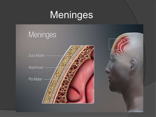
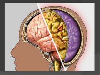
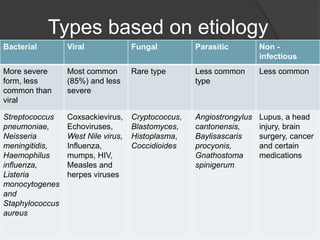
INTRODUCTION
 Meningitis is a devastating disease and
remains a major public health challenge.
 Meningitis can be caused by many different
pathogens including viruses and fungi but
the highest global burden is seen with
bacterial meningitis.
 Together with sepsis, meningitis is
estimated to cause more deaths in children
under 5 years of age than malaria.
 Survivors can suffer severe sequelae with
considerable social and economic costs
Meninges
Definition
 It is defined as an acute inflammation of
the protective layer - the meninges
covering brain and the spinal cord.
 The arachnoid and pia mater become
inflamed and opaque along with the first
two layers of the cortex and the spinal
cord.
 Many complications can result from this
inflammation such as the increased risk of
infarctions leading to blockage of cerebral
spinal fluid flow, thromboses in the cortical
veins and additional clinical symptoms
Types of Meningitis based on
duration
1. Acute
 It is a life-threatening inflammation often caused by a
bacterial or viral infection which develop over the course
of a few hours to days
2. Subacute
 Develops over days to a few weeks – mainly bacterial
3. Chronic
 Persists for at least 1 month without spontaneous
resolution. The most common etiologies of chronic
meningitis fall into 3 broad categories: fungal and
tuberculous infectious, autoimmune, and neoplastic
Types based on etiology
Bacterial Viral Fungal Parasitic Non -
infectious
More severe
form, less
common than
viral
Most common
(85%) and less
severe
Rare type Less common
type
Less common
Streptococcus
pneumoniae,
Neisseria
meningitidis,
Haemophilus
influenza,
Listeria
monocytogenes
and
Staphylococcus
aureus
Coxsackievirus,
Echoviruses,
West Nile virus,
Influenza,
mumps, HIV,
Measles and
herpes viruses
Cryptococcus,
Blastomyces,
Histoplasma,
Coccidioides
Angiostrongylus
cantonensis,
Baylisascaris
procyonis,
Gnathostoma
spinigerum
Lupus, a head
injury, brain
surgery, cancer
and certain
medications
Risk factors
 Age- children younger than 5 years
 Use of immunosuppressive drugs
 Chronic malnutrition
 AIDS
 CSF Shunt
 Chronic alcoholism
 Diabetes
 Pneumonia
Pyogenic Meningitis
 Other wise called as acute bacterial
meningitis, a life-threatening CNS
infectious disease with elevated mortality
and disability rates.
 Acute bacterial meningitis is rapidly
developing inflammation of the meninges
that cover the brain and spinal cord and of
the fluid-filled space between the meninges
(subarachnoid space) when it is caused by
bacteria.
 Infection causing meningitis arises in the
nasopharynx
Prevalance
 Bacterial meningitis appears more
frequently in populations that are in close
living quarter
 Although the prevalence of meningitis has
decreased, it is believed that many cases
go unreported.
 The incidence of meningitis is 2 of 6 per
10,000 adults per year in developed
countries and is up to ten times higher in
less-developed countries.
 Can develop in infants and children,
particularly in geographic areas where children
are not vaccinated. As people age, acute
bacterial meningitis becomes more common
 If meningitis develops within the first 48 hours
after birth, it is usually acquired from the
mother. It may be transmitted from mother to
newborn as the newborn passes through the
birth canal. In these cases, meningitis is often
part of a serious bloodstream infection (sepsis)
Etiology
 In neonates: Gram –ve bacilli, e.g. E. coli, Klebsiella.
Haemophilus influenzae.
 In children: Haemophilus influenzae. Pneumococcus
(Strep. pneumoniae). Meningococcus. (Neisseria
meningitidis).
 In adults: Pneumococcus. Meningococcus.
 Other bacteria: Listeria monocytogenes, Streptococcus
pyogenes and Staphylococcus aureus are occasionally
responsible.
 Infections of mixed aetiology (two or more bacteria)
may occur following head injury, mastoiditis or
iatrogenically after lumbar puncture.
Route of entry
 Spread through the bloodstream from an infection in another part
of the body (the most common route)
 Spread from another infection in the head, such as sinusitis or
an ear infection (often caused by Streptococcus pneumoniae)
 After a wound penetrates the skull or meninges (often caused
by Staphylococcus aureus)
 Surgery is done of the brain or spinal cord (often caused by
gram-negative bacteria)
 When a drain (shunt), placed in the brain to relieve increased
pressure in the skull, becomes infected
 When bacteria enter through a birth defect in the skull or spine
(such as spina bifida)
Pathology
 Causative organism enters into CSF
through blood brain barrier
 A purulent exudate most evident in the
basal cisterns extends throughout the
subarachnoid space.
 The underlying brain becomes
congested, oedematous and ischaemic.
 The integrity of the pia mater normally
protects against brain abscess formation
 The cytokines, interleukin, tumour necrosis factor,
and prostaglandin E2 are released as part of an
acute inflammatory response.
 They increase vascular permeability, cause a loss of
cerebrovascular autoregulation and exacerbate
neuronal injury.
 The inflammatory exudate may also affect vascular
structures crossing the subarachnoid space
producing an arteritis or venous thrombophlebitis
with resultant infarction.
 Similarly, cranial nerves may suffer direct damage.
 Hydrocephalus can result from CSF obstruction.
Clinical presentation
 The classical clinical triad is fever,
headache and neck stiffness.
 Prodromal features (variable)
 A respiratory infection otitis media or
pneumonia associated with muscle pain.
 Meningitic symptoms
 Severe frontal/occipital headache
 Stiff neck
 Photophobia
 Systemic signs:
High fever, Transient purpuric or petechial
skin rash in meningococcal meningitis.
 Meningitic signs:
1. Brudzinki’s sign - is caused by passive neck
flexion producing flexion of the hips or
knees.
2. Kernig’s sign presents, as restrictive passive
extension of the knee while the hip is flexed.[
Associated neurological
signs
1. Impaired conscious level
2. Focal or generalised seizures are
frequent.
3. Cranial nerve signs occur in 15% of
patients.
4. Sensorineural deafness (not due to
concurrent otitis media but to direct
cochlear involvement) – 20%
5. Focal neurological signs – hemiparesis,
dysphasia, hemianopia, papilloedema –
occur in 10%.
Non-neurological complications
 Shock ← Meningitis →
Arthritis
↓ Septic complications
Inappropriate secretion of ADH
Acute bacterial endocarditis
Coagulation disorders:
Thrombocytopenia – disseminated intravascular
coagulation
Features specific to causative bacteria
Haemophilus meningitis Meningococcal meningitis Pneumococcal meningitis
Generally occurs
in small children.
Preceding upper
respiratory tract
infection. Onset
abrupt with a brief
prodrome.
Organism is carried in the
nasopharynx. Septicaemia
can occur with arthralgia;
purpuric skin rash. When
overwhelming, confluent
haemorrhages appear in
the skin due to
disseminated intravascular
coagulation.
Predominantly an adult
disorder. Often associated
with debilitation, e.g.
alcoholism.
May result from pneumonia,
middle ear, sinus infection
or follow splenectomy.
Onset may be explosive,
progressing to death within
a few hours.
Outcome
Generally good
Less than 5%
mortality.
Gradual onset – good
prognosis.
Sudden onset with
septicaemia –
poor outcome.
Overall mortality – 10%.
Mortality – 20%.
Poor prognostic signs –
coma,
seizures, increased protein
in
CSF.
Investigation
 CT/MRI
Typically shows thin and linear leptomeningeal
enhancement
 CSF examination
Moderate increase in pressure < 300 mm CSF, Gram stain of
spun-down sediment
 Serological/immunological tests
The latex particle agglutination (LA) test, for the detection of
bacteria antigen in CSF
 Blood cultures
Organism isolated in 80% of cases of Haemophilus meningitis.
Pneumococcus and meningococcus in less than 50% of
patients.
 Check serum electrolytes.
important in view of the frequency of
inappropriate antidiuretic hormone
secretion.
 Detect the source of infection.
Chest X-ray – pneumonia
Skull X-ray – fracture
Sinus X-ray – sinusitis
Petrous views – mastoiditis
Management
 Initial therapy
 Neonates (above 1 month) – ampicillin, +
aminoglycoside and cephalosporin
 Children (under 5 years) – vancomycin + 3rd
generation cephalosporin
 Adults – vancomycin + 3rd generation
cephalosporin
 Immunocompromised patient – vancomycin +
ampicillin + cephalosporin
 Steroids
A four-day regimen of dexamethasone,
starting before or with the first dose of
antibiotics, is now recommended in
children with haemophilus and adults
with bacterial meningitis likely to be
pneumococcal.
Prevention
 Haemophilus vaccine (HiB vaccine) in
children.
 The pneumococcal conjugate vaccine is
now a routine childhood immunization
and is very effective at preventing
pneumococcal meningitis.
 Household members and others in close
contact with people who have
meningococcal meningitis should
receive preventive antibiotics
VIRAL MENINGITS
 Meningitis is the commonest type of viral
infection of the central nervous system.
 The term aseptic meningitis includes
viral meningitis as well as other forms of
meningitis where routine culture reveals
no other organisms.
ETIOLOGY
 Common causal viruses
1. Enteroviruses
2. Mumps virus
3. Herpes simplex (subtype 2)
4. Epstein-barr virus (ebv)
 Rare causal viruses
1. Lymphocytic choriomeningitis
2. Human immunodeficiency virus
3. West nile virus
 Enterovirus infection - affects children/young adults and
occurs seasonally in late summer. Spread is by the faecal/oral
route.
 Mumps – affects children/young adults. Winter/spring
incidence.
 Herpes simplex (type 2) – accounts for 5% of viral
meningitis. Develops in 25% of patients with primary genital
infection (suspect in sexually active adults). Can cause a
recurrent meningitis (Mollaret’s meningitis).
 Lymphocytic choriomeningitis – affects any age and is a
consequence of airborne spread from rodent droppings.
 Human Immunodeficiency Virus (HIV) – suspect in high risk
groups. HIV antibodies are often absent and develop 1–3
months later during convalescence.
Transmission of Viral Meningitis
 Spread through the bloodstream from an infection in another part of
the body (the most common way)
 Contact with contaminated stool, which may occur when infected
people do not wash their hands after a bowel movement or when
they swim in a public swimming pool (for enteroviruses)
 Sexual intercourse or other genital contact with an infected person
(for HSV-2 and HIV)
 A bite of an insect, such as a mosquito (for West Nile virus, St.
Louis virus, Zika virus, or Chikungunya virus)
 Spread through the air by inhaling the virus (for varicella-zoster
virus)
 Contact with dust or food contaminated by the urine or stool of
infected mice or pet hamsters (for lymphocytic choriomeningitis
virus)
 Use of infected needles to inject drugs (for HIV)
CLINICAL PRESENTATION
 PRODROMAL PHASE
1. Fever
2. Malaise
3. Sore throat
 MENINGEAL PHASE
1. Headache
2. Photophobia
3. Drowiseness
 RECOVERY PHASE
1. 7- 14 days
Signs
 Mild meningism
 Neck stiffness
 Kernig’s sign +ve
 No focal signs
 Skin rashes, parotitis, diarrohea,
myalgia may or may not present
Complications
 Febrile seizures
 Inappropriate ADH secretion.
Investigations
 The CSF cell count is elevated
(lymphocytes or monocytes) with a normal
glucose and protein.
 PCR detection of viral DNA/RNA in CSF
though diagnostic
 Virus may be cultured from throat swabs or
stool.
 Serological tests on serum in acute and
convalescent phases are especially
valuable in detecting mumps and herpes
simplex (type 2).
Differential diagnosis
 Tuberculous or fungal meningitis
 Leptospirosis
 Sarcoidosis
 Carcinomatous meningitis
 Partially treated bacterial meningitis
 Parameningeal chronic infection which
evokes a meningeal response, e.g.
mastoiditis.
Treatment
 Treatment for viral meningitis is mostly
supportive.
 Rest, hydration, antipyretics, and pain or
anti-inflammatory medications may be
given as needed
 Prognosis is excellent
THANK YOU

Meningitis

  • 2.
    INTRODUCTION  Meningitis isa devastating disease and remains a major public health challenge.  Meningitis can be caused by many different pathogens including viruses and fungi but the highest global burden is seen with bacterial meningitis.  Together with sepsis, meningitis is estimated to cause more deaths in children under 5 years of age than malaria.  Survivors can suffer severe sequelae with considerable social and economic costs
  • 3.
  • 4.
    Definition  It isdefined as an acute inflammation of the protective layer - the meninges covering brain and the spinal cord.  The arachnoid and pia mater become inflamed and opaque along with the first two layers of the cortex and the spinal cord.  Many complications can result from this inflammation such as the increased risk of infarctions leading to blockage of cerebral spinal fluid flow, thromboses in the cortical veins and additional clinical symptoms
  • 6.
    Types of Meningitisbased on duration 1. Acute  It is a life-threatening inflammation often caused by a bacterial or viral infection which develop over the course of a few hours to days 2. Subacute  Develops over days to a few weeks – mainly bacterial 3. Chronic  Persists for at least 1 month without spontaneous resolution. The most common etiologies of chronic meningitis fall into 3 broad categories: fungal and tuberculous infectious, autoimmune, and neoplastic
  • 7.
    Types based onetiology Bacterial Viral Fungal Parasitic Non - infectious More severe form, less common than viral Most common (85%) and less severe Rare type Less common type Less common Streptococcus pneumoniae, Neisseria meningitidis, Haemophilus influenza, Listeria monocytogenes and Staphylococcus aureus Coxsackievirus, Echoviruses, West Nile virus, Influenza, mumps, HIV, Measles and herpes viruses Cryptococcus, Blastomyces, Histoplasma, Coccidioides Angiostrongylus cantonensis, Baylisascaris procyonis, Gnathostoma spinigerum Lupus, a head injury, brain surgery, cancer and certain medications
  • 8.
    Risk factors  Age-children younger than 5 years  Use of immunosuppressive drugs  Chronic malnutrition  AIDS  CSF Shunt  Chronic alcoholism  Diabetes  Pneumonia
  • 9.
    Pyogenic Meningitis  Otherwise called as acute bacterial meningitis, a life-threatening CNS infectious disease with elevated mortality and disability rates.  Acute bacterial meningitis is rapidly developing inflammation of the meninges that cover the brain and spinal cord and of the fluid-filled space between the meninges (subarachnoid space) when it is caused by bacteria.  Infection causing meningitis arises in the nasopharynx
  • 10.
    Prevalance  Bacterial meningitisappears more frequently in populations that are in close living quarter  Although the prevalence of meningitis has decreased, it is believed that many cases go unreported.  The incidence of meningitis is 2 of 6 per 10,000 adults per year in developed countries and is up to ten times higher in less-developed countries.
  • 11.
     Can developin infants and children, particularly in geographic areas where children are not vaccinated. As people age, acute bacterial meningitis becomes more common  If meningitis develops within the first 48 hours after birth, it is usually acquired from the mother. It may be transmitted from mother to newborn as the newborn passes through the birth canal. In these cases, meningitis is often part of a serious bloodstream infection (sepsis)
  • 12.
    Etiology  In neonates:Gram –ve bacilli, e.g. E. coli, Klebsiella. Haemophilus influenzae.  In children: Haemophilus influenzae. Pneumococcus (Strep. pneumoniae). Meningococcus. (Neisseria meningitidis).  In adults: Pneumococcus. Meningococcus.  Other bacteria: Listeria monocytogenes, Streptococcus pyogenes and Staphylococcus aureus are occasionally responsible.  Infections of mixed aetiology (two or more bacteria) may occur following head injury, mastoiditis or iatrogenically after lumbar puncture.
  • 13.
    Route of entry Spread through the bloodstream from an infection in another part of the body (the most common route)  Spread from another infection in the head, such as sinusitis or an ear infection (often caused by Streptococcus pneumoniae)  After a wound penetrates the skull or meninges (often caused by Staphylococcus aureus)  Surgery is done of the brain or spinal cord (often caused by gram-negative bacteria)  When a drain (shunt), placed in the brain to relieve increased pressure in the skull, becomes infected  When bacteria enter through a birth defect in the skull or spine (such as spina bifida)
  • 14.
    Pathology  Causative organismenters into CSF through blood brain barrier  A purulent exudate most evident in the basal cisterns extends throughout the subarachnoid space.  The underlying brain becomes congested, oedematous and ischaemic.  The integrity of the pia mater normally protects against brain abscess formation
  • 15.
     The cytokines,interleukin, tumour necrosis factor, and prostaglandin E2 are released as part of an acute inflammatory response.  They increase vascular permeability, cause a loss of cerebrovascular autoregulation and exacerbate neuronal injury.  The inflammatory exudate may also affect vascular structures crossing the subarachnoid space producing an arteritis or venous thrombophlebitis with resultant infarction.  Similarly, cranial nerves may suffer direct damage.  Hydrocephalus can result from CSF obstruction.
  • 17.
    Clinical presentation  Theclassical clinical triad is fever, headache and neck stiffness.  Prodromal features (variable)  A respiratory infection otitis media or pneumonia associated with muscle pain.  Meningitic symptoms  Severe frontal/occipital headache  Stiff neck  Photophobia
  • 18.
     Systemic signs: Highfever, Transient purpuric or petechial skin rash in meningococcal meningitis.  Meningitic signs: 1. Brudzinki’s sign - is caused by passive neck flexion producing flexion of the hips or knees. 2. Kernig’s sign presents, as restrictive passive extension of the knee while the hip is flexed.[
  • 20.
    Associated neurological signs 1. Impairedconscious level 2. Focal or generalised seizures are frequent. 3. Cranial nerve signs occur in 15% of patients. 4. Sensorineural deafness (not due to concurrent otitis media but to direct cochlear involvement) – 20% 5. Focal neurological signs – hemiparesis, dysphasia, hemianopia, papilloedema – occur in 10%.
  • 21.
    Non-neurological complications  Shock← Meningitis → Arthritis ↓ Septic complications Inappropriate secretion of ADH Acute bacterial endocarditis Coagulation disorders: Thrombocytopenia – disseminated intravascular coagulation
  • 22.
    Features specific tocausative bacteria Haemophilus meningitis Meningococcal meningitis Pneumococcal meningitis Generally occurs in small children. Preceding upper respiratory tract infection. Onset abrupt with a brief prodrome. Organism is carried in the nasopharynx. Septicaemia can occur with arthralgia; purpuric skin rash. When overwhelming, confluent haemorrhages appear in the skin due to disseminated intravascular coagulation. Predominantly an adult disorder. Often associated with debilitation, e.g. alcoholism. May result from pneumonia, middle ear, sinus infection or follow splenectomy. Onset may be explosive, progressing to death within a few hours. Outcome Generally good Less than 5% mortality. Gradual onset – good prognosis. Sudden onset with septicaemia – poor outcome. Overall mortality – 10%. Mortality – 20%. Poor prognostic signs – coma, seizures, increased protein in CSF.
  • 23.
    Investigation  CT/MRI Typically showsthin and linear leptomeningeal enhancement  CSF examination Moderate increase in pressure < 300 mm CSF, Gram stain of spun-down sediment  Serological/immunological tests The latex particle agglutination (LA) test, for the detection of bacteria antigen in CSF  Blood cultures Organism isolated in 80% of cases of Haemophilus meningitis. Pneumococcus and meningococcus in less than 50% of patients.
  • 24.
     Check serumelectrolytes. important in view of the frequency of inappropriate antidiuretic hormone secretion.  Detect the source of infection. Chest X-ray – pneumonia Skull X-ray – fracture Sinus X-ray – sinusitis Petrous views – mastoiditis
  • 25.
    Management  Initial therapy Neonates (above 1 month) – ampicillin, + aminoglycoside and cephalosporin  Children (under 5 years) – vancomycin + 3rd generation cephalosporin  Adults – vancomycin + 3rd generation cephalosporin  Immunocompromised patient – vancomycin + ampicillin + cephalosporin
  • 26.
     Steroids A four-dayregimen of dexamethasone, starting before or with the first dose of antibiotics, is now recommended in children with haemophilus and adults with bacterial meningitis likely to be pneumococcal.
  • 27.
    Prevention  Haemophilus vaccine(HiB vaccine) in children.  The pneumococcal conjugate vaccine is now a routine childhood immunization and is very effective at preventing pneumococcal meningitis.  Household members and others in close contact with people who have meningococcal meningitis should receive preventive antibiotics
  • 28.
    VIRAL MENINGITS  Meningitisis the commonest type of viral infection of the central nervous system.  The term aseptic meningitis includes viral meningitis as well as other forms of meningitis where routine culture reveals no other organisms.
  • 29.
    ETIOLOGY  Common causalviruses 1. Enteroviruses 2. Mumps virus 3. Herpes simplex (subtype 2) 4. Epstein-barr virus (ebv)  Rare causal viruses 1. Lymphocytic choriomeningitis 2. Human immunodeficiency virus 3. West nile virus
  • 30.
     Enterovirus infection- affects children/young adults and occurs seasonally in late summer. Spread is by the faecal/oral route.  Mumps – affects children/young adults. Winter/spring incidence.  Herpes simplex (type 2) – accounts for 5% of viral meningitis. Develops in 25% of patients with primary genital infection (suspect in sexually active adults). Can cause a recurrent meningitis (Mollaret’s meningitis).  Lymphocytic choriomeningitis – affects any age and is a consequence of airborne spread from rodent droppings.  Human Immunodeficiency Virus (HIV) – suspect in high risk groups. HIV antibodies are often absent and develop 1–3 months later during convalescence.
  • 31.
    Transmission of ViralMeningitis  Spread through the bloodstream from an infection in another part of the body (the most common way)  Contact with contaminated stool, which may occur when infected people do not wash their hands after a bowel movement or when they swim in a public swimming pool (for enteroviruses)  Sexual intercourse or other genital contact with an infected person (for HSV-2 and HIV)  A bite of an insect, such as a mosquito (for West Nile virus, St. Louis virus, Zika virus, or Chikungunya virus)  Spread through the air by inhaling the virus (for varicella-zoster virus)  Contact with dust or food contaminated by the urine or stool of infected mice or pet hamsters (for lymphocytic choriomeningitis virus)  Use of infected needles to inject drugs (for HIV)
  • 32.
    CLINICAL PRESENTATION  PRODROMALPHASE 1. Fever 2. Malaise 3. Sore throat  MENINGEAL PHASE 1. Headache 2. Photophobia 3. Drowiseness  RECOVERY PHASE 1. 7- 14 days
  • 33.
    Signs  Mild meningism Neck stiffness  Kernig’s sign +ve  No focal signs  Skin rashes, parotitis, diarrohea, myalgia may or may not present
  • 34.
    Complications  Febrile seizures Inappropriate ADH secretion.
  • 35.
    Investigations  The CSFcell count is elevated (lymphocytes or monocytes) with a normal glucose and protein.  PCR detection of viral DNA/RNA in CSF though diagnostic  Virus may be cultured from throat swabs or stool.  Serological tests on serum in acute and convalescent phases are especially valuable in detecting mumps and herpes simplex (type 2).
  • 36.
    Differential diagnosis  Tuberculousor fungal meningitis  Leptospirosis  Sarcoidosis  Carcinomatous meningitis  Partially treated bacterial meningitis  Parameningeal chronic infection which evokes a meningeal response, e.g. mastoiditis.
  • 37.
    Treatment  Treatment forviral meningitis is mostly supportive.  Rest, hydration, antipyretics, and pain or anti-inflammatory medications may be given as needed  Prognosis is excellent
  • 38.