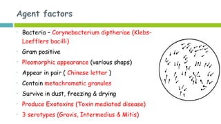
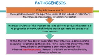
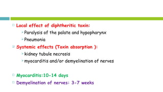
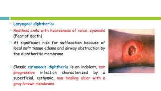
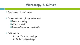
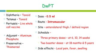
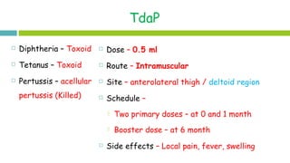
Call Girls Ludhiana Just Call 9907093804 Top Class Call Girl Service Available
Diptheria & pertussis
1. 100 days Cough
Dr Deepak UpadhyayDr Deepak Upadhyay
Assistant Professor
Department of Community Medicine
2. Agent factors
• Bacteria – Bordetelle Pertussis
• Gram negative
• Small, ovoid, coco-bacillus
• Non motile, Non sporing
• Survival in environment – very short
• Produce Exotoxins (Toxin mediated
disease)
3. Agent factors (cont.)
Reservoir – Human are only known reservoir
Source of Infection – Cases (Clinical + Subclinical)
Infective Material – Nasopharyngeal secretions
Mode of transmission - Airborne droplets
Period of Communicability – In the catarrhal stage and 3 weeks
after the onset of cough
4. Host Factors
Females > Male
Age Incidence
Pre vaccination era - children birth to 5 years
of age (Preschool Children)
Post vaccination introduction - in School going
children (5 - 10 yr)
Immunity –
Maternal antibodies – no role / no protection
One attack – 90% immunity
Secondary attack – 10% cases
5. Environmental factors
Endemic disease with epidemic potential
Epidemics during winter season.
Incubation period – 7 – 14 days
8. Clinical Manifestations (cont.)
Paroxysmal cough stage
Cough increases – distinctive bouts
Violent spasms of continuous coughing (Paroxysm)
At the end of paroxysm - long inspiratory effort (whoop)
Whoop end with vomiting and occasional aspiration
During episode of cough - Cyanosis followed by vomiting,
exhaustion and seizures
9. Clinical Manifestations (cont.)
In between episodes child look well
Cough increase for next 2-3 weeks and decreases over next 10
weeks
Paroxysmal cough>2 weeks with or without whoop and/or post-
tussive vomiting is the hallmark feature of pertussis
Convalescence stage
period of gradual recovery even up to 6 months
10. Complications
Due to increase intra abdominal pressure
Hernias (inguinal / umbilical)
Rectal prolapse
Sub-conjuctival hemorrhage
Subcutaneous emphysema
Bronchopneumonia
Atelectasis
Neurological complications (due to the toxin or hypoxia or cerebral
hemorrhage)
Seizures (1 in 100)
Encephalopathy (1 in 300)
11. DIAGNOSIS
Suspected on the basis of history and clinical examination
Confirmed by culture, genomics or serology
1.Blood investigations
Elevated WBC count with lymphocytosis.
The absolute lymphocyte count of ≥20,000 is highly suggestive
1.Direct fluorescent antibody testing
IgA antibodies in nasal secretions
IgM antibodies against toxin
12. DIAGNOSIS
3. Culture and Microscopy:
Gold standard specially in the
catarrhal stage.
A saline nasal swab
Swab from the nasopharynx
Direct plate method
4. PCR:
most sensitive
can be done even after antibiotic
exposure.
13. TREATMENT
1. Avoidance of irritants, smoke and other cough promoting factors
2.2. Antibiotics:Antibiotics:
Erythromycin orally for 2 weeks
or
Azithromycin orally for 5 days
or
Clarithromycin orally for 7 days
3.3. Supportive treatmentSupportive treatment -Supplemental oxygen, hydration, cough
mixtures and bronchodilators (in individual cases)
14. Active Prevention
Children < 7 years
DwPT (whole cell pertussis)
Children > 7 years
TdaP (acellular pertussis)
Passive prevention
By immunoglobulin
Chemoprophylaxis
Erythromycin
15. Dr Deepak UpadhyayDr Deepak Upadhyay
Assistant Professor
Department of Community Medicine
17. Agent factors (cont.)
Reservoir – Human are only
known reservoir
Source of Infection – Cases
(Clinical) + Carriers
Infective Material
Nasopharyngeal secretions
Discharge from cutaneous
lesions
Mode of transmission –
Airborne droplets
Direct contact to skin lesion
Fomites
Period of Communicability
Cases – during clinical disease
Carrier – remain infectious
up to 1 year
18. Host Factors
Females > Male
Age Incidence
Pre vaccination era - children 6 months to 5 years of age
(Preschool Children)
Post vaccination introduction - in School going children (5 -
10 yr)
Immunity –
Maternal antibodies – through milk
70 % children develop immunity through subclinical
infection
19. Environmental factors
Endemic disease with epidemic potential
Epidemics during winter season.
Incubation period – 7 – 14 days
21. Local effect of diphtheritic toxin:
Paralysis of the palate and hypopharynx
Pneumonia
Systemic effects (Toxin absorption ):
kidney tubule necrosis
myocarditis and/or demyelination of nerves
Myocarditis:10-14 days
Demyelination of nerves: 3-7 weeks
22. Clinical Manifestations
Classification ( depend upon location):
Nasal
Pharyngeal (Faucial Diptheria)
tonsilar
laryngeal or laryngo-tracheal
skin, eye or genitalia
23. Nasal Diphtheria
Infection of the anterior nares
More common among infants,
Causes serosanguineous, purulent, erosive rhinitis with membrane
formation
Shallow ulceration of the external nares and upper lip is
characteristic
Unilateral nasal discharge is quite pathognomic of nasal diphtheria
24. Pharyngeal & Tonsilar diptheria
Sore throat is the universal early symptom
Only half of patients have fever
Dysphagia, hoarseness, malaise, or headache
On examination
Foul smell
A yellowish membrane (Pseudomemdrane) on tonsils / pharyngeal
wall may extend to uvula, soft palate, posterior oropharynx,
hypopharynx, or glottic areas
Erythema in adjacent areas
Enlarged cervical lymph nodes: bull-neck appearance
27. Laryngeal diphtheria:
Restless child with hoarseness of voice, cyanosis
(Fear of death)
At significant risk for suffocation because of
local soft tissue edema and airway obstruction by
the diphtheritic membrane
Classic cutaneous diphtheria is an indolent, non
progressive infection characterized by a
superficial, ecthymic, non healing ulcer with a
gray-brown membrane
28. COMPLICATIONS
1.1. Respiratory tract obstructionRespiratory tract obstruction by pseudomembranes
1.1. Toxic CardiomyopathyToxic Cardiomyopathy:
1. In 10-25% of patients
2. Responsible for 50-60% of deaths
3. Tachycardia out of proportion to fever
4. Prolonged PR interval and changes in the ST-T wave
5. Elevation of the serum aspartate aminotransferase
concentration closely parallels the severity of myonecrosis
29. 3.3. Toxic NeuropathyToxic Neuropathy:
Hypoesthesia and soft palate paralysis
Afterwards weakness of the posterior pharyngeal, laryngeal, and
facial nerves : a nasal quality in the voice, difficulty in swallowing and
risk for aspiration
Cranial neuropathies (5th wk): oculomotor and ciliary paralysis-
strabismus, blurred vision, or difficulty with accommodation
Symmetric polyneuropathy (10 days to 3 mo): motor deficits with
diminished deep tendon reflexes
Monitoring for paralysis of the diaphragm muscle
Recovery from the neuritis is often slow but usually complete.
Corticosteroids are not recommended.
30. DIAGNOSIS
Suspected on the basis of history and clinical examination
Confirmed by culture, toxigenicity
1.Blood investigations
Elevated WBC count with lymphocytosis.
1.Toxigenicity testing
•Elek s Gel precipitation testing
•Shick test
32. TREATMENT
1. Antitoxin:
Mainstay of therapy
Neutralizes only free toxin, efficacy diminishes with elapsed
time
2. Antimicrobial therapy
Halt toxin production, treat localized infection and prevent
transmission of the organism to contacts
Erythromycin
Or
aqueous crystalline penicillin G / procaine penicillin
33. Active Prevention
Children < 7 years
DwPT (whole cell pertussis)
Children > 7 years
TdaP (acellular pertussis)
Passive prevention
By immunoglobulin
Chemoprophylaxis
Erythromycin (all contact)
34. DwPT
Dose – 0.5 ml
Route – Intramuscular
Site – anterolateral thigh / deltoid region
Schedule –
Three primary doses – at 6, 10, 14 weeks
Two booster doses – at 18 months & 5 years
Side effects – Local pain, fever, swelling
Diphtheria – Toxoid
Tetanus – Toxoid
Pertussis – Live whole
cell vaccine
Adjuvant – Aluminum
Phosphate
Preservative –
Thiomersal
35. TdaP
Dose – 0.5 ml
Route – Intramuscular
Site – anterolateral thigh / deltoid region
Schedule –
Two primary doses – at 0 and 1 month
Booster dose – at 6 month
Side effects – Local pain, fever, swelling
Diphtheria – Toxoid
Tetanus – Toxoid
Pertussis – acellular
pertussis (Killed)
Editor's Notes
Due to difficulty in expelling the thick mucous form the tracheobronchial tree
Erythromycin (40-50 mg/kg/day 6 hrly orally for 2 weeks
or
Azithromycin 10 mg/kg for 5 days in children&lt;6 months and for children&gt;6 months 10 mg/kg on day 1, followed by 5mg/kg from day2-5
or
Clarithromycin 15 mg/kg 12 hrly for 7 days
The vaccine is live and attenuated and confers lifelong immunity.
Given to children 12 and 15 months and again between 3-6 years of age
All types of carriers are seen
Even cattle act as carrior
The risk for significant complications correlates directly with the extent and severity of exudative local oropharyngeal disease as well as delay in administration of antitoxin
Antitoxin is administered as a single empirical dose of 20,000-120,000 U based on the degree of toxicity, site and size of the membrane, and duration of illness
Elimination of the organism should be documented by negative results of at least 2 successive cultures of specimens from the nose and throat (or skin) obtained 24 hr apart after completion of therapy
Prognosis: depends on the virulence of the organism (subspecies gravis), patient age, immunization status, site of infection and speed of administration of the antitoxin
The case fatality rate of almost 10% for respiratory tract diphtheria
At recovery, administration of diphtheria toxoid is indicated to complete the primary series or booster doses of immunization, because not all patients develop antibodies to diphtheritic toxin after infection
The vaccine is live and attenuated and confers lifelong immunity.
Given to children 12 and 15 months and again between 3-6 years of age