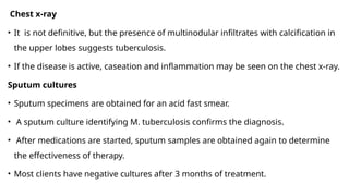
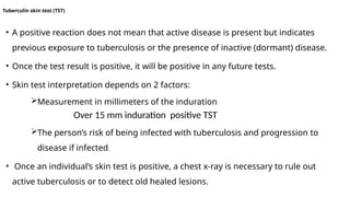
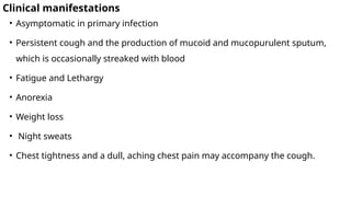
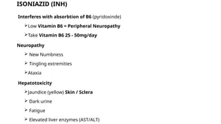
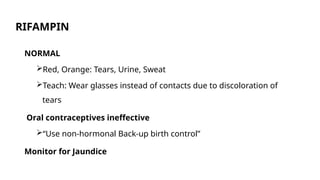
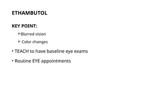
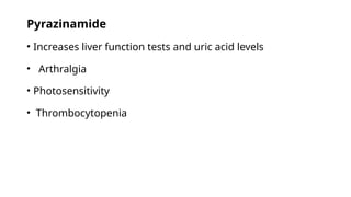
UNIT THREE
Air-borne Diseases
Air-borne Diseases
 The organisms causing the diseases in the air-borne group enter the body via
the respiratory tract.
 When a patient or carrier of pathogens talks, coughs, laughs, or sneezes,
he/she discharges fluid droplets.
 The smallest of these remain up in the air for some time and may be inhaled
by a new host.
 Droplets with a size of 1-5 microns are quite easily drawn in to the lungs and
retained there.
 Droplets that are bigger in size will not remain air-borne for long but will fall
to the ground. Here, however, they dry and mix with dusare able to survive
1. Tuberculosis
Overview
• Tuberculosis is the world’s foremost cause of death from a single infectious
agent.
• It causing 26% of avoidable deaths in developing countries.
 TB affects individuals of all ages and both sexes
 It usually affects economically and culturally disadvantaged segment of a
population where access to health services is often limited.
• It is estimated that each year more than 8 million new cases of tuberculosis
occur worldwide and approximately 3 million persons die of the disease.
• The rate of infection has increased, particularly among HIV-infected people
Introduction
• Tuberculosis is an infectious disease caused by the mycobacterium, M.
tuberculosis.
Characteristics of the Bacteria
• It is aerobic bacteria.
• It similar to other bacterial organisms except for an outer waxy
capsule that makes them more resistant to destruction.
• The waxy coat also causes the organism to retain red dye when
treated with acid in acid-fast staining.
• Thus, the mycobacteria are often referred to as acid-fast bacilli.
Cont…
• Although M. tuberculosis can infect practically any organ of the body,
the lungs are most frequently involved.
• The tubercle bacilli are strict aerobes that thrive in an oxygen-rich
environment.
• This explains their tendency to cause disease in the upper lobe or
upper parts of the lower lobe of the lung, where the ventilation and
oxygen content are greatest.
Two forms of tuberculosis pose a particular threat to humans:
A. Human tuberculosis
 Airborne infection spread by minute, invisible particles, called droplet nuclei,
that are harbored in the respiratory secretions of persons with active
tuberculosis.
 Mode of transmission directly from person to person
B. Bovine tuberculosis
 Transmitted from infected animal to Person.
Acquired by drinking milk from infected cows, and it initially affects the
gastrointestinal tract.
Primary TB
• Primary tuberculosis occurs in a person lacking previous contact with the tubercle
bacillus.
• It typically is initiated as a result of inhaling droplet nuclei that contain the
tubercle bacillus.
• Inhaled droplet nuclei pass down the bronchial tree without settling on the
epithelium and implant in a respiratory bronchiole.
• After entering the lung, the bacilli are surrounded and engulfed by macrophages.
• M. tuberculosis has no known endotoxins or exotoxins; therefore, there is no early
immunoglobulin response to infection.
• The tubercle bacillus grows slowly, dividing every 25 to 32 hours in the
macrophage.
• As the bacilli multiply, the macrophages degrade some mycobacteria and
present antigen to the T lymphocytes for development of a cell-mediated
immune response.
• The organisms grow for 2 to 12 weeks until they reach sufficient numbers
to elicit a cellular immune response.
• In persons with intact cellmediated immunity, this action is followed by the
development of a single, gray-white, circumscribed granulomatous lesion.
• Within 2 to 3 weeks, the central portion of the granloma undergoes soft,
caseous (cheeselike) necrosis.
 Latent TB infection: Individuals with latent TB infection do not have
symptoms as there is no tissue destruction by the bacilli and are not
infectious.
 In immunocompetent individuals, only 5-10% of infected persons
develop active disease in their lifetime.
 Active TB disease may arise from progression of the primary lesion
after infection (Primary TB), or from endogenous reactivation of latent
foci.
 latent tuberculosis infection has the potential to develop into active
Secondary Tuberculosis
• Latent TB change to Active TB
• Secondary tuberculosis represents either reinfection from inhaled
droplet nuclei or reactivation of a previously healed primary lesion.
• It often occurs in situations of impaired body defense mechanisms.
• The partial immunity that follows primary tuberculosis affords
protection against reinfection.
• The cell-mediated hypersensitivity reaction can bean aggravating
factor
• The progression from LTBI to Active TB disease may occur at any
time, from soon to many years later.
• primary/secondary TB usually affects the lungs (Pulmonary TB)
though any body part can be affected after haematogenous and/or
lymphatic spread of the bacilli (extra-pulmonary TB).
• If massive haematogenic dissemination occurs, all organs can be
affected (miliary TB).
Risk factors for Tuberculosis
• HIV patients
• Child younger than 5 years of age
• Drinking unpasteurized milk if the cow is infected with bovine tuberculosis
• Homeless individuals or those from a lower socioeconomic group, minority
group, or refugee group.
• Individuals in constant, frequent contact with an untreated or undiagnosed
individual.
• Health care workers
• Individuals living in crowded areas, such as long-term care facilities, prisons,
and mental health facilities
• Older client
• Individuals with malnutrition, infection, immune dysfunction.
• Immunosuppressed as a result of medication therapy
• Individuals who abuse alcohol or are intravenous drug users
Diagnosis of TB
Client history
• Past exposure to tuberculosis
• Client’s country of origin and travel to foreign countries in which the incidence
of tuberculosis is high.
• Recent history of influenza, pneumonia, febrile illness, cough, or foul-smelling
sputum production
• Previous tests for tuberculosis; results of the testing
• Recent BCG vaccine
Chest x-ray
• It is not definitive, but the presence of multinodular infiltrates with calcification in
the upper lobes suggests tuberculosis.
• If the disease is active, caseation and inflammation may be seen on the chest x-ray.
Sputum cultures
• Sputum specimens are obtained for an acid fast smear.
• A sputum culture identifying M. tuberculosis confirms the diagnosis.
• After medications are started, sputum samples are obtained again to determine
the effectiveness of therapy.
• Most clients have negative cultures after 3 months of treatment.
• A positive reaction does not mean that active disease is present but indicates
previous exposure to tuberculosis or the presence of inactive (dormant) disease.
• Once the test result is positive, it will be positive in any future tests.
• Skin test interpretation depends on 2 factors:
Measurement in millimeters of the induration
Over 15 mm induration positive TST
The person’s risk of being infected with tuberculosis and progression to
disease if infected.
• Once an individual’s skin test is positive, a chest x-ray is necessary to rule out
active tuberculosis or to detect old healed lesions.
Tuberculin skin test (TST)
Clinical manifestations
• Asymptomatic in primary infection
• Persistent cough and the production of mucoid and mucopurulent sputum,
which is occasionally streaked with blood
• Fatigue and Lethargy
• Anorexia
• Weight loss
• Night sweats
• Chest tightness and a dull, aching chest pain may accompany the cough.
Group Discussion
Discuss about the treatment and prevention of
Tuberculosis?
Treatment of TB
• Treatment of identified lesions depends on whether the individual has active
disease or has only been exposed to the disease.
• Treatment is difficult because the bacterium has a waxy substance on the
capsule that makes penetration and destruction difficult.
• The use of a multidrug regimen destroys organisms as quickly as possible and
minimizes the emergence of drug-resistant organisms.
• Active tuberculosis is treated with a combination of medications to which the
organism is susceptible.
• Individuals with active tuberculosis are treated for 6 to 9 months
• clients with human immunodeficiency virus (HIV) infection are treated
for a longer period of time.
• After the infected individual has received medication for 2 to 3 weeks,
the risk of transmission is greatly reduced.
• Most clients have negative sputum cultures after 3 months of
compliance with medication therapy.
• Individuals who have been exposed to active tuberculosis are treated
with preventive isoniazid for 9 to 12 months.
• Two groups meet the criteria established for the use of anti mycobacterial
therapy for tuberculosis:
Persons with active tuberculosis
Risk for development of an active form of the disease
• The primary drugs used in the treatment of tuberculosis are
1. Isoniazid (INH)
2. Rifampin
3. Pyrazinamide (PZA)
4. Ethambutol
5. Streptomycin
ISONIAZID (INH)
Interferes with absorbtion of B6 (pyridoxinde)
Low Vitamin B6 = Peripheral Neuropathy
Take Vitamin B6 25 - 50mg/day
Neuropathy
 New Numbness
 Tingling extremities
Ataxia
Hepatotoxicity
Jaundice (yellow) Skin / Sclera
 Dark urine
 Fatigue
 Elevated liver enzymes (AST/ALT)
RIFAMPIN
NORMAL
Red, Orange: Tears, Urine, Sweat
Teach: Wear glasses instead of contacts due to discoloration of
tears
Oral contraceptives ineffective
“Use non-hormonal Back-up birth control”
Monitor for Jaundice
ETHAMBUTOL
KEY POINT:
Blurred vision
 Color changes
• TEACH to have baseline eye exams
• Routine EYE appointments
Pyrazinamide
• Increases liver function tests and uric acid levels
• Arthralgia
• Photosensitivity
• Thrombocytopenia
Health education
• Instruct the client to follow the medication regimen exactly as
prescribed and always to have a supply of the medication on hand.
• Advise the client that the medication regimen is continued up to 12
months depending on the situation.
• Advise the client of the side and adverse effects of the medication and
ways of minimizing them to ensure compliance.
• Reassure the client that after 2 to 3 weeks of medication therapy, it is
unlikely that the client will infect anyone.
• Advise the client to resume activities gradually.
• Instruct the client about the need for adequate nutrition and a well-
balanced diet (foods rich in iron, protein, and vitamin C) to promote
healing and to prevent recurrence of the infection.
• Inform the client and family that respiratory isolation is not necessary
because family members already have been exposed.
• Instruct the client to cover the mouth and nose when coughing or
sneezing and to put used tissues into plastic bags.
Reading Assignment
MDR-TB

Communicable Disease prevention and Control .pptx

  • 1.
  • 2.
    Air-borne Diseases  Theorganisms causing the diseases in the air-borne group enter the body via the respiratory tract.  When a patient or carrier of pathogens talks, coughs, laughs, or sneezes, he/she discharges fluid droplets.  The smallest of these remain up in the air for some time and may be inhaled by a new host.  Droplets with a size of 1-5 microns are quite easily drawn in to the lungs and retained there.  Droplets that are bigger in size will not remain air-borne for long but will fall to the ground. Here, however, they dry and mix with dusare able to survive
  • 3.
    1. Tuberculosis Overview • Tuberculosisis the world’s foremost cause of death from a single infectious agent. • It causing 26% of avoidable deaths in developing countries.  TB affects individuals of all ages and both sexes  It usually affects economically and culturally disadvantaged segment of a population where access to health services is often limited. • It is estimated that each year more than 8 million new cases of tuberculosis occur worldwide and approximately 3 million persons die of the disease. • The rate of infection has increased, particularly among HIV-infected people
  • 4.
    Introduction • Tuberculosis isan infectious disease caused by the mycobacterium, M. tuberculosis. Characteristics of the Bacteria • It is aerobic bacteria. • It similar to other bacterial organisms except for an outer waxy capsule that makes them more resistant to destruction. • The waxy coat also causes the organism to retain red dye when treated with acid in acid-fast staining. • Thus, the mycobacteria are often referred to as acid-fast bacilli.
  • 5.
    Cont… • Although M.tuberculosis can infect practically any organ of the body, the lungs are most frequently involved. • The tubercle bacilli are strict aerobes that thrive in an oxygen-rich environment. • This explains their tendency to cause disease in the upper lobe or upper parts of the lower lobe of the lung, where the ventilation and oxygen content are greatest.
  • 6.
    Two forms oftuberculosis pose a particular threat to humans: A. Human tuberculosis  Airborne infection spread by minute, invisible particles, called droplet nuclei, that are harbored in the respiratory secretions of persons with active tuberculosis.  Mode of transmission directly from person to person B. Bovine tuberculosis  Transmitted from infected animal to Person. Acquired by drinking milk from infected cows, and it initially affects the gastrointestinal tract.
  • 7.
    Primary TB • Primarytuberculosis occurs in a person lacking previous contact with the tubercle bacillus. • It typically is initiated as a result of inhaling droplet nuclei that contain the tubercle bacillus. • Inhaled droplet nuclei pass down the bronchial tree without settling on the epithelium and implant in a respiratory bronchiole. • After entering the lung, the bacilli are surrounded and engulfed by macrophages. • M. tuberculosis has no known endotoxins or exotoxins; therefore, there is no early immunoglobulin response to infection.
  • 8.
    • The tuberclebacillus grows slowly, dividing every 25 to 32 hours in the macrophage. • As the bacilli multiply, the macrophages degrade some mycobacteria and present antigen to the T lymphocytes for development of a cell-mediated immune response. • The organisms grow for 2 to 12 weeks until they reach sufficient numbers to elicit a cellular immune response. • In persons with intact cellmediated immunity, this action is followed by the development of a single, gray-white, circumscribed granulomatous lesion. • Within 2 to 3 weeks, the central portion of the granloma undergoes soft, caseous (cheeselike) necrosis.
  • 9.
     Latent TBinfection: Individuals with latent TB infection do not have symptoms as there is no tissue destruction by the bacilli and are not infectious.  In immunocompetent individuals, only 5-10% of infected persons develop active disease in their lifetime.  Active TB disease may arise from progression of the primary lesion after infection (Primary TB), or from endogenous reactivation of latent foci.  latent tuberculosis infection has the potential to develop into active
  • 10.
    Secondary Tuberculosis • LatentTB change to Active TB • Secondary tuberculosis represents either reinfection from inhaled droplet nuclei or reactivation of a previously healed primary lesion. • It often occurs in situations of impaired body defense mechanisms. • The partial immunity that follows primary tuberculosis affords protection against reinfection. • The cell-mediated hypersensitivity reaction can bean aggravating factor
  • 11.
    • The progressionfrom LTBI to Active TB disease may occur at any time, from soon to many years later. • primary/secondary TB usually affects the lungs (Pulmonary TB) though any body part can be affected after haematogenous and/or lymphatic spread of the bacilli (extra-pulmonary TB). • If massive haematogenic dissemination occurs, all organs can be affected (miliary TB).
  • 12.
    Risk factors forTuberculosis • HIV patients • Child younger than 5 years of age • Drinking unpasteurized milk if the cow is infected with bovine tuberculosis • Homeless individuals or those from a lower socioeconomic group, minority group, or refugee group. • Individuals in constant, frequent contact with an untreated or undiagnosed individual. • Health care workers
  • 13.
    • Individuals livingin crowded areas, such as long-term care facilities, prisons, and mental health facilities • Older client • Individuals with malnutrition, infection, immune dysfunction. • Immunosuppressed as a result of medication therapy • Individuals who abuse alcohol or are intravenous drug users
  • 14.
    Diagnosis of TB Clienthistory • Past exposure to tuberculosis • Client’s country of origin and travel to foreign countries in which the incidence of tuberculosis is high. • Recent history of influenza, pneumonia, febrile illness, cough, or foul-smelling sputum production • Previous tests for tuberculosis; results of the testing • Recent BCG vaccine
  • 15.
    Chest x-ray • Itis not definitive, but the presence of multinodular infiltrates with calcification in the upper lobes suggests tuberculosis. • If the disease is active, caseation and inflammation may be seen on the chest x-ray. Sputum cultures • Sputum specimens are obtained for an acid fast smear. • A sputum culture identifying M. tuberculosis confirms the diagnosis. • After medications are started, sputum samples are obtained again to determine the effectiveness of therapy. • Most clients have negative cultures after 3 months of treatment.
  • 16.
    • A positivereaction does not mean that active disease is present but indicates previous exposure to tuberculosis or the presence of inactive (dormant) disease. • Once the test result is positive, it will be positive in any future tests. • Skin test interpretation depends on 2 factors: Measurement in millimeters of the induration Over 15 mm induration positive TST The person’s risk of being infected with tuberculosis and progression to disease if infected. • Once an individual’s skin test is positive, a chest x-ray is necessary to rule out active tuberculosis or to detect old healed lesions. Tuberculin skin test (TST)
  • 17.
    Clinical manifestations • Asymptomaticin primary infection • Persistent cough and the production of mucoid and mucopurulent sputum, which is occasionally streaked with blood • Fatigue and Lethargy • Anorexia • Weight loss • Night sweats • Chest tightness and a dull, aching chest pain may accompany the cough.
  • 18.
    Group Discussion Discuss aboutthe treatment and prevention of Tuberculosis?
  • 19.
    Treatment of TB •Treatment of identified lesions depends on whether the individual has active disease or has only been exposed to the disease. • Treatment is difficult because the bacterium has a waxy substance on the capsule that makes penetration and destruction difficult. • The use of a multidrug regimen destroys organisms as quickly as possible and minimizes the emergence of drug-resistant organisms. • Active tuberculosis is treated with a combination of medications to which the organism is susceptible.
  • 20.
    • Individuals withactive tuberculosis are treated for 6 to 9 months • clients with human immunodeficiency virus (HIV) infection are treated for a longer period of time. • After the infected individual has received medication for 2 to 3 weeks, the risk of transmission is greatly reduced. • Most clients have negative sputum cultures after 3 months of compliance with medication therapy. • Individuals who have been exposed to active tuberculosis are treated with preventive isoniazid for 9 to 12 months.
  • 21.
    • Two groupsmeet the criteria established for the use of anti mycobacterial therapy for tuberculosis: Persons with active tuberculosis Risk for development of an active form of the disease • The primary drugs used in the treatment of tuberculosis are 1. Isoniazid (INH) 2. Rifampin 3. Pyrazinamide (PZA) 4. Ethambutol 5. Streptomycin
  • 22.
    ISONIAZID (INH) Interferes withabsorbtion of B6 (pyridoxinde) Low Vitamin B6 = Peripheral Neuropathy Take Vitamin B6 25 - 50mg/day Neuropathy  New Numbness  Tingling extremities Ataxia Hepatotoxicity Jaundice (yellow) Skin / Sclera  Dark urine  Fatigue  Elevated liver enzymes (AST/ALT)
  • 23.
    RIFAMPIN NORMAL Red, Orange: Tears,Urine, Sweat Teach: Wear glasses instead of contacts due to discoloration of tears Oral contraceptives ineffective “Use non-hormonal Back-up birth control” Monitor for Jaundice
  • 24.
    ETHAMBUTOL KEY POINT: Blurred vision Color changes • TEACH to have baseline eye exams • Routine EYE appointments
  • 25.
    Pyrazinamide • Increases liverfunction tests and uric acid levels • Arthralgia • Photosensitivity • Thrombocytopenia
  • 26.
    Health education • Instructthe client to follow the medication regimen exactly as prescribed and always to have a supply of the medication on hand. • Advise the client that the medication regimen is continued up to 12 months depending on the situation. • Advise the client of the side and adverse effects of the medication and ways of minimizing them to ensure compliance. • Reassure the client that after 2 to 3 weeks of medication therapy, it is unlikely that the client will infect anyone.
  • 27.
    • Advise theclient to resume activities gradually. • Instruct the client about the need for adequate nutrition and a well- balanced diet (foods rich in iron, protein, and vitamin C) to promote healing and to prevent recurrence of the infection. • Inform the client and family that respiratory isolation is not necessary because family members already have been exposed. • Instruct the client to cover the mouth and nose when coughing or sneezing and to put used tissues into plastic bags.
  • 28.