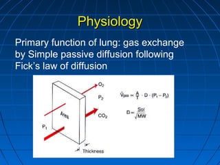
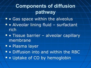
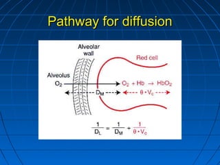
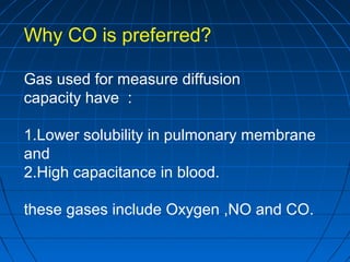
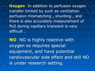
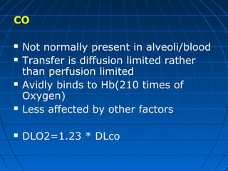
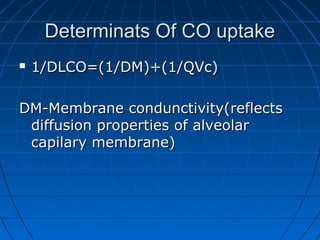
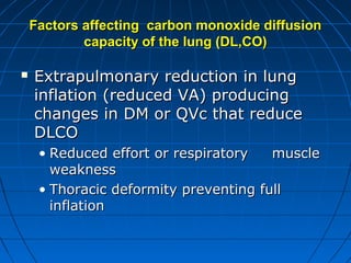
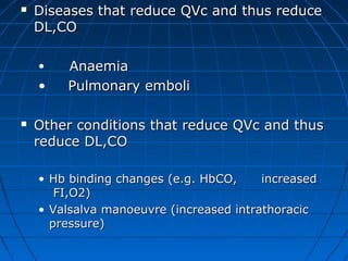
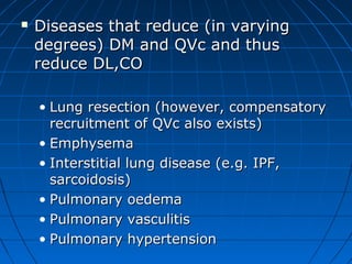
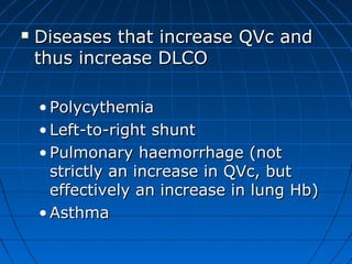
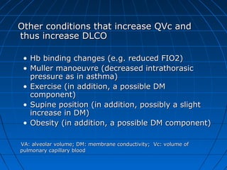
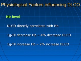
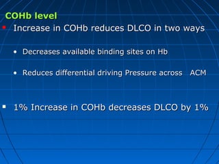
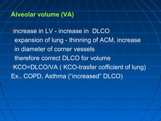
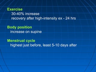
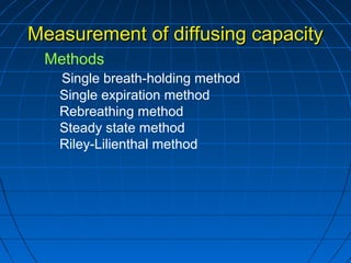
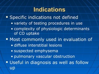
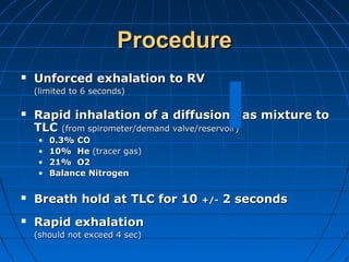
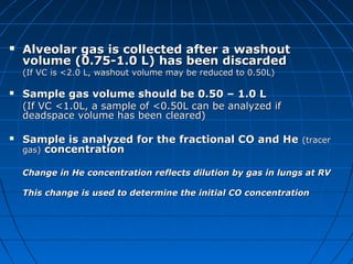
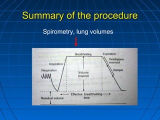
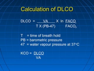
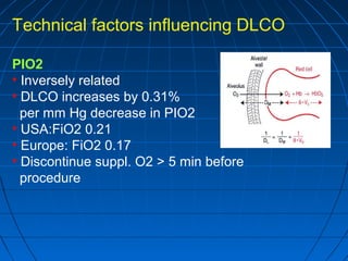
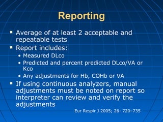
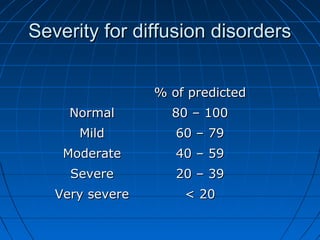
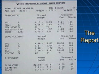
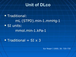
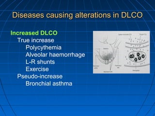
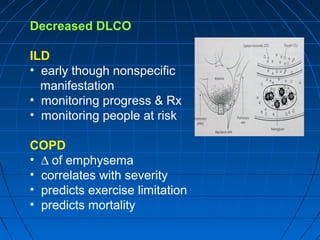
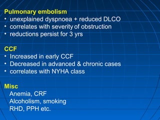
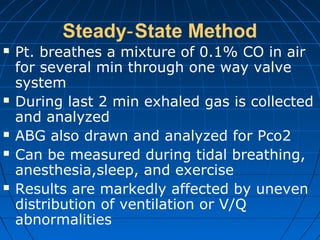
4. TerminologyTerminology
•North America: “Diffusing capacity”historical
term
•Europe: “Transfer factor” beacause
measurment of CO uptake reflects a no. of
process(not just diffusion ) and its submaximal
value and thus,not truly a capacity.
5. Components of diffusion
pathway
• Gas space within the alveolus
• Alveolar lining fluid – surfactant
rich
• Tissue barrier – alveolar capillary
membrane
• Plasma layer
• Diffusion into and within the RBC
• Uptake of CO by hemoglobin
7. Why CO is preferred?
Gas used for measure diffusion
capacity have :
1.Lower solubility in pulmonary membrane
and
2.High capacitance in blood.
these gases include Oxygen ,NO and CO.
8. • Oxygen In addition to perfusion oxygen
transfer limited by such as ventilation
perfusion mismatching , shunting , and
there is also accurately measurement of
Po2 during capillary transient is very
difficult .
• NO NO is highly reactive with
oxygen so requires special
equipment, and have potential
cardiovascular side effect and still NO
is under research setting.
9. CO
Not normally present in alveoli/blood
Transfer is diffusion limited rather
than perfusion limited
Avidly binds to Hb(210 times of
Oxygen)
Less affected by other factors
DLO2=1.23 * DLco
10. Determinats Of CO uptakeDeterminats Of CO uptake
1/DLCO=(1/DM)+(1/QVc)1/DLCO=(1/DM)+(1/QVc)
DM-Membrane condunctivity(reflectsDM-Membrane condunctivity(reflects
diffusion properties of alveolardiffusion properties of alveolar
capilary membrane)capilary membrane)
11. Factors affecting carbon monoxide diffusionFactors affecting carbon monoxide diffusion
capacity of the lung (DL,CO)capacity of the lung (DL,CO)
Extrapulmonary reduction in lungExtrapulmonary reduction in lung
inflation (reduced VA) producinginflation (reduced VA) producing
changes in DM or QVc that reducechanges in DM or QVc that reduce
DLCODLCO
• Reduced effort or respiratory muscleReduced effort or respiratory muscle
weaknessweakness
• Thoracic deformity preventing fullThoracic deformity preventing full
inflationinflation
12. Diseases that reduce QVc and thus reduceDiseases that reduce QVc and thus reduce
DL,CODL,CO
• AnaemiaAnaemia
• Pulmonary emboliPulmonary emboli
Other conditions that reduce QVc and thusOther conditions that reduce QVc and thus
reduce DL,COreduce DL,CO
• Hb binding changes (e.g. HbCO, increasedHb binding changes (e.g. HbCO, increased
FI,O2)FI,O2)
• Valsalva manoeuvre (increased intrathoracicValsalva manoeuvre (increased intrathoracic
pressure)pressure)
13. Diseases that reduce (in varyingDiseases that reduce (in varying
degrees) DM and QVc and thusdegrees) DM and QVc and thus
reduce DL,COreduce DL,CO
• Lung resection (however, compensatoryLung resection (however, compensatory
recruitment of QVc also exists)recruitment of QVc also exists)
• EmphysemaEmphysema
• Interstitial lung disease (e.g. IPF,Interstitial lung disease (e.g. IPF,
sarcoidosis)sarcoidosis)
• Pulmonary oedemaPulmonary oedema
• Pulmonary vasculitisPulmonary vasculitis
• Pulmonary hypertensionPulmonary hypertension
14. Diseases that increase QVc andDiseases that increase QVc and
thus increase DLCOthus increase DLCO
• PolycythemiaPolycythemia
• Left-to-right shuntLeft-to-right shunt
• Pulmonary haemorrhage (notPulmonary haemorrhage (not
strictly an increase in QVc, butstrictly an increase in QVc, but
effectively an increase in lung Hb)effectively an increase in lung Hb)
• AsthmaAsthma
15. Other conditions that increase QVc andOther conditions that increase QVc and
thus increase DLCOthus increase DLCO
• Hb binding changes (e.g. reduced FIO2)Hb binding changes (e.g. reduced FIO2)
• Muller manoeuvre (decreased intrathorasicMuller manoeuvre (decreased intrathorasic
pressure as in asthma)pressure as in asthma)
• Exercise (in addition, a possible DMExercise (in addition, a possible DM
component)component)
• Supine position (in addition, possibly a slightSupine position (in addition, possibly a slight
increase in DM)increase in DM)
• Obesity (in addition, a possible DM component)Obesity (in addition, a possible DM component)
VA: alveolar volume; DM: membrane conductivity; Vc: volume ofVA: alveolar volume; DM: membrane conductivity; Vc: volume of
pulmonary capillary bloodpulmonary capillary blood
17. COHb levelCOHb level
Increase in COHb reduces DLCO in two waysIncrease in COHb reduces DLCO in two ways
• Decreases available binding sites on HbDecreases available binding sites on Hb
• Reduces differential driving Pressure across ACMReduces differential driving Pressure across ACM
1% Increase in COHb decreases DLCO by 1%1% Increase in COHb decreases DLCO by 1%
18. Alveolar volume (VA)
•increase in LV - increase in DLCO
expansion of lung - thinning of ACM, increase
in diameter of corner vessels
therefore correct DLCO for volume
•KCO=DLCO/VA ( KCO-trasfer cofficient of lung)
Ex.. COPD, Asthma (“increased” DLCO)
19. Circadian rhythm
DLCO drops 1-2%/hr between 9.30am-9.30pm
Gender & Ethnicity
lower in women for a given height
lower in African-Americans, Asians
Body size: DLco varies with BSA
better predicted by height & weight
2
DLcO=BSA(M) (18.84) -6.8
Age: DLco declines in linear fashion with
20. Exercise
30-40% increase
recovery after high-intensity ex - 24 hrs
Body position
increase on supine
Menstrual cycle
highest just before, least 5-10 days after
21. Measurement of diffusing capacityMeasurement of diffusing capacity
Methods
Single breath-holding method
Single expiration method
Rebreathing method
Steady state method
Riley-Lilienthal method
22. Indications
Specific indications not defined
• variety of testing procedures in use
• complexity of physiologic determinants
of CO uptake
Most commonly used in evaluation of
• diffuse interstitial lesions
• suspected emphysema
• pulmonary vascular obstruction
Useful in diagnosis as well as follow
up
25. ProcedureProcedure
Unforced exhalation to RVUnforced exhalation to RV
(limited to 6 seconds)(limited to 6 seconds)
Rapid inhalation of a diffusion gas mixture toRapid inhalation of a diffusion gas mixture to
TLCTLC (from spirometer/demand valve/reservoir)(from spirometer/demand valve/reservoir)
• 0.3% CO0.3% CO
• 10% He10% He (tracer gas)(tracer gas)
• 21% O221% O2
• Balance NitrogenBalance Nitrogen
Breath hold at TLC for 10Breath hold at TLC for 10 +/-+/- 2 seconds2 seconds
Rapid exhalationRapid exhalation
(should not exceed 4 sec)(should not exceed 4 sec)
26. Alveolar gas is collected after a washoutAlveolar gas is collected after a washout
volume (0.75-1.0 L) has been discardedvolume (0.75-1.0 L) has been discarded
(If VC is <2.0 L, washout volume may be reduced to 0.50L)(If VC is <2.0 L, washout volume may be reduced to 0.50L)
Sample gas volume should be 0.50 – 1.0 LSample gas volume should be 0.50 – 1.0 L
(If VC <1.0L, a sample of <0.50L can be analyzed if(If VC <1.0L, a sample of <0.50L can be analyzed if
deadspace volume has been cleared)deadspace volume has been cleared)
Sample is analyzed for the fractional CO and HeSample is analyzed for the fractional CO and He (tracer(tracer
gas)gas) concentrationconcentration
Change in He concentration reflects dilution by gas in lungs at RVChange in He concentration reflects dilution by gas in lungs at RV
This change is used to determine the initial CO concentrationThis change is used to determine the initial CO concentration
27. Summary of the procedureSummary of the procedure
Spirometry, lung volumes
28. Acceptability CriteriaAcceptability Criteria
Volume-Time tracing should show smooth,Volume-Time tracing should show smooth,
rapid inspirationrapid inspiration (<4 sec) from RV to TLC(<4 sec) from RV to TLC
Expiration should be rapidExpiration should be rapid but not forced; 4but not forced; 4
seconds or lessseconds or less
Dead space washout should be 0.75 – 1.00 LDead space washout should be 0.75 – 1.00 L
(0.5 L if VC is less than 2.0 L)(0.5 L if VC is less than 2.0 L)
Alveolar sample volume should be 0.5 to 1.0 LAlveolar sample volume should be 0.5 to 1.0 L
29. Inspired volume should be at leastInspired volume should be at least 85%85% ofof
previously recorded best VCpreviously recorded best VC
Breath hold time should 10 sec +/- 2 sec (NoBreath hold time should 10 sec +/- 2 sec (No
Valsalva or Mueller maneuver)Valsalva or Mueller maneuver)
Expiration in <4 s (and sample collection timeExpiration in <4 s (and sample collection time
<3s)#, with appropriate clearance of VD and<3s)#, with appropriate clearance of VD and
proper sampling/analysis of alveolar gasproper sampling/analysis of alveolar gas
The average of two or more acceptable testThe average of two or more acceptable test
should be reported. Duplicate determinationsshould be reported. Duplicate determinations
should be within 10% of highest value or 3 mlshould be within 10% of highest value or 3 ml
CO/min/mm HgCO/min/mm Hg
30. Repeatability and Number of
Tests
Obtain at least 2 acceptable tests
Repeatability requirement – 2 acceptable
tests
• within 3 units, OR
• 10 % of the highest value
Report the average of 2 acceptable tests
that meet repeatability requirement
More than 5 tests are not recommended
Eur Respir J 2005; 26: 720–735
31. • Average DLAverage DLcocosb valuesb value
25 ml CO/min/mm Hg (STPD)25 ml CO/min/mm Hg (STPD)
32. Inspiratory maneuver
14%He, 18%O2, 0.27%CO)
breathhold
Deadspace washout(0.75
L)
If VC<2L, reduce to 0.5L
Sample collection
volume
0.5-1LIf VC<2L, reduce
to 0.5L
33. AdvantagesAdvantages
No invasive measuring proceduresNo invasive measuring procedures
Analysis of only two gases is requiredAnalysis of only two gases is required
Test is easily and rapidly performedTest is easily and rapidly performed
34. DisadvantagesDisadvantages
Difficult breathing maneuverDifficult breathing maneuver
Not practical during exercise testingNot practical during exercise testing
Less than maximal inspired VCLess than maximal inspired VC
volumes affect measurementvolumes affect measurement
accuracyaccuracy
V/Q mismatches can affect theV/Q mismatches can affect the
resultsresults
35. Calculation of DLCOCalculation of DLCO
DLCO = VA X ln FACOi
T X (PB-47) FACOF
T = time of breath hold
PB = barometric pressure
47 = water vapour pressure at 37o
C
KCO = DLCO
VA
36. Technical factors influencing DLCO
PIO2
• Inversely related
• DLCO increases by 0.31%
per mm Hg decrease in PIO2
• USA:FiO2 0.21
• Europe: FiO2 0.17
• Discontinue suppl. O2 > 5 min before
procedure
37. Other Technical variables
•Inspired volume
•Duration and condition of breath hold
•Deadspace washout volume
•Method of gas analysis
•Method of measuring VA
38. Equipment quality controlEquipment quality control
Gas-analyser zeroing DoneGas-analyser zeroing Done
before/after each testbefore/after each test
Volume accuracy Tested dailyVolume accuracy Tested daily
Standard subject or simulator testingStandard subject or simulator testing
Tested at least weeklyTested at least weekly
Gas-analyser linearity Tested every 3Gas-analyser linearity Tested every 3
monthsmonths
Timer Tested every 3 monthsTimer Tested every 3 months
39. Reporting
Average of at least 2 acceptable and
repeatable tests
Report includes:
• Measured DLco
• Predicted and percent predicted DLco/VA or
Kco
• Any adjustments for Hb, COHb or VA
If using continuous analyzers, manual
adjustments must be noted on report so
interpreter can review and verify the
adjustments
Eur Respir J 2005; 26: 720–735
40. Severity for diffusion disordersSeverity for diffusion disorders
% of predicted% of predicted
NormalNormal 80 – 10080 – 100
MildMild 60 – 7960 – 79
ModerateModerate 40 – 5940 – 59
SevereSevere 20 – 3920 – 39
Very severeVery severe < 20< 20
44. Decreased DLCO
ILD
• early though nonspecific
manifestation
• monitoring progress & Rx
• monitoring people at risk
COPD
• ∆ of emphysema
• correlates with severity
• predicts exercise limitation
• predicts mortality
45. Pulmonary embolism
• unexplained dyspnoea + reduced DLCO
• correlates with severity of obstruction
• reductions persist for 3 yrs
CCF
• Increased in early CCF
• Decreased in advanced & chronic cases
• correlates with NYHA class
Misc
Anemia, CRF
Alcoholism, smoking
RHD, PPH etc.
46. Steady State Method‐
Pt. breathes a mixture of 0.1% CO in air
for several min through one way valve
system
During last 2 min exhaled gas is collected
and analyzed
ABG also drawn and analyzed for Pco2
Can be measured during tidal breathing,
anesthesia,sleep, and exercise
Results are markedly affected by uneven
distribution of ventilation or V/Q
abnormalities
47. AdvantagesAdvantages
• Natural breathing maneuverNatural breathing maneuver
• Allow greater variety of clinicalAllow greater variety of clinical
conditionsconditions
DisadvantagesDisadvantages
• More complex and difficult to performMore complex and difficult to perform
(PACO)(PACO)
• PPCCCO back pressureCO back pressure
• More affected by V/Q abnormalitiesMore affected by V/Q abnormalities
48. Rebreathing Method
Pt rebreathes the test gas from reservoir,
the volume of which equals pt’s FEV1
Rebreathing continues for 30 45 s, at‐
controlled rate of 30 per min
More variable
Requires considerable patient cooperation
to attain rapid respiratory rate required
49. AdvantageAdvantage
Least affected byLeast affected by
• V / Q abnormalitiesV / Q abnormalities
• Changes in the subject’s lung volume at theChanges in the subject’s lung volume at the
time of measurementtime of measurement
50. DisadvantagesDisadvantages
Complexity of the instrumentationComplexity of the instrumentation
and equations requiredand equations required
Affected by PAffected by PCCCO buildupCO buildup
Need for subject cooperation withNeed for subject cooperation with
breathingbreathing
PPCCCO back pressureCO back pressure
51. Interpretation
• Relationship between DLCO and lung volume is
not linear, so DLco/VA or DLco/TLC do not
provide an appropriate way to normalize DLco
for lung volume
• Conceptually, low DLco but high DLco/VA:
extraparenchymal abnormality (e.g.
pneumonectomy or chest wall restriction)
• Low DLco and low DLco/VA: parenchymal abnorm