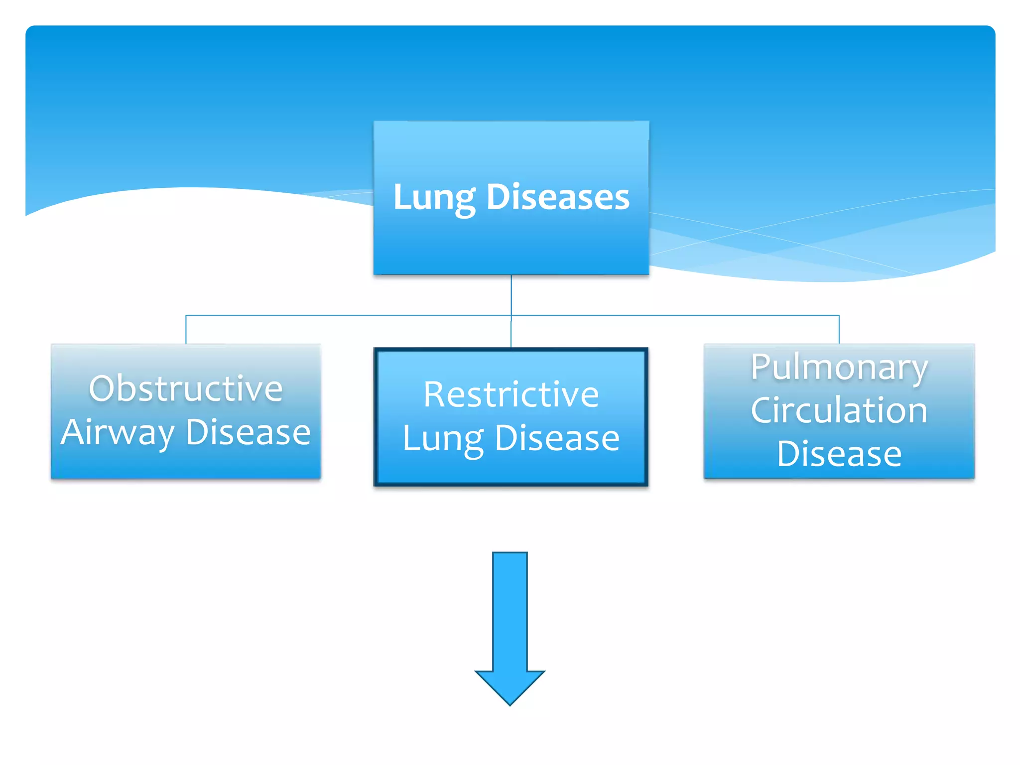
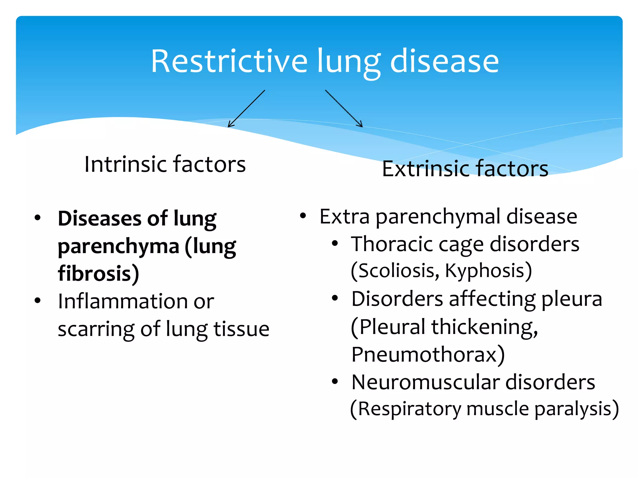
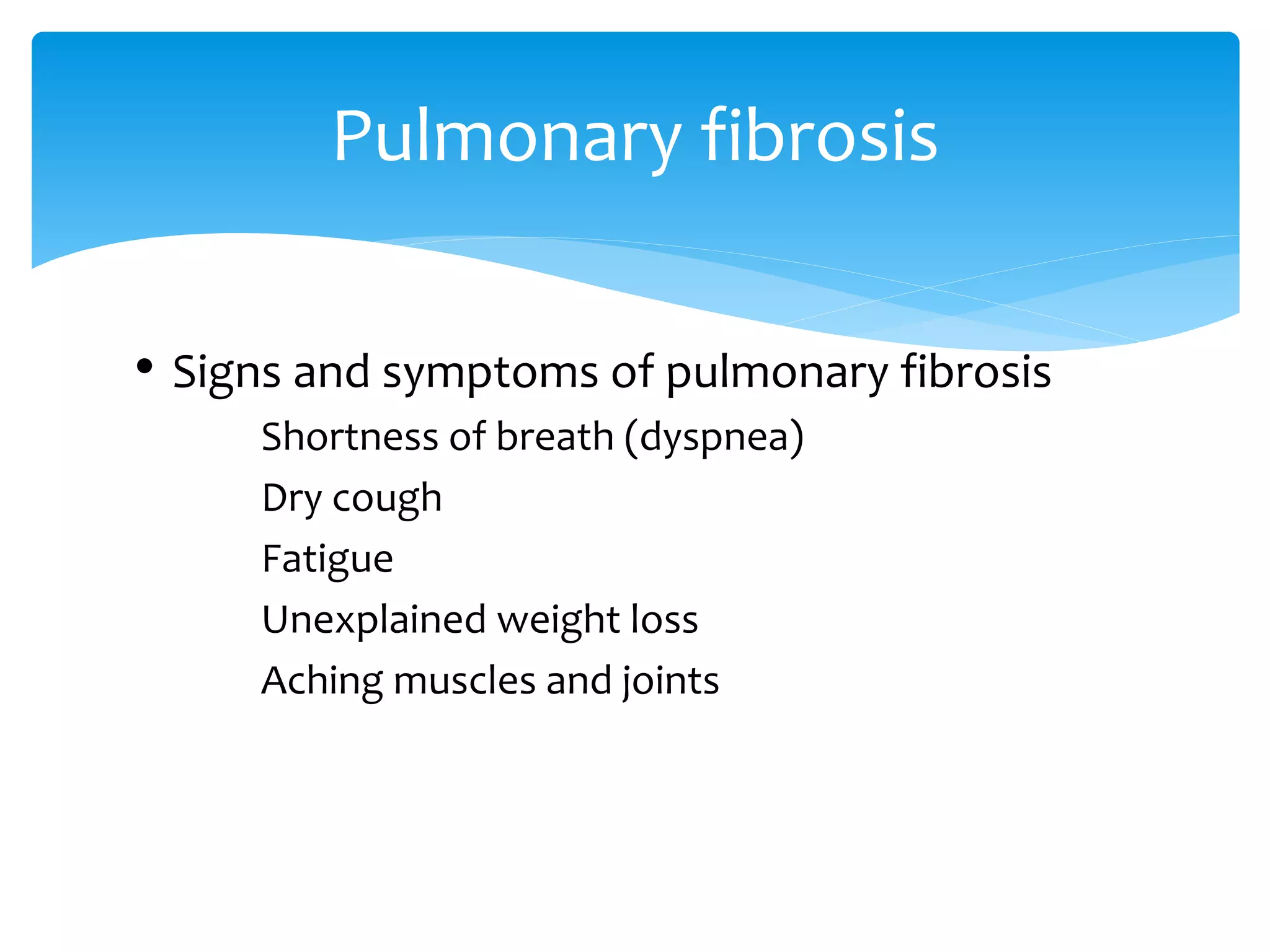
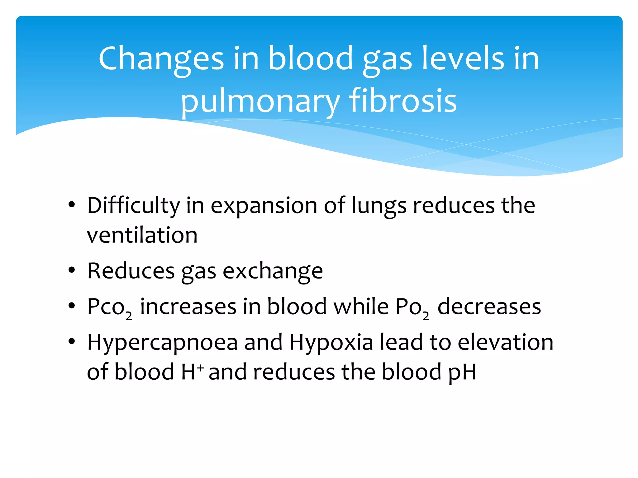
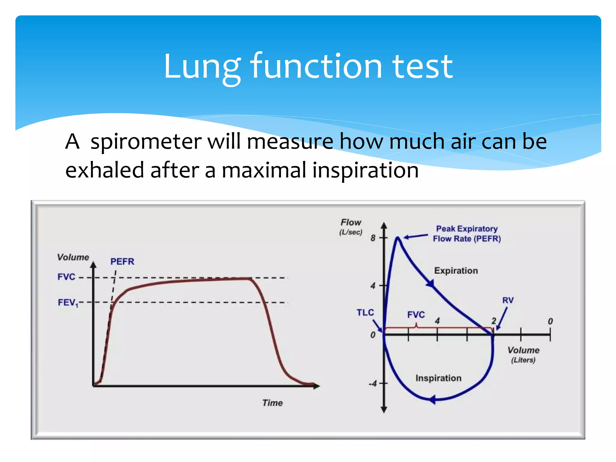
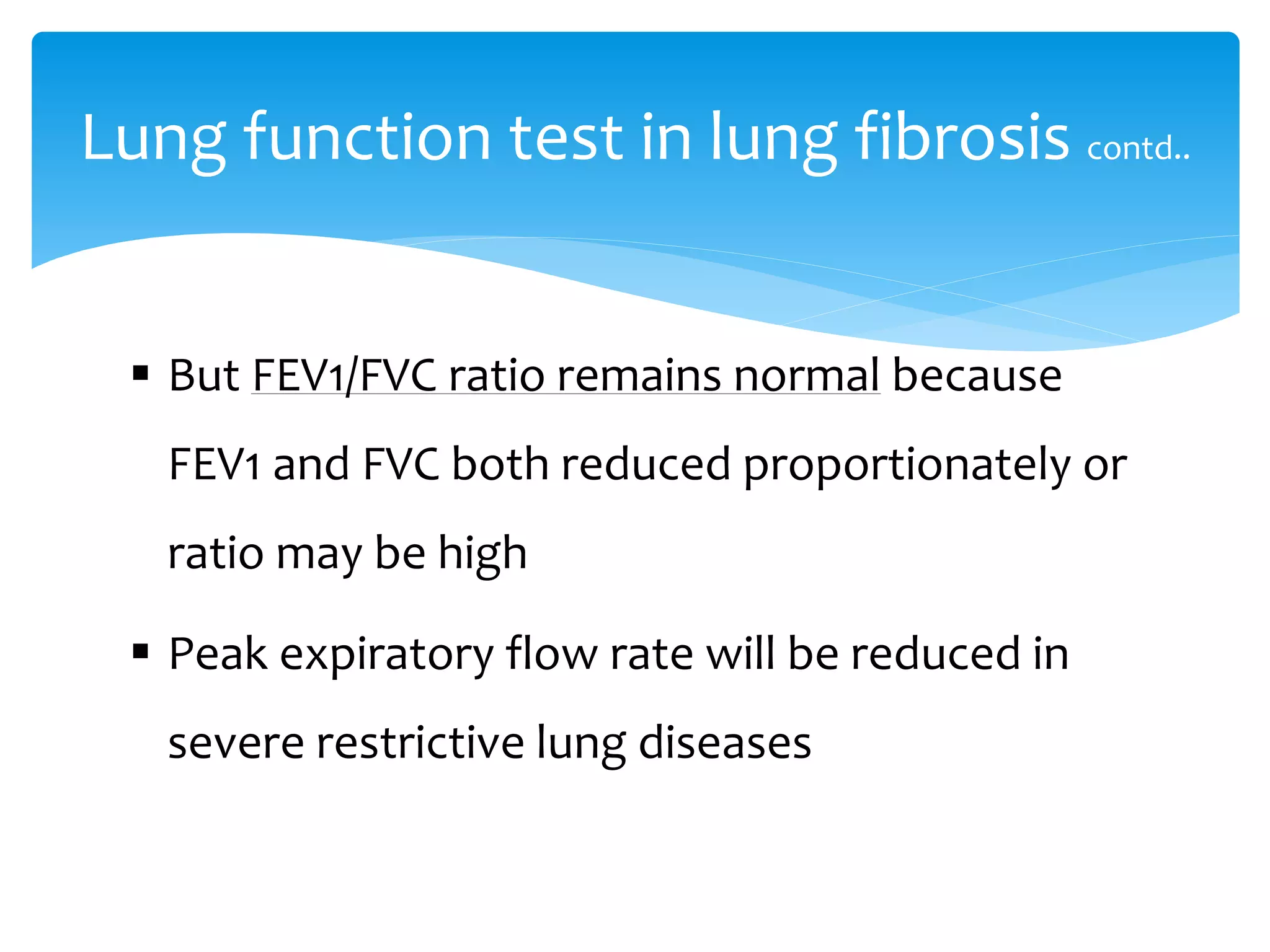
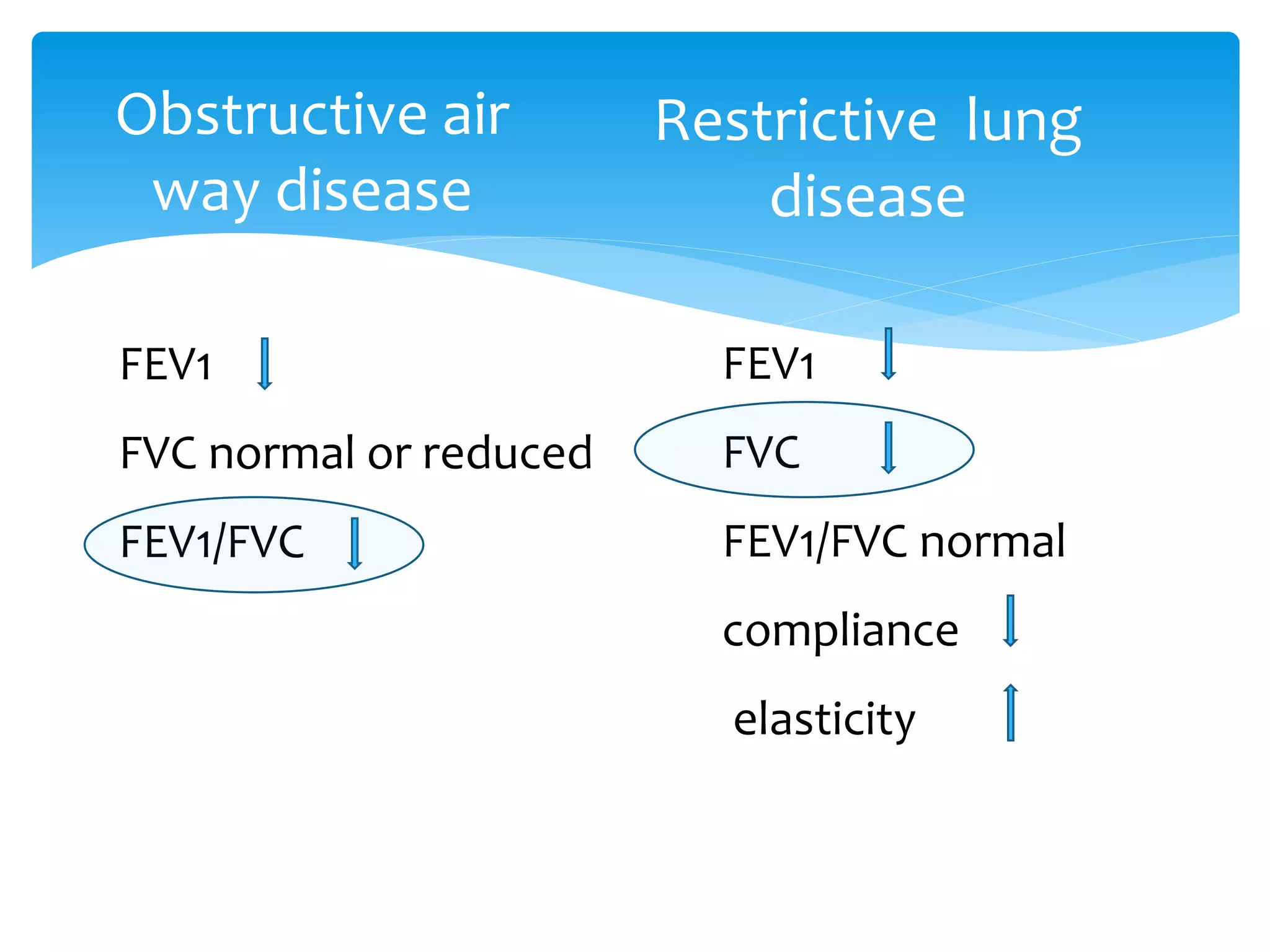
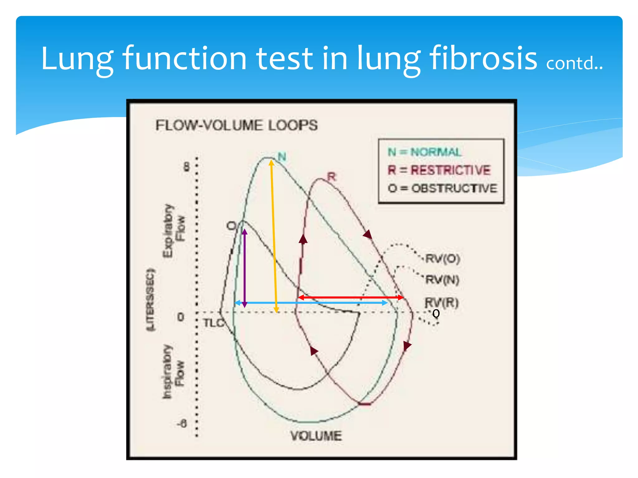
Small group presentation which was done during our physiology days under the guidance of Prof. Sampath Gunawardena senior lecturer in department of Physiology, Faculty of Medicine University of Ruhuna.
This document discusses restrictive lung disease, which is defined by reduced lung volumes. It can be caused by intrinsic lung diseases that affect the lung parenchyma through inflammation or scarring, or extrinsic disorders of the chest wall, pleura, or respiratory muscles. Common intrinsic lung diseases include idiopathic pulmonary fibrosis (IPF), sarcoidosis, hypersensitivity pneumonitis, and interstitial lung disease caused by drugs. Extrinsic disorders involve diseases of the pleura, chest wall, or neuromuscular system. Restrictive lung disease results in hypoxemia, reduced diffusion capacity, and impaired gas exchange. Evaluation involves pulmonary function tests and imaging, and treatment depends on the underlying cause.
Restrictive lung diseases can be caused by chest wall disorders like kyphoscoliosis or interstitial lung diseases such as pneumoconiosis from inhaling inorganic dusts. Pneumoconiosis includes conditions like coal worker's pneumoconiosis from coal dust inhalation, silicosis from silica exposure in occupations like mining, and asbestosis from asbestos exposure. These diseases are characterized by nodular scarring in the lungs visible on x-ray. Long-term inhalation of very small dust particles can lead to fibrotic nodule formation and restricted lung function over time. Silicosis presents as small fibrotic nodules throughout the lungs and is associated with occupations involving silica exposure
1. The document discusses several obstructive lung diseases including emphysema, chronic bronchitis, asthma, and bronchiectasis.
2. Emphysema is characterized by destruction of alveolar walls without significant fibrosis leading to enlarged air spaces. Chronic bronchitis involves thickening of bronchial walls and excess mucus production.
3. Asthma is a chronic inflammatory disease involving recurrent airway obstruction, inflammation, and hyperresponsiveness triggered by various stimuli. Bronchiectasis permanently dilates the bronchi and bronchioles due to destruction of muscles and tissues often caused by infection or obstruction.
This document discusses aerosol therapy and nebulizers. It defines aerosols and outlines factors that influence aerosol deposition in the lungs such as particle size, respiratory anatomy, and breathing patterns. It describes different aerosol delivery devices including metered dose inhalers, dry powder inhalers, and nebulizers. The document focuses on nebulizers, outlining their types (jet and ultrasonic), workings, indications, drugs used, and proper technique for administration. Nebulizers are indicated when precise dosing is needed for critically ill, young, elderly or handicapped patients. Proper technique and positioning can optimize drug deposition in the lungs.
Chronic bronchitis is defined as a productive cough that lasts for three months or more per year for at least two years. Tobacco smoking is the primary cause and leads to chronic obstructive pulmonary disease in most cases. Symptoms include a chronic cough with mucus, wheezing, and shortness of breath. Treatment focuses on quitting smoking, using inhaled bronchodilators and steroids, and addressing exacerbating factors through medications, oxygen therapy, or lung transplantation in severe cases.
Pulmonary rehabilitation is a comprehensive intervention based on a thorough patient assessment followed by patient tailored therapies that include, but are not limited to, exercise training, education, and behavior change, designed to improve the physical and psychological condition of people with chronic respiratory disease and to promote the long-term adherence to health-enhancing behaviors”
The document provides information about exercise tolerance tests. Key points include:
1) Exercise tolerance tests evaluate the cardiovascular system's response to exercise under controlled conditions and can detect issues like coronary artery disease.
2) The tests have several purposes like detecting coronary artery disease, evaluating physical capacity, and assessing response to medical interventions.
3) There are different types of exercises used in the tests, including isometric, dynamic, and combinations of the two. Dynamic exercise is considered most appropriate for evaluating cardiovascular response.
A thoracotomy is a surgical procedure where an incision is made through the chest wall to access the organs within the chest cavity like the lungs and heart. It is performed by a thoracic surgeon or emergency physician in a hospital operating room or emergency department. Reasons for thoracotomy include diagnosing and treating conditions like lung cancer or injuries from the chest. Risks include bleeding, infection and pain during a long recovery period.
Humidity therapy adds moisture to air delivered to patients and is used to overcome humidity deficits when the upper airway is bypassed or for humidifying dry medical gases. It helps maintain normal humidity levels in the airways and can assist in managing conditions like thick secretions or hypothermia. Various humidifiers actively add heat or water to air or passively recycle exhaled heat and moisture, with different types suited to invasive or non-invasive ventilation.
This document describes the procedure of pneumonectomy and the role of physiotherapy both before and after the surgery. Pneumonectomy involves complete removal of a lung, usually done to treat lung cancer, infections, or other lung diseases. Physiotherapy before surgery focuses on teaching exercises and breathing techniques to prepare the patient. After surgery, physiotherapy aims to clear secretions, expand the remaining lung, prevent complications, and restore movement and exercise tolerance through a gradual recovery program over 2-3 weeks before discharge.
Pulmonary function tests (PFTs) measure how well the lungs work by assessing lung volumes, air flow, and gas exchange. PFTs can help diagnose various respiratory diseases by detecting abnormalities. The tests involve spirometry to measure volumes of air inhaled and exhaled during normal and forced breathing maneuvers. Obstructive diseases reduce air flow, shown by a decreased FEV1 and FEV1/FVC ratio. Restrictive diseases limit lung expansion, shown by a reduced FVC but normal or increased FEV1/FVC ratio. PFTs are useful for evaluating respiratory function and monitoring treatment effects.
This document discusses various chest wall deformities and conditions that affect the structure and function of the chest wall. It covers scoliosis, pectus excavatum, ankylosing spondylitis, flail chest, thoracoplasty, pectus carinatum, Poland Syndrome, and chest wall tumors. For each condition, it describes the presentation, pathophysiology, clinical features, treatment options, and relevant anatomy and imaging findings.
physiotherapy management for chronic obstructive pulmonary disease Sunil kumar
role of physiotherapy in chronic obstructive pulmonary disease, principles of physical therapy management in copd, physiotherapy assessing and treatment for copd
Chronic obstructive pulmonary disease (COPD) is defined as a lung condition characterized by airflow limitation that is not fully reversible. The document discusses COPD, including its types (chronic bronchitis and emphysema), pathology, evaluation, diagnosis, and physiotherapy management. Physiotherapy management focuses on removing secretions, improving breathing patterns, increasing exercise tolerance, and preventing dyspnea through techniques like breathing exercises, postural drainage, inspiratory muscle training, and ergonomic advice.
This document summarizes the role of physiotherapy in treating respiratory conditions. It discusses treatments to increase ventilation and oxygenation such as breathing exercises and positioning techniques. It also covers treatments to improve secretion clearance like postural drainage and cough techniques. Physiotherapy aims to improve patients' exercise tolerance through endurance training and uses electrotherapy to reduce pain.
Obstructive vs. Restrictive Lung diseaseFatima Awadh
This document discusses the differences between restrictive and obstructive lung diseases. Restrictive lung diseases are characterized by a reduction in total lung capacity below 80% of predicted value, while obstructive lung diseases are characterized by a reduction in airflow, seen through a decreased forced expiratory volume in 1 second and ratio of forced expiratory volume to forced vital capacity. Key lung volumes and capacities such as tidal volume, inspiratory reserve volume, and residual volume are also defined.
Bronchial hygiene therapy involves noninvasive techniques to clear secretions and improve lung function. It includes techniques like positioning, coughing, breathing exercises, and chest manipulation. The goals are to prevent accumulation and promote removal of secretions to improve respiratory status. Indications are excessive sputum production and ineffective cough. Contraindications include conditions that increase risk of aspiration or compromise hemodynamics.
Pleurodesis is a procedure to induce adhesion of the pleural layers to treat recurrent pneumothorax or malignant pleural effusion. It involves using sclerosing agents or surgical abrasion. Talc, tetracycline derivatives like doxycycline, and minocycline are common sclerosing agents used. The procedure involves draining the pleural fluid then injecting the sclerosing agent through a chest tube while the lung is expanded to cause an inflammatory response and formation of fibrous adhesions between the pleural layers.
Coughing is a normal protective mechanism that acts to clear secretions and foreign materials from the tracheobronchial tree. It is initiated by irritation of the sensitive bronchi and trachea, and can occur voluntarily or reflexively. Coughing involves both afferent and efferent pathways - afferent pathways include sensory nerves, while efferent pathways involve the recurrent laryngeal nerve and spinal nerves. The coughing mechanism involves inspiration of air, followed by forced contraction of abdominal and intercostal muscles to increase lung pressure and expel air rapidly through the vocal cords to shear and remove mucus or foreign materials from the airways.
This document provides information on auscultating breath sounds and other body sounds. It describes how to properly auscultate breath sounds by placing the stethoscope on landmarks of the chest wall. Normal breath sounds are described as well as adventitious sounds such as crackles, wheezes, and rhonchi. The causes and characteristics of different breath sound types are explained. Proper cardiac auscultation is also briefly mentioned.
Pectus carinatum, also known as pigeon chest, is a rare chest wall malformation where the sternum and ribs protrude abnormally. It develops during childhood and worsens during puberty, causing symptoms like shortness of breath, chest pain, and asthma. It can be caused by genetic factors or abnormalities in cartilage development. Diagnosis involves x-rays, CT scans, or MRI. Treatment options include bracing or Ravitch surgery to remove abnormal cartilage and place the breastbone in a normal position.
1. Interstitial lung diseases (ILDs) involve the lung parenchyma including the alveoli, capillaries, and spaces between.
2. ILDs are classified based on known causes, idiopathic forms, and granulomatous types. Idiopathic pulmonary fibrosis is the most common idiopathic form.
3. Clinical presentation involves breathlessness, cough, and reduced lung function. Investigations include chest imaging showing infiltrates and fibrosis, and lung biopsies to determine classification. Treatment focuses on removing exposures, suppressing inflammation, and palliating symptoms primarily using corticosteroids.
This document summarizes several types of chest wall deformities. It describes pectus excavatum, or funnel chest, which is characterized by a depression of the sternum. It affects about 1 in 400 births and is more common in boys. Pectus carinatum, or pigeon chest, involves a protrusion of the sternum. The document also discusses Poland's syndrome, cleft sternum, ectopia cordis (where the heart is located outside the chest), and thoracic deformities seen in diffuse skeletal disorders like Jeune's syndrome and Jarcho-Levin syndrome. It provides details on symptoms, causes, evaluation, and surgical repair options for each condition.
The 6-minute walk test (6MWT) is an easy to perform and practical test that has been used in the assessment of patients with a variety of cardiopulmonary diseases including pulmonary arterial hypertension (PAH). It simply measures the distance that a patient can walk on a flat, hard surface in a period of 6 minutes.
Physiotherapy management of chronic obstructive pulmonary disease ppt by Oluw...OluwadamilareAkinwan
This document presents an overview of physiotherapy management for chronic obstructive pulmonary disease (COPD). It discusses the epidemiology, pathophysiology, clinical features, diagnosis, stages, and medical management of COPD. It then describes the role of physiotherapy during acute exacerbations, including techniques to reduce work of breathing and secretion removal. Physiotherapy is also involved in pulmonary rehabilitation to improve patient function and management through exercise training and education. Physiotherapy aims to prevent exacerbations and optimize lung function in stable COPD patients.
Acute respiratory distress syndrome (ARDS) is a sudden and progressive form of acute respiratory failure in which the alveolar capillary membrane becomes damaged and more permeable to intravascular fluid resulting in severe dyspnoea, hypoxemia and diffuse pulmonary infiltrates.
This document defines COPD and discusses its epidemiology, risk factors, pathology, clinical features, investigations, management, and treatment. COPD is a common lung disease characterized by persistent airflow limitation associated with an enhanced inflammatory response in the airways. It is usually caused by significant exposure to noxious particles or gases, most commonly from cigarette smoke. Management involves smoking cessation, bronchodilators, inhaled corticosteroids, oxygen therapy, vaccines, and other drugs to reduce symptoms and exacerbations.
This document defines COPD and discusses its epidemiology, risk factors, pathology, clinical features, investigations, management, and treatment. COPD is a common lung disease characterized by persistent airflow limitation associated with an enhanced inflammatory response in the airways. It is usually caused by significant exposure to noxious particles or gases, most commonly from cigarette smoke. Management involves smoking cessation, bronchodilators, inhaled corticosteroids, oxygen therapy, vaccines, and addressing exacerbations.
This document provides an overview of chronic obstructive pulmonary disease (COPD). It defines COPD as a preventable and treatable disease characterized by airflow limitation caused by cigarette smoking and exposure to other noxious particles. The document outlines the risk factors, pathogenesis, pathophysiology, symptoms, diagnosis, classification, and management of COPD. Spirometry is a key test for COPD diagnosis and assessment. The goals of treatment are to prevent progression, relieve symptoms, improve exercise tolerance, and prevent exacerbations.
This document provides an overview of interstitial lung disease (ILD). ILD encompasses over 200 lung disorders that involve scarring or damage to the lungs' interstitium. Progressive fibrosis can occur in some ILDs and is associated with worse outcomes. Idiopathic pulmonary fibrosis is the most common progressive ILD and is characterized by lung scarring. Progressive-fibrosing ILD describes patients with fibrotic ILDs that may deteriorate despite treatment. Diagnosis involves evaluating symptoms, imaging, pulmonary function tests, biopsies and labs to identify the specific ILD and develop a treatment plan which may include immunosuppressants or removing environmental exposures.
This document discusses atelectasis, which is the collapse or closure of alveoli in the lungs. It defines atelectasis and reviews its causes, types, symptoms, diagnosis, and treatment. Atelectasis can be obstructive or non-obstructive, acute or chronic. Risk factors include smoking and general anesthesia. Diagnosis involves chest x-ray, pulse oximetry, and arterial blood gas analysis. Treatment focuses on treating the underlying cause, chest physiotherapy, bronchodilators, surgery if needed, and preventing complications like pneumonia. Nursing care involves airway clearance techniques and strategies to improve ventilation and gas exchange.
Respiratory Complication Of Rheumatic Diseasedrmomusa
This document discusses respiratory complications of rheumatic diseases. It covers causes of diffuse parenchymal lung disease including infections, drugs, and connective tissue diseases. Clinical evaluation involves assessing symptoms, signs, imaging like HRCT, lung function tests, and biopsies. Specific lung manifestations are discussed for diseases like rheumatoid arthritis, SLE, and scleroderma. Drugs that can cause interstitial lung disease or other pulmonary complications are also outlined. Future areas of research are mentioned.
This document provides information on interstitial lung disease from a seminar presented by Ms. Saheli Chakraborty. It defines interstitial lung disease as progressive scarring of lung tissue. It discusses the objectives, introduction, definition, etiology, risk factors, pathophysiology, clinical manifestations, diagnostic evaluations, complications, management, nursing management, nursing diagnoses and common types of interstitial lung diseases including sarcoidosis, idiopathic pulmonary fibrosis, interstitial pneumonia, asbestosis and acute interstitial pneumonitis.
This document discusses interstitial lung diseases (ILD), also known as diffuse parenchymal lung diseases (DPLD). It provides the following key points:
1. ILD can be caused by over 200 diseases that result in damage to the lung interstitium. Common causes include occupational exposures, collagen vascular diseases, drugs, infections, and idiopathic interstitial pneumonias.
2. Accurately diagnosing ILD requires a multidisciplinary approach including clinical evaluation, radiology such as high-resolution CT, and pathology including surgical lung biopsy.
3. Idiopathic pulmonary fibrosis (IPF) is the most common idiopathic interstitial pneumonia and
Chronic obstructive pulmonary disease (COPD) is a chronic inflammatory lung disease that causes obstructed airflow from the lungs. Symptoms include breathing difficulty, cough, mucus (sputum) production and wheezing.15/04/2020
Acute respiratory failure can be caused by either problems with oxygenation (Type I) or ventilation (Type II). Type I is characterized by a ventilation-perfusion (V/Q) mismatch and includes conditions like ARDS, pulmonary edema, and pneumonia. Type II involves decreased respiratory drive or respiratory muscle weakness and includes neuromuscular disorders or COPD. ARDS is diagnosed using a P/F ratio below 150 and involves three pathological phases over weeks - exudative, proliferative, and fibrotic. Prone positioning improves outcomes in ARDS by redistributing ventilation and perfusion as well as reducing lung compression.
Chronic obstructive pulmonary disease (COPD) refers to a group of lung diseases including chronic bronchitis and emphysema that are characterized by persistent airflow limitation. The main causes of COPD are tobacco smoking, exposure to secondhand smoke, and air pollution. Symptoms include cough, sputum production, and shortness of breath. Diagnosis involves assessing symptoms, lung function tests, and chest imaging. Treatment focuses on smoking cessation, medications to relieve symptoms and prevent exacerbations, pulmonary rehabilitation, and managing complications.
Emphysema is an obstructive lung disease characterized by permanent enlargement of the air spaces distal to the terminal bronchioles without significant fibrosis. There are three main types: centriacinar, panacinar, and paraseptal emphysema. The predominant symptoms are dyspnea and cough. Emphysema is usually caused by cigarette smoking and air pollution and presents in older adults. Treatment focuses on smoking cessation, medications, oxygen therapy, surgery, and replacement therapies. The main causes of death relate to respiratory failure and heart disease.
The document discusses Chronic Obstructive Pulmonary Disease (COPD). It defines COPD as a common lung disease characterized by airflow limitation caused by exposure to noxious particles or gases. The main symptoms include dyspnea, cough, and sputum production. Smoking is the primary risk factor. The pathology involves chronic inflammation in the lungs leading to airflow limitation. Diagnosis is confirmed by spirometry showing persistent airflow limitation. Management involves smoking cessation, bronchodilators, anti-inflammatories, oxygen therapy, pulmonary rehabilitation, and treatment of exacerbations and comorbidities.
This document discusses restrictive pulmonary disorders, which are lung diseases that result in decreased lung expansion. It focuses on disorders of the lung parenchyma, including diffuse interstitial lung diseases like idiopathic pulmonary fibrosis, sarcoidosis, and hypersensitivity pneumonitis. It describes the pathogenesis, clinical manifestations, diagnosis, and treatment of these conditions. Occupational lung diseases caused by inhalation of toxic particles like coal dust, silica, and asbestos are also summarized.
The document provides information about a pulmonary function test (PFT), including the physiology, types of tests, purposes, indications, and contraindications. A PFT can evaluate lung volumes, capacities, flows, and gas exchange. It helps diagnose and classify pulmonary diseases as obstructive or restrictive based on abnormalities in flows versus volumes. Key tests described are closed-circuit helium dilution, open-circuit nitrogen washout, and body plethysmography. PFTs are used to identify lung impairment, determine its severity, and evaluate response to treatment.
Approach To Diffuse Parenchymal Lung DiseasesGamal Agmy
This document provides an overview of interstitial lung diseases (ILD), also known as diffuse parenchymal lung diseases (DPLD). It begins by defining the pulmonary interstitium and reviewing the spectrum of ILD. Common clinical presentations are discussed. The document then reviews approaches to diagnosis, including history, physical exam, imaging like chest x-ray and CT, pulmonary function tests, and lung sampling. Common radiographic patterns seen in ILD like ground glass, reticulation, nodules and cysts are also summarized.
Muktapishti is a traditional Ayurvedic preparation made from Shoditha Mukta (Purified Pearl), is believed to help regulate thyroid function and reduce symptoms of hyperthyroidism due to its cooling and balancing properties. Clinical evidence on its efficacy remains limited, necessitating further research to validate its therapeutic benefits.
Osteoporosis - Definition , Evaluation and Management .pdfJim Jacob Roy
Osteoporosis is an increasing cause of morbidity among the elderly.
In this document , a brief outline of osteoporosis is given , including the risk factors of osteoporosis fractures , the indications for testing bone mineral density and the management of osteoporosis
- Video recording of this lecture in English language: https://youtu.be/kqbnxVAZs-0
- Video recording of this lecture in Arabic language: https://youtu.be/SINlygW1Mpc
- Link to download the book free: https://nephrotube.blogspot.com/p/nephrotube-nephrology-books.html
- Link to NephroTube website: www.NephroTube.com
- Link to NephroTube social media accounts: https://nephrotube.blogspot.com/p/join-nephrotube-on-social-media.html
Histololgy of Female Reproductive System.pptxAyeshaZaid1
Dive into an in-depth exploration of the histological structure of female reproductive system with this comprehensive lecture. Presented by Dr. Ayesha Irfan, Assistant Professor of Anatomy, this presentation covers the Gross anatomy and functional histology of the female reproductive organs. Ideal for students, educators, and anyone interested in medical science, this lecture provides clear explanations, detailed diagrams, and valuable insights into female reproductive system. Enhance your knowledge and understanding of this essential aspect of human biology.
Integrating Ayurveda into Parkinson’s Management: A Holistic ApproachAyurveda ForAll
Explore the benefits of combining Ayurveda with conventional Parkinson's treatments. Learn how a holistic approach can manage symptoms, enhance well-being, and balance body energies. Discover the steps to safely integrate Ayurvedic practices into your Parkinson’s care plan, including expert guidance on diet, herbal remedies, and lifestyle modifications.
Promoting Wellbeing - Applied Social Psychology - Psychology SuperNotesPsychoTech Services
A proprietary approach developed by bringing together the best of learning theories from Psychology, design principles from the world of visualization, and pedagogical methods from over a decade of training experience, that enables you to: Learn better, faster!
TEST BANK For Community Health Nursing A Canadian Perspective, 5th Edition by...Donc Test
TEST BANK For Community Health Nursing A Canadian Perspective, 5th Edition by Stamler, Verified Chapters 1 - 33, Complete Newest Version Community Health Nursing A Canadian Perspective, 5th Edition by Stamler, Verified Chapters 1 - 33, Complete Newest Version Community Health Nursing A Canadian Perspective, 5th Edition by Stamler Community Health Nursing A Canadian Perspective, 5th Edition TEST BANK by Stamler Test Bank For Community Health Nursing A Canadian Perspective, 5th Edition Pdf Chapters Download Test Bank For Community Health Nursing A Canadian Perspective, 5th Edition Pdf Download Stuvia Test Bank For Community Health Nursing A Canadian Perspective, 5th Edition Study Guide Test Bank For Community Health Nursing A Canadian Perspective, 5th Edition Ebook Download Stuvia Test Bank For Community Health Nursing A Canadian Perspective, 5th Edition Questions and Answers Quizlet Test Bank For Community Health Nursing A Canadian Perspective, 5th Edition Studocu Test Bank For Community Health Nursing A Canadian Perspective, 5th Edition Quizlet Test Bank For Community Health Nursing A Canadian Perspective, 5th Edition Stuvia Community Health Nursing A Canadian Perspective, 5th Edition Pdf Chapters Download Community Health Nursing A Canadian Perspective, 5th Edition Pdf Download Course Hero Community Health Nursing A Canadian Perspective, 5th Edition Answers Quizlet Community Health Nursing A Canadian Perspective, 5th Edition Ebook Download Course hero Community Health Nursing A Canadian Perspective, 5th Edition Questions and Answers Community Health Nursing A Canadian Perspective, 5th Edition Studocu Community Health Nursing A Canadian Perspective, 5th Edition Quizlet Community Health Nursing A Canadian Perspective, 5th Edition Stuvia Community Health Nursing A Canadian Perspective, 5th Edition Test Bank Pdf Chapters Download Community Health Nursing A Canadian Perspective, 5th Edition Test Bank Pdf Download Stuvia Community Health Nursing A Canadian Perspective, 5th Edition Test Bank Study Guide Questions and Answers Community Health Nursing A Canadian Perspective, 5th Edition Test Bank Ebook Download Stuvia Community Health Nursing A Canadian Perspective, 5th Edition Test Bank Questions Quizlet Community Health Nursing A Canadian Perspective, 5th Edition Test Bank Studocu Community Health Nursing A Canadian Perspective, 5th Edition Test Bank Quizlet Community Health Nursing A Canadian Perspective, 5th Edition Test Bank Stuvia
One health condition that is becoming more common day by day is diabetes.
According to research conducted by the National Family Health Survey of India, diabetic cases show a projection which might increase to 10.4% by 2030.
2. What are the main types of lung diseases?
Characteristic of the restrictive lung disease
Different causes for restrictive lung disease
Identification of restrictive lung disease by
lung function test
Comparison between obstructive airway
disease and restrictive lung disease
Objectives
6. • Medications
chemotherapy drugs - methotrexate
antiarrhythmic drug - amiodarone
• Medical conditions
Pneumonia
Tuberculosis
Rheumatoid arthritis
Factors that affect lung tissue contd.
7. Pulmonary fibrosis occurs when lung tissue
becomes damaged and scarred. This thickened,
stiff tissue makes it more difficult for lungs to
expand
Because of lung fibrosis
elasticity
compliance
Intrinsic factors
9. • Signs and symptoms of pulmonary fibrosis
Shortness of breath (dyspnea)
Dry cough
Fatigue
Unexplained weight loss
Aching muscles and joints
Pulmonary fibrosis
11. • Difficulty in expansion of lungs reduces the
ventilation
• Reduces gas exchange
• Pco2 increases in blood while Po2 decreases
• Hypercapnoea and Hypoxia lead to elevation
of blood H+ and reduces the blood pH
Changes in blood gas levels in
pulmonary fibrosis
13. A spirometer will measure how much air can be
exhaled after a maximal inspiration
Lung function test
14. Restricted lung expansion effectively reduce
Total lung capacity (TLC)
Vital capacity (VC)
Forced vital capacity (FVC)
Forced expiratory volume in 1 second (FEV1)
Lung function test in lung fibrosis
15. But FEV1/FVC ratio remains normal because
FEV1 and FVC both reduced proportionately or
ratio may be high
Peak expiratory flow rate will be reduced in
severe restrictive lung diseases
Lung function test in lung fibrosis contd..
19. • Pulmonary fibrosis cannot be reversed
• Some treatments reduce the symptoms and
slow the progress of the disease
Treatments and drugs for lung
fibrosis
20. • Treatment
Initially treated with a corticosteroid
Sometimes in combination with other drugs that
suppress the immune system
e.g. - cyclosporine
Oxygen therapy (not very effective if the
pulmonary ventilation is significantly reduced)
Treatments and drugs for lung
fibrosis contd.
21. Ganong’s Review of Medical Physiology 24th
Edition
Hutchison’s clinical methods 23rd Edition
Kumar & Clarks Clinical Medicine
Guyton and Hall Textbook of Medical Physiology
http://www.mayoclinic.org/diseases-conditions
http://www.nlm.nih.gov/pubs/factsheets/pubmed
.html
References