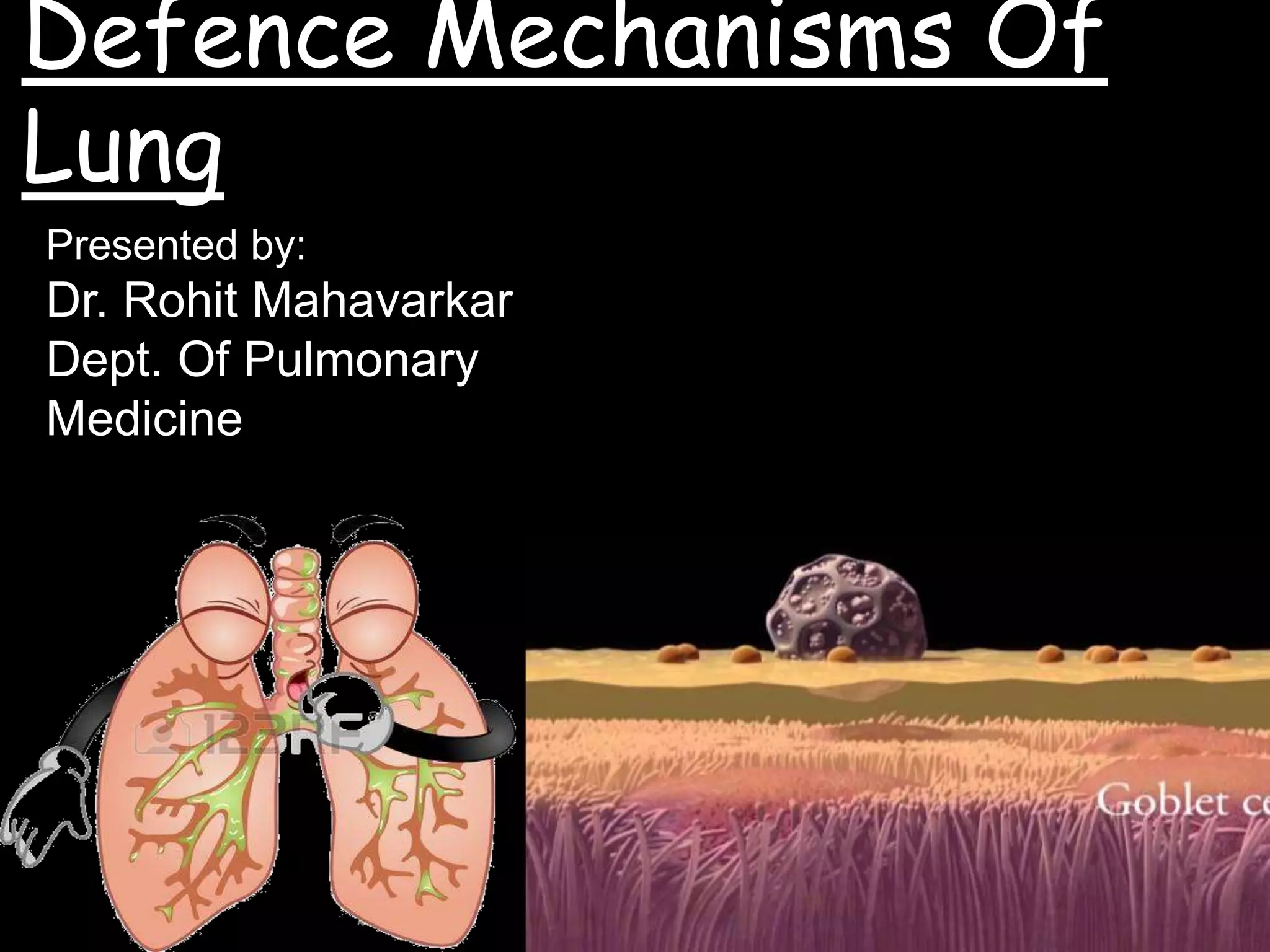
This document discusses the defence mechanisms of the lungs. It outlines several lines of defence, including physical barriers like mucus and cilia, surfactant, immunoglobulins, antimicrobial peptides, complement proteins, and cellular defences such as alveolar macrophages. Alveolar macrophages patrol the lungs and destroy pathogens via phagocytosis. They also initiate and control inflammatory responses by secreting chemokines and cytokines and help repair tissue damage. The lungs have robust protective mechanisms to filter pathogens from the air we breathe daily.
Overview of lung defense mechanisms against various pollutants and pathogens, including cough reflex, mucociliary clearance, mucus properties, and immune responses.
• Everyday ourlungs is exposed to 7000 litres of
air.
• It is exposed to dust, pollen, bacteria etc.
• Despite these exposures, pathology does not
occur always. This is due to local primary
protective mechanism.
• If infections penetrate primary defences, then
secondary responses including inflammatory and
classical immune responses come to action.
3.
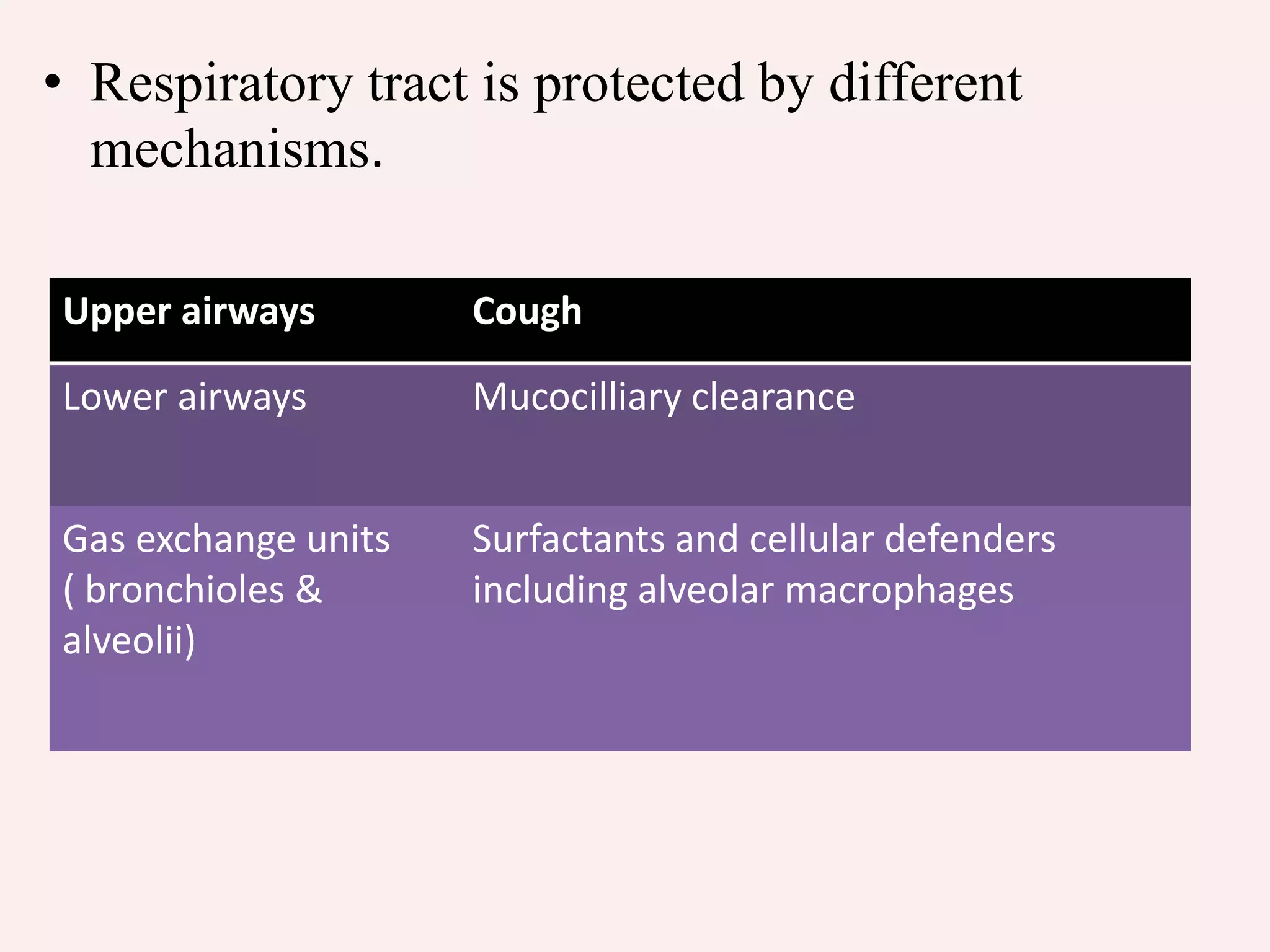
• Respiratory tractis protected by different
mechanisms.
Upper airways Cough
Lower airways Mucocilliary clearance
Gas exchange units
( bronchioles &
alveolii)
Surfactants and cellular defenders
including alveolar macrophages
4.
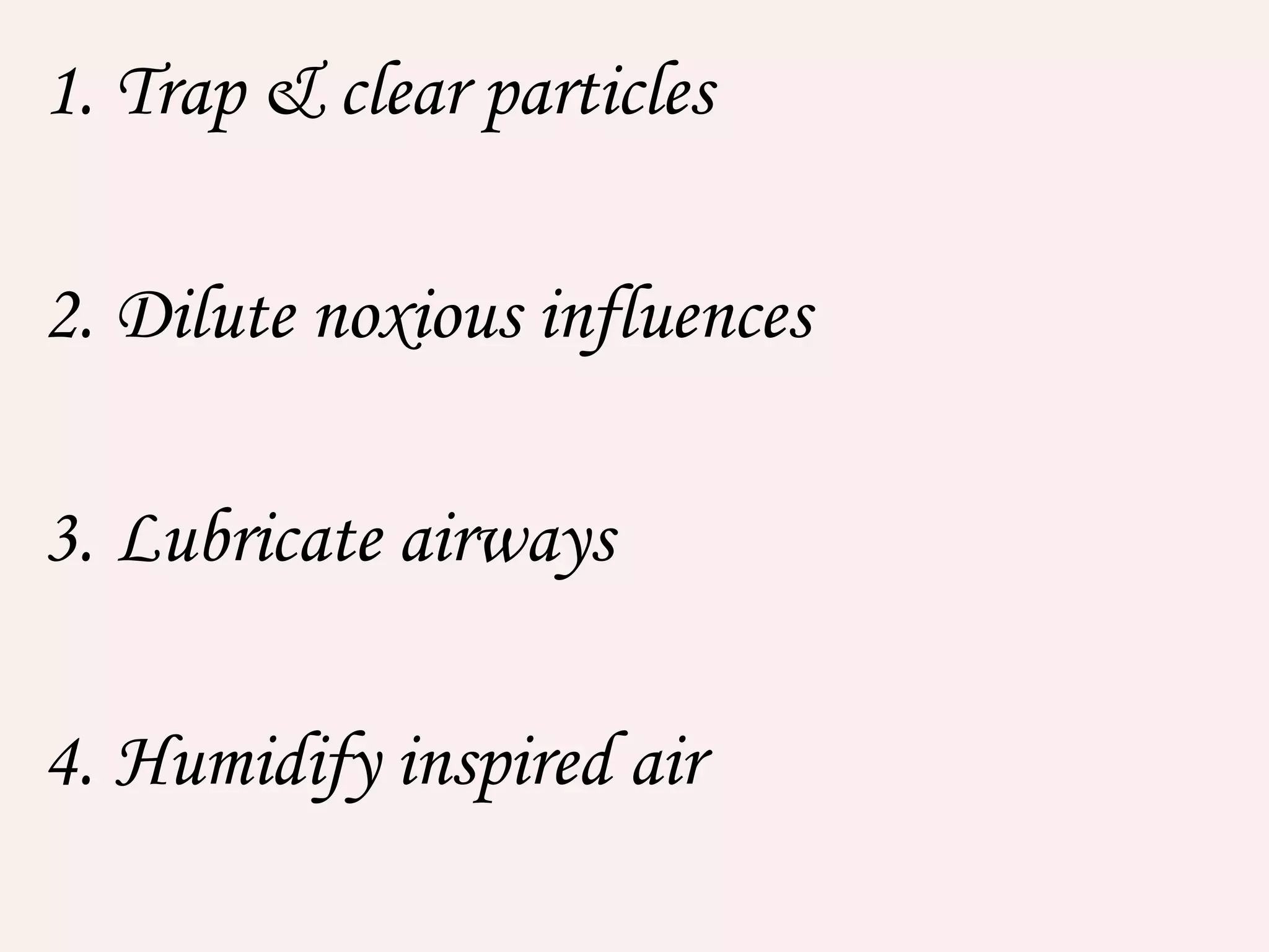
• Mucus inupper airways & surfactant in gas
exchange, contains variety of proteins with
defence properties against the infection.
• Cells also have important cytoprotective
antioxidant & anti-proteinase mechanisms.
5.
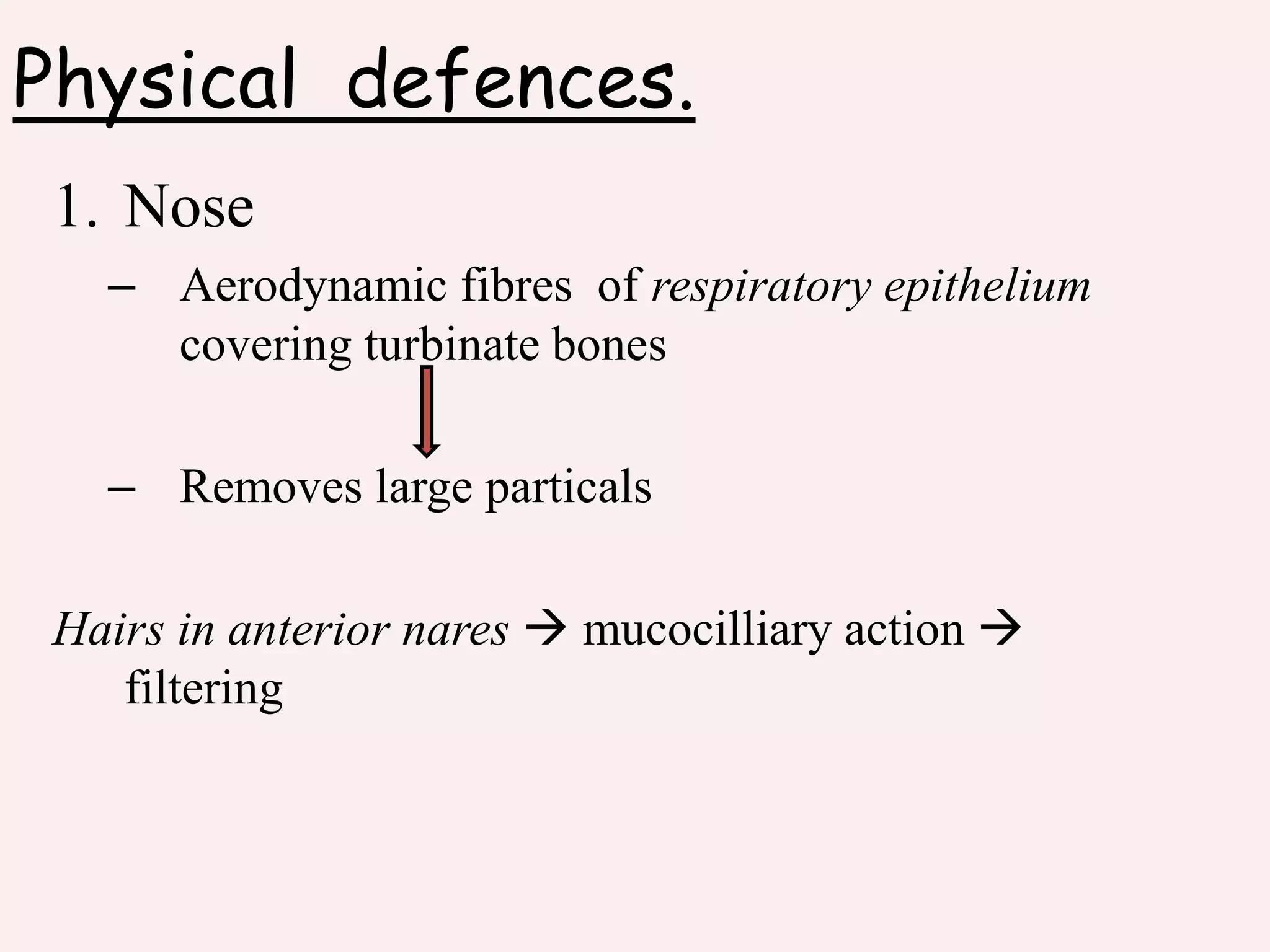
Physical defences.
1. Nose
–Aerodynamic fibres of respiratory epithelium
covering turbinate bones
– Removes large particals
Hairs in anterior nares mucocilliary action
filtering
6.
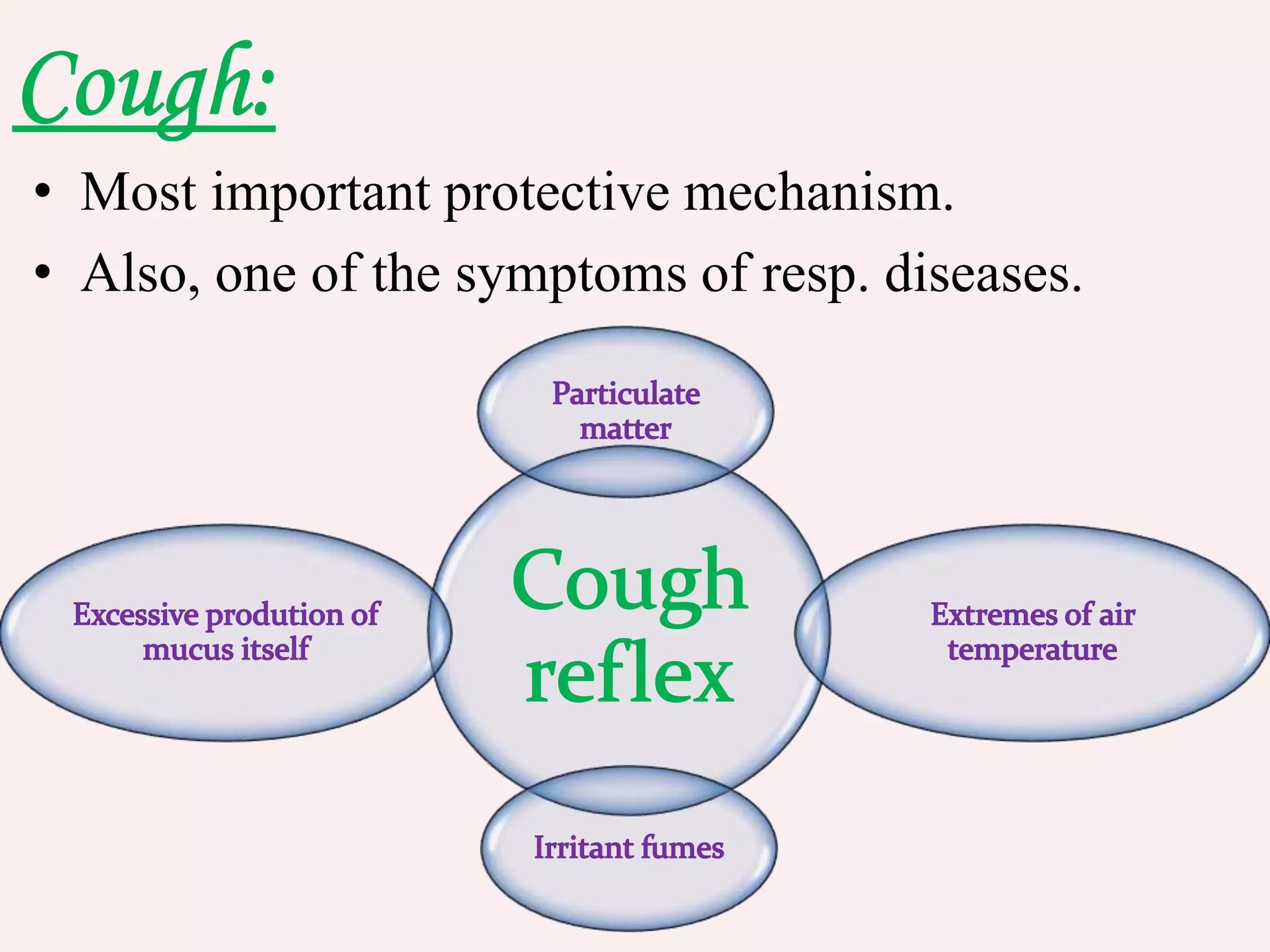
• Most importantprotective mechanism.
• Also, one of the symptoms of resp. diseases.
7.
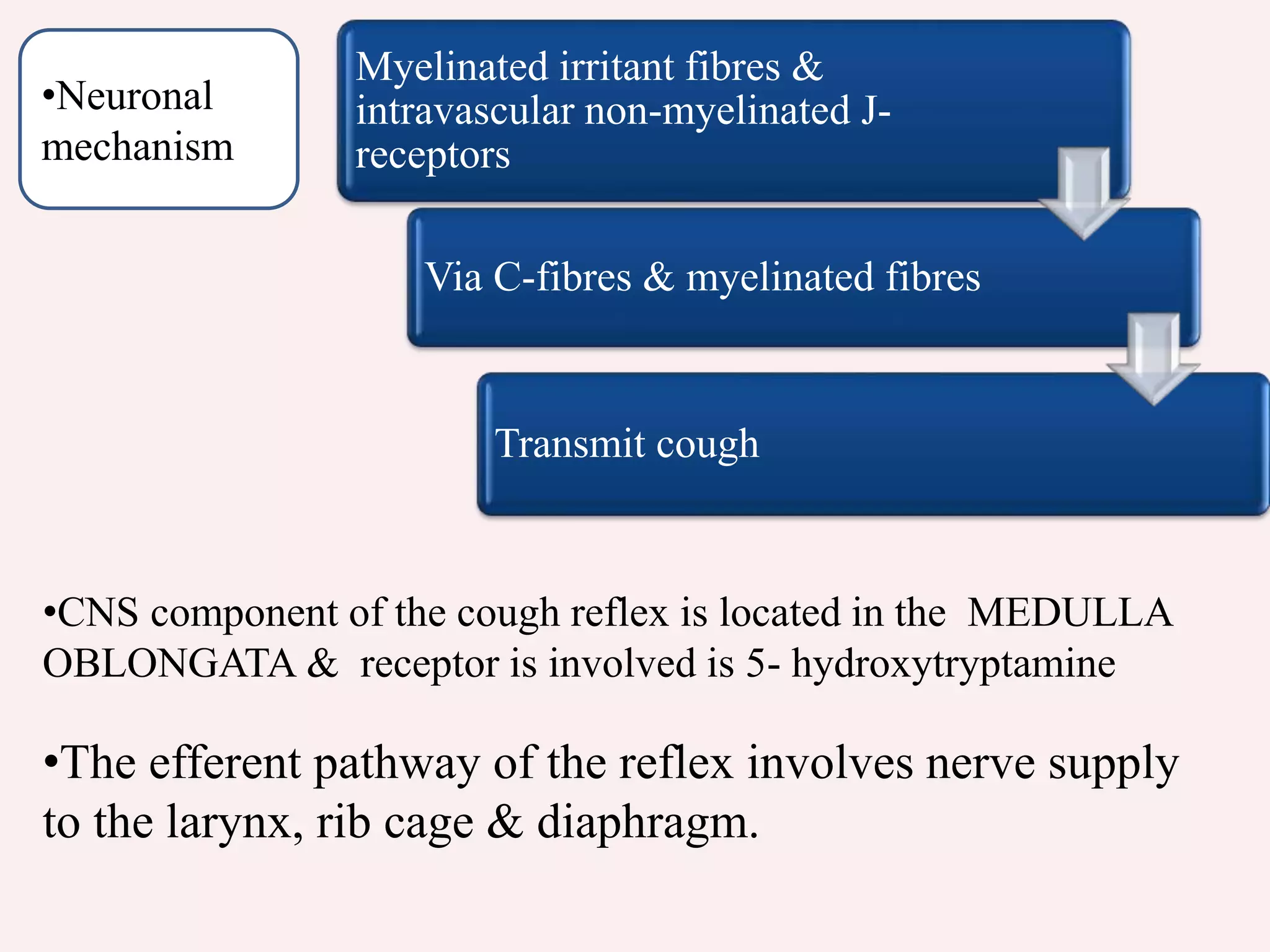
•The efferent pathwayof the reflex involves nerve supply
to the larynx, rib cage & diaphragm.
•CNS component of the cough reflex is located in the MEDULLA
OBLONGATA & receptor is involved is 5- hydroxytryptamine
Myelinated irritant fibres &
intravascular non-myelinated J-
receptors
Via C-fibres & myelinated fibres
Transmit cough
•Neuronal
mechanism
8.
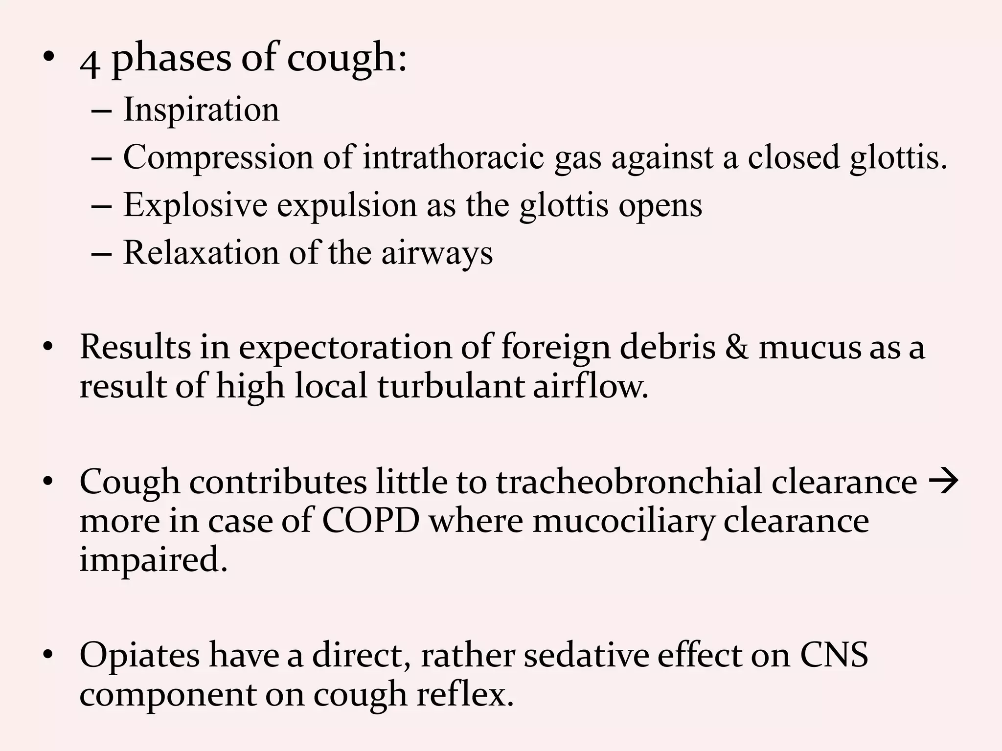
• 4 phasesof cough:
– Inspiration
– Compression of intrathoracic gas against a closed glottis.
– Explosive expulsion as the glottis opens
– Relaxation of the airways
• Results in expectoration of foreign debris & mucus as a
result of high local turbulant airflow.
• Cough contributes little to tracheobronchial clearance
more in case of COPD where mucociliary clearance
impaired.
• Opiates have a direct, rather sedative effect on CNS
component on cough reflex.
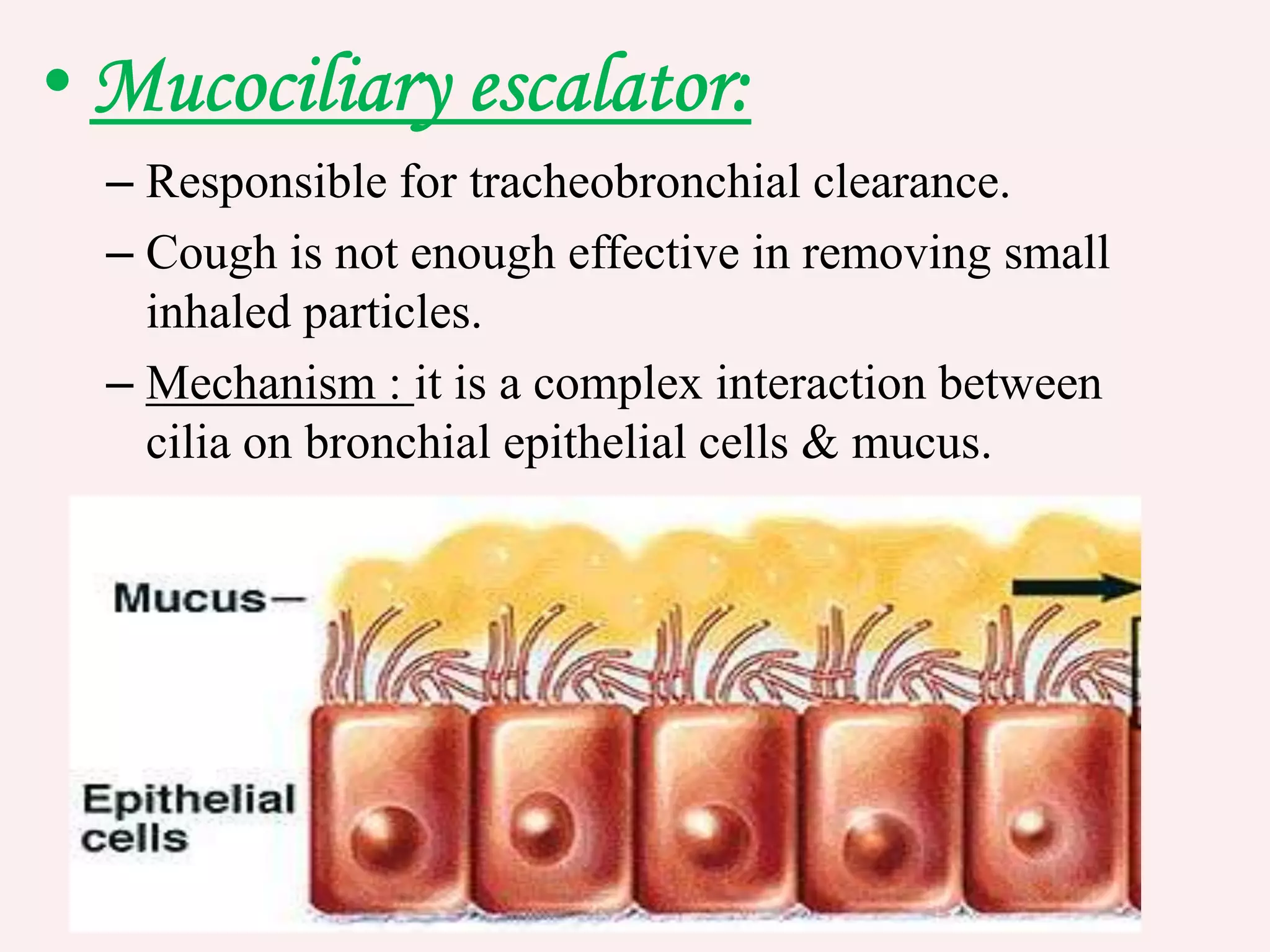
9.
– Responsible fortracheobronchial clearance.
– Cough is not enough effective in removing small
inhaled particles.
– Mechanism : it is a complex interaction between
cilia on bronchial epithelial cells & mucus.
10.
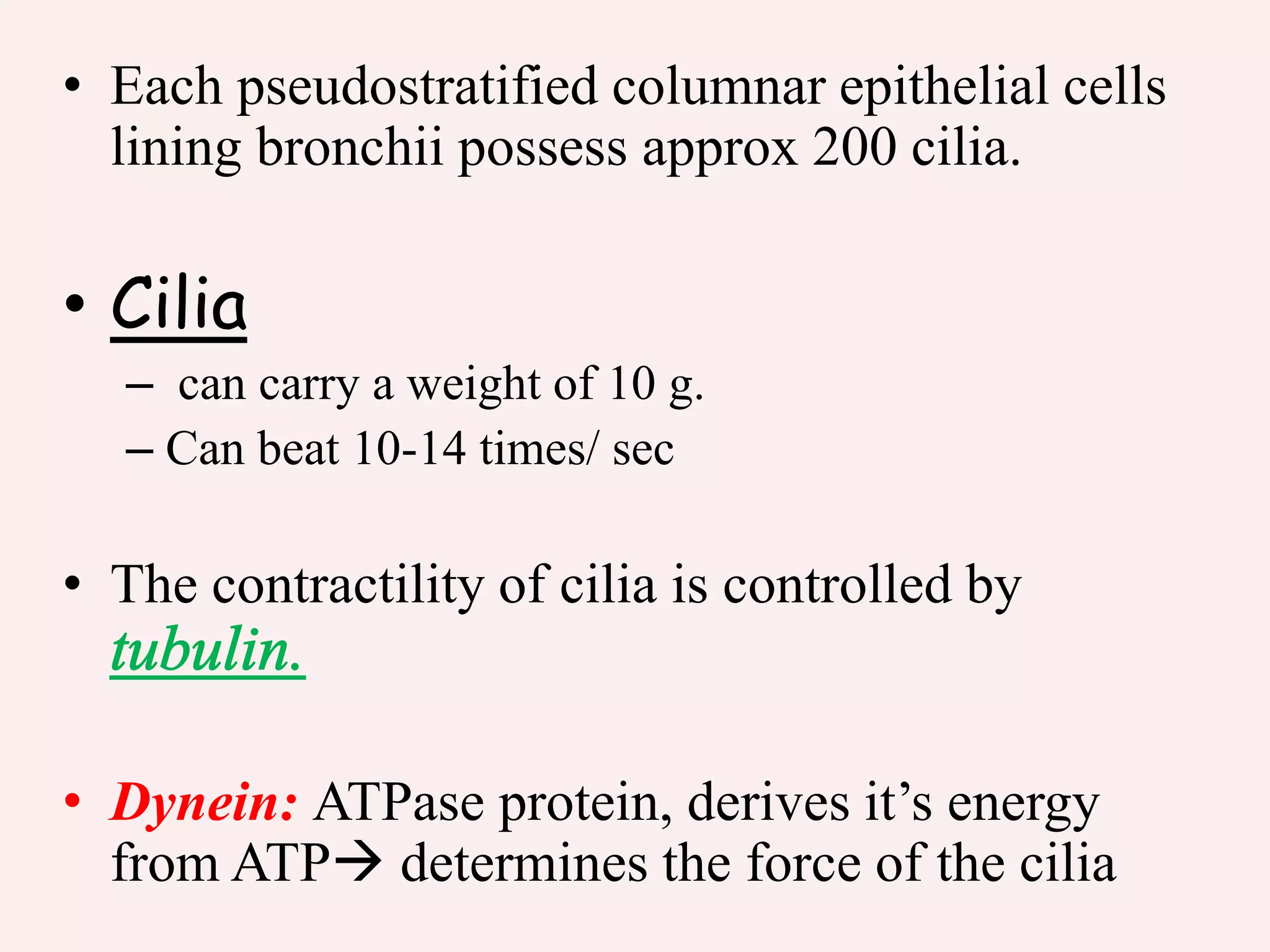
• Each pseudostratifiedcolumnar epithelial cells
lining bronchii possess approx 200 cilia.
• Cilia
– can carry a weight of 10 g.
– Can beat 10-14 times/ sec
• The contractility of cilia is controlled by
• Dynein: ATPase protein, derives it’s energy
from ATP determines the force of the cilia
11.
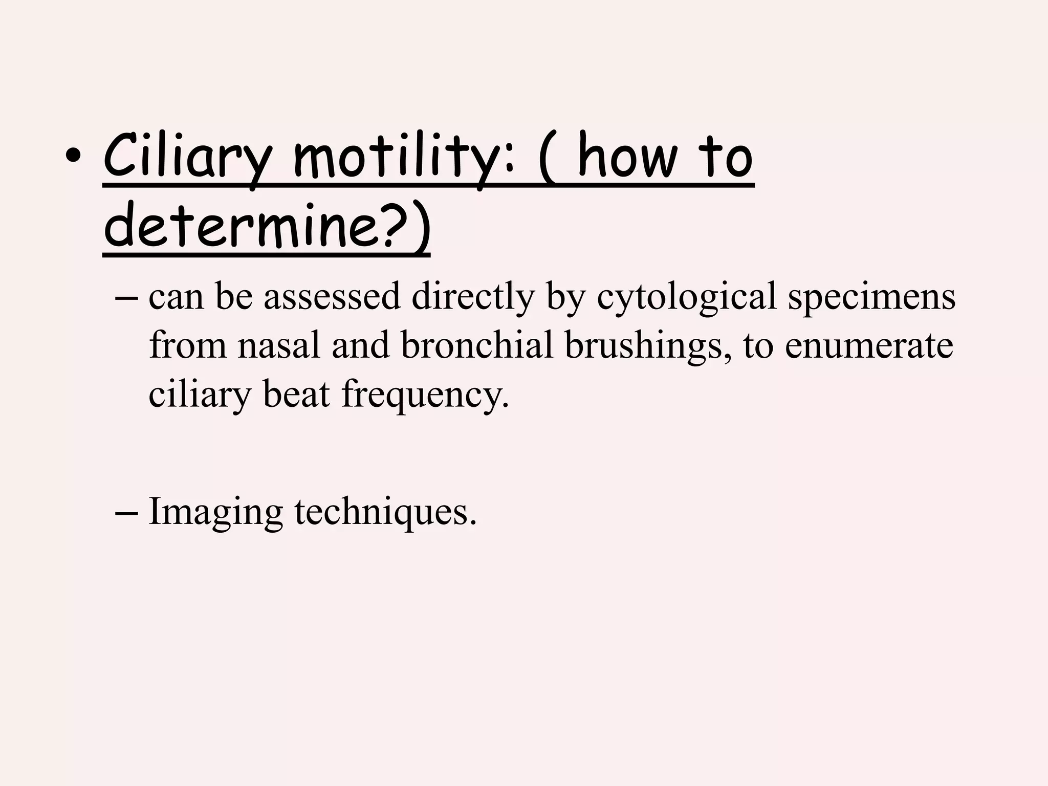
• Ciliary motility:( how to
determine?)
– can be assessed directly by cytological specimens
from nasal and bronchial brushings, to enumerate
ciliary beat frequency.
– Imaging techniques.
12.
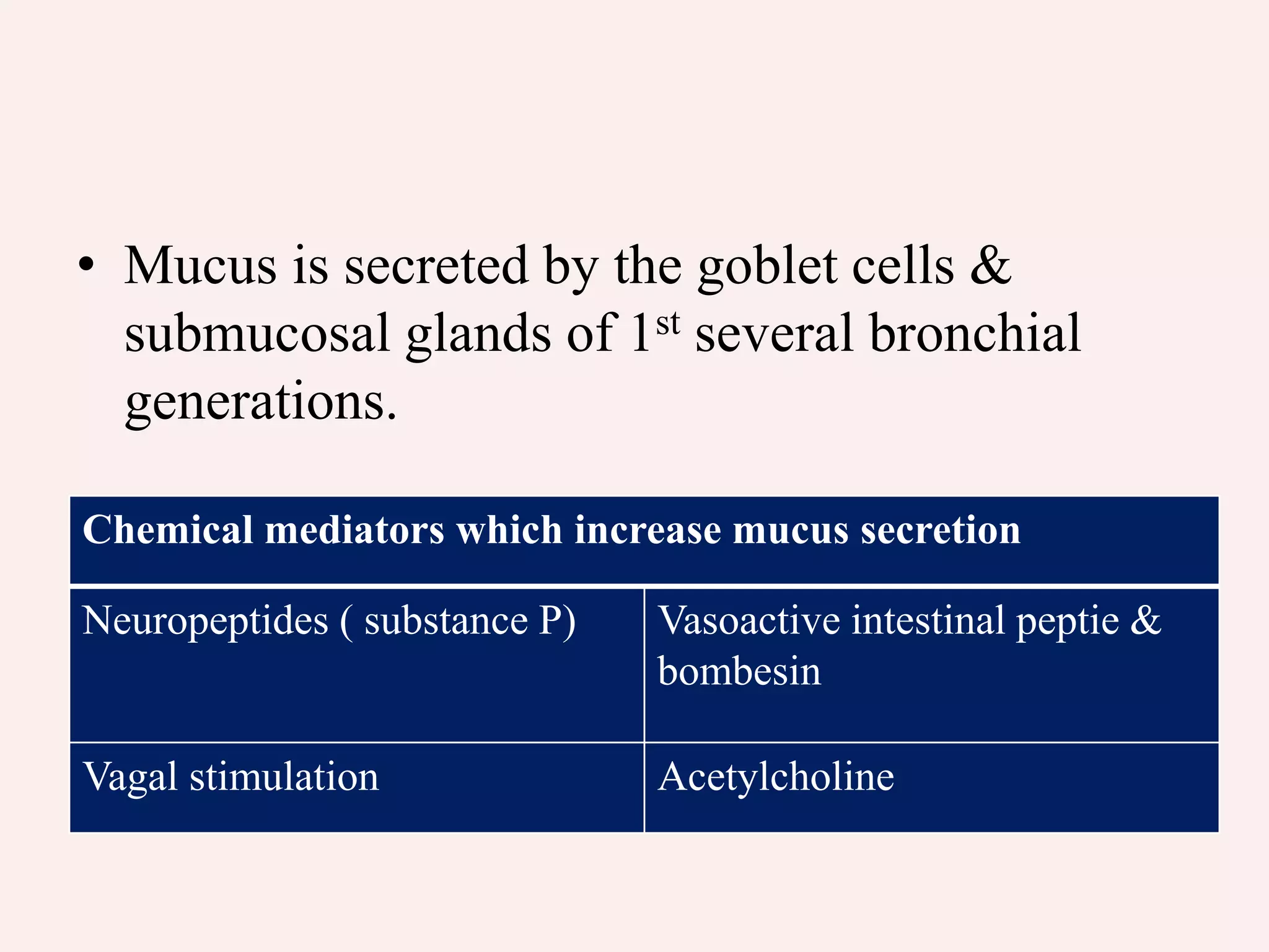
• Mucus issecreted by the goblet cells &
submucosal glands of 1st several bronchial
generations.
Chemical mediators which increase mucus secretion
Neuropeptides ( substance P) Vasoactive intestinal peptie &
bombesin
Vagal stimulation Acetylcholine
13.
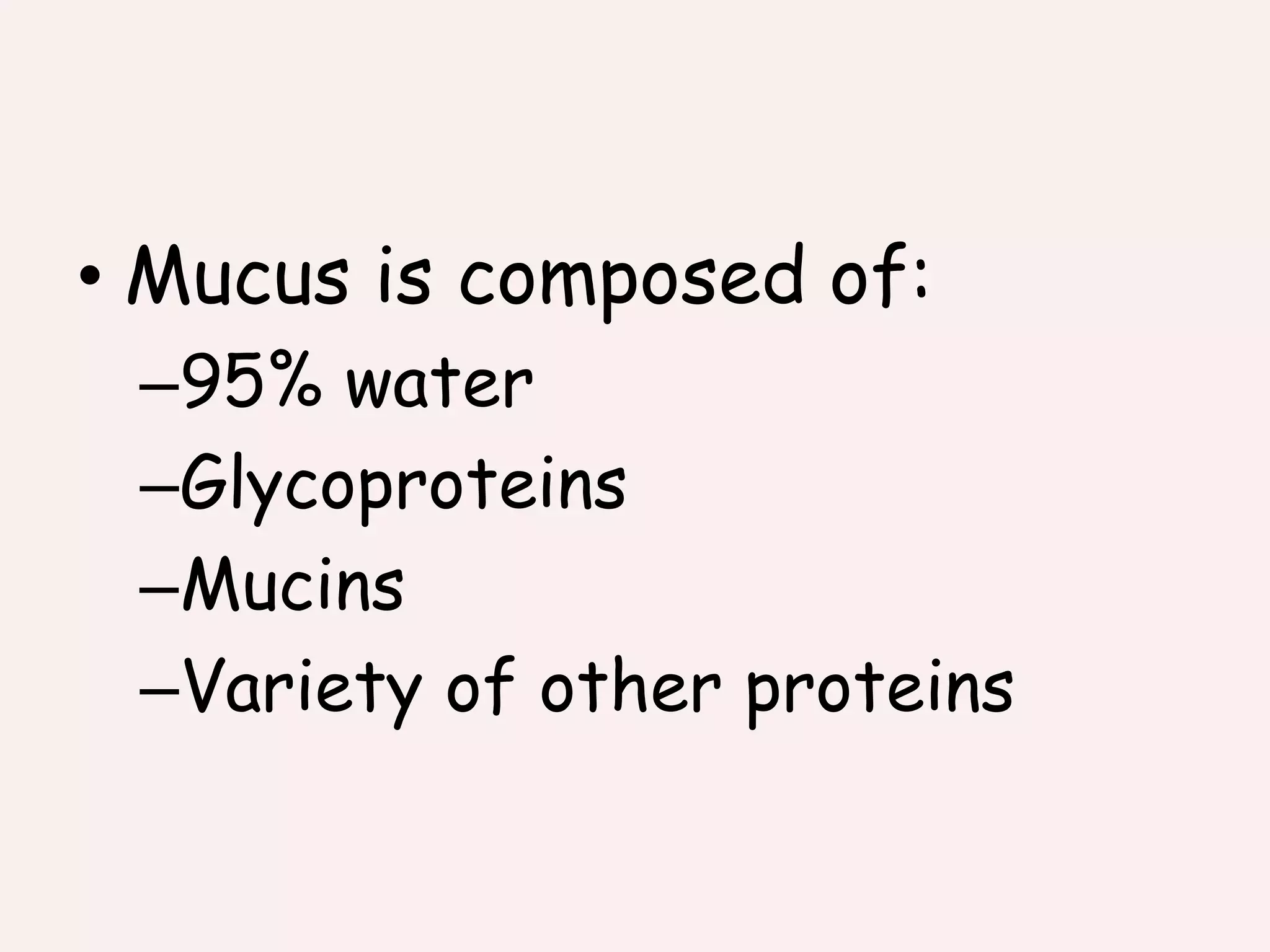
• Mucus iscomposed of:
–95% water
–Glycoproteins
–Mucins
–Variety of other proteins
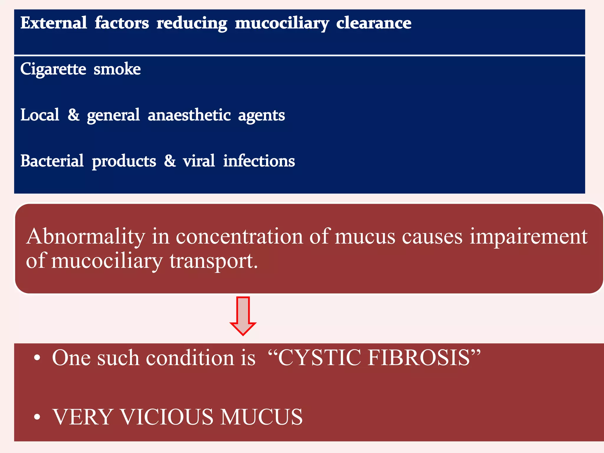
Abnormality in concentrationof mucus causes impairement
of mucociliary transport.
• One such condition is “CYSTIC FIBROSIS”
• VERY VICIOUS MUCUS
17.
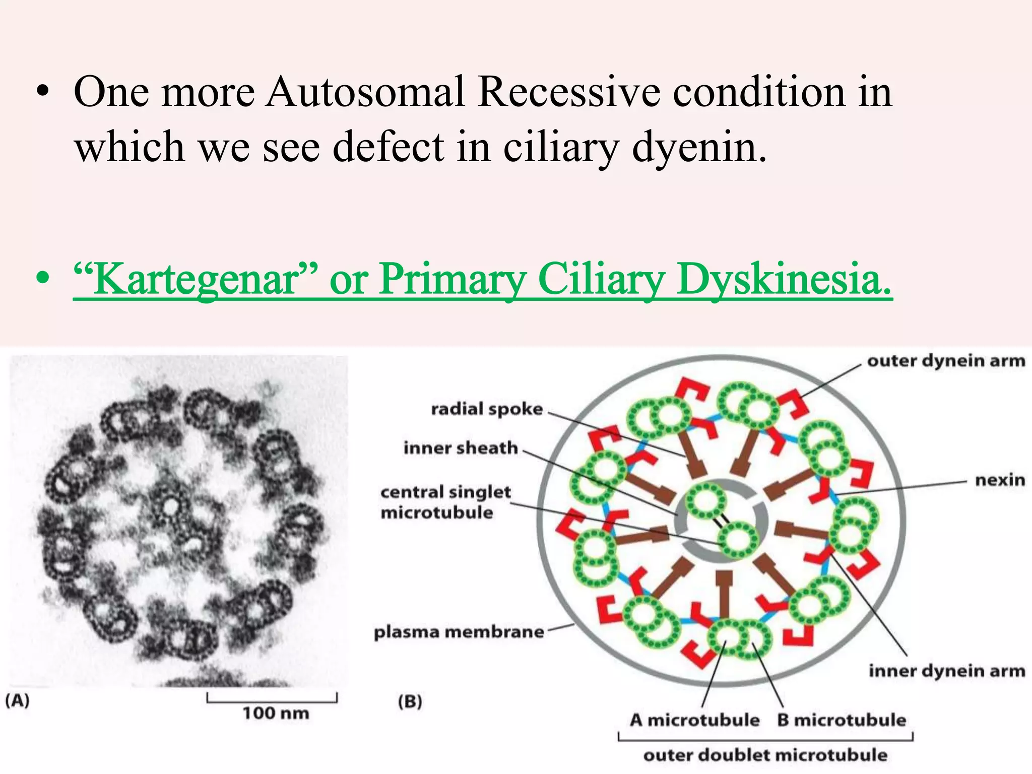
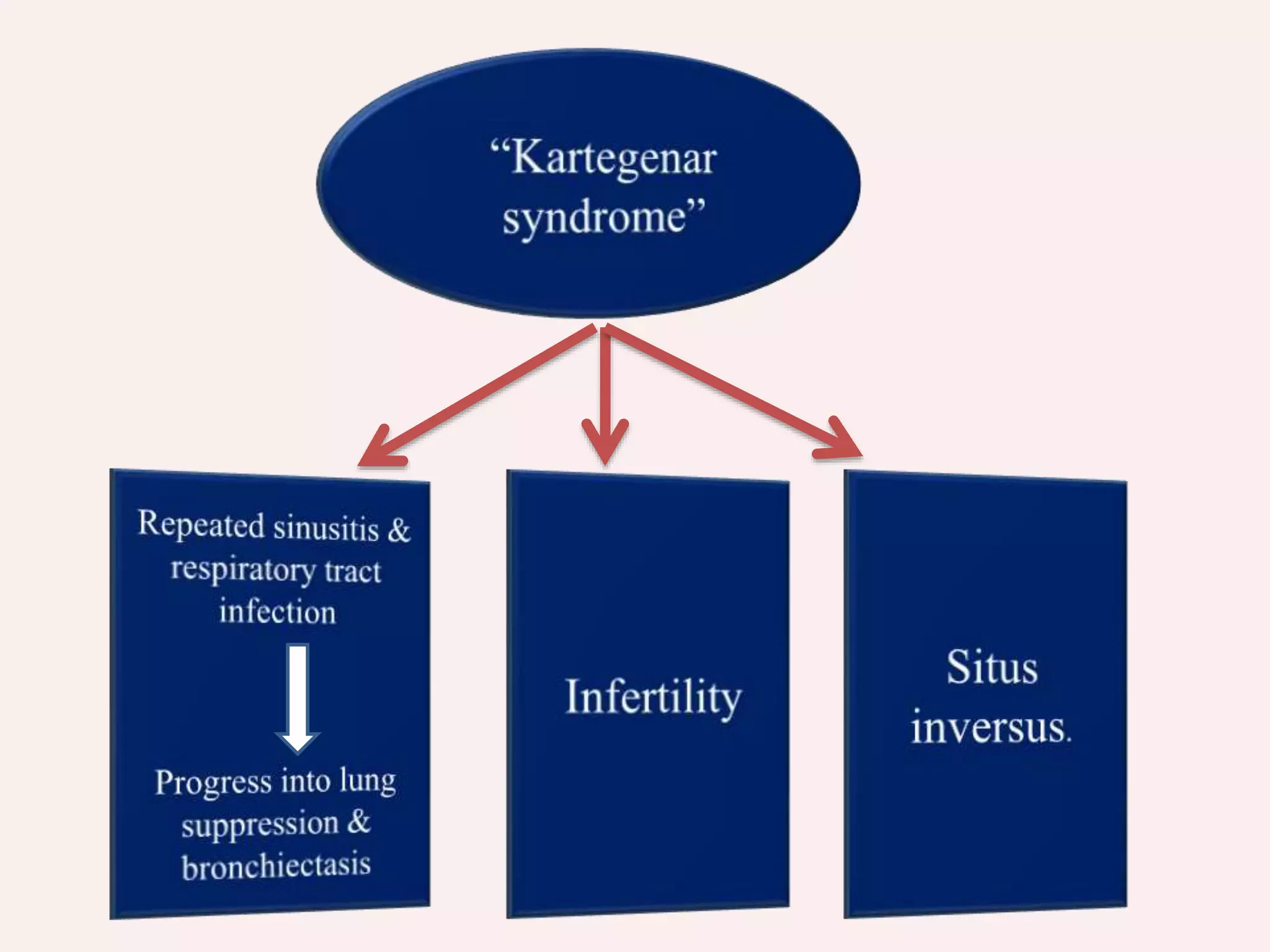
• One moreAutosomal Recessive condition in
which we see defect in ciliary dyenin.
19.
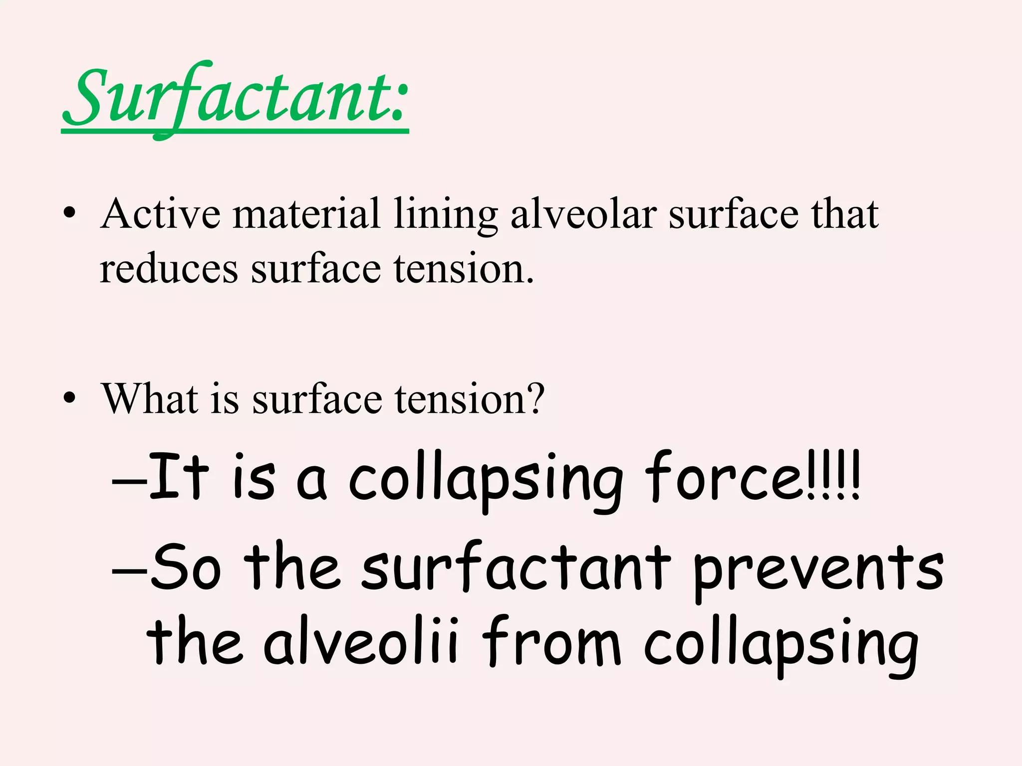
• Active materiallining alveolar surface that
reduces surface tension.
• What is surface tension?
–It is a collapsing force!!!!
–So the surfactant prevents
the alveolii from collapsing
21.
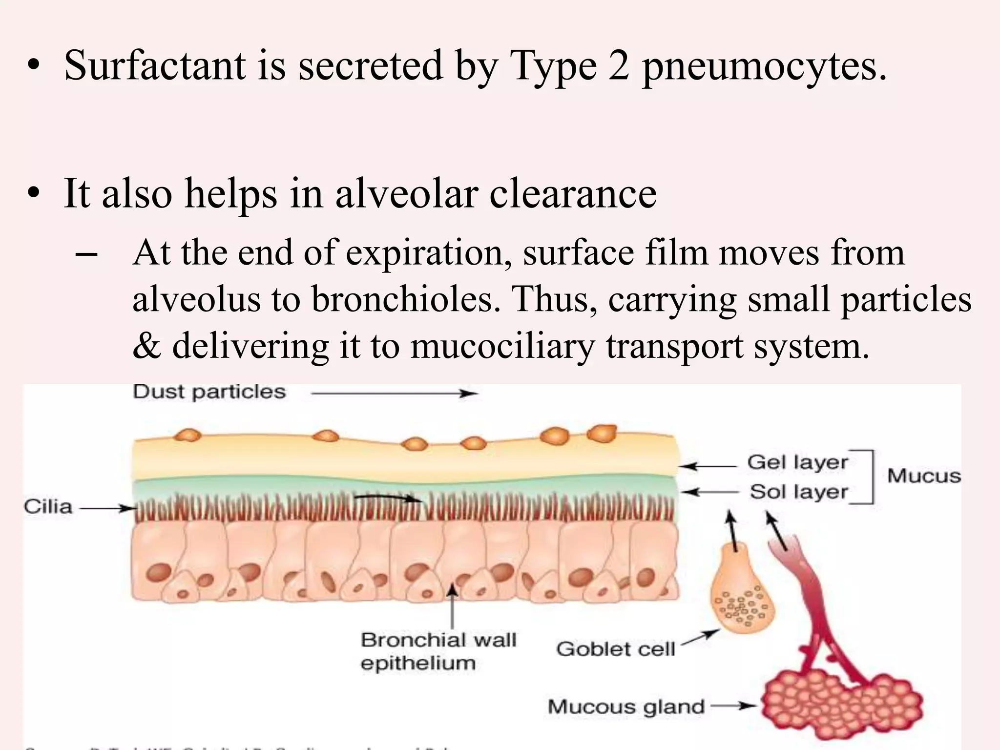
• Surfactant issecreted by Type 2 pneumocytes.
• It also helps in alveolar clearance
– At the end of expiration, surface film moves from
alveolus to bronchioles. Thus, carrying small particles
& delivering it to mucociliary transport system.
22.
• The compositionof surfactant also contains
surfactant proteins
– SP-A
– SP-B
– SP-C
– SP-D
• Surfactant also enhances local non-specific
pulmonary immune defence mechanisms.
• It exerts influence on neutrophils function which
include neutrophil adhesion.
23.
Surfactant proteins:
– Mostabundant
– Closely resemble C1q.
– Enhances alveolar macrophages phagocytosis.
, may also share same effects of SP-A on
inflammatory cells & macrophages.
• Surfactant can be damaged by a number of noxious
stimuli. Alteration in surfactant is important in
pathogenesis of ARDS.
24.
• Apart fromsurfactant proteins, many other
proteins are important in lung defences. Such
proteins are derived from plasma.
25.
Immunoglobulins
• Normally allsecretions contain immunoglobulins but in
different proportions.
• IgA, is in excess as compared to IgG & IgM.
• Immunoglobulins produced by a local lung tissue – from
plasma cells & B-lymphocytes
• IgA is secreted maximum in the upper airways.
• Deficiency oF IgA is associated with bacterial infections.
26.
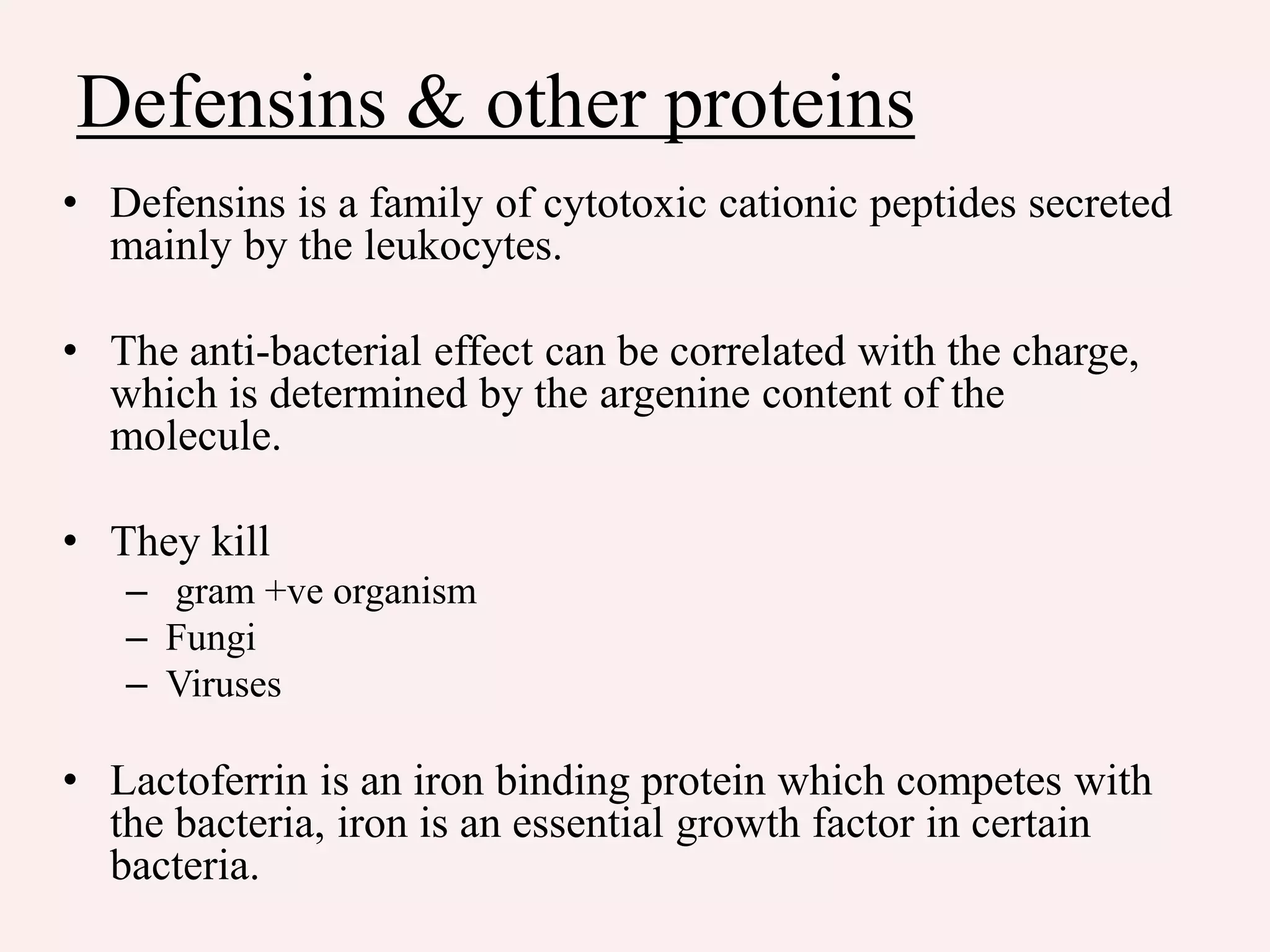
Defensins & otherproteins
• Defensins is a family of cytotoxic cationic peptides secreted
mainly by the leukocytes.
• The anti-bacterial effect can be correlated with the charge,
which is determined by the argenine content of the
molecule.
• They kill
– gram +ve organism
– Fungi
– Viruses
• Lactoferrin is an iron binding protein which competes with
the bacteria, iron is an essential growth factor in certain
bacteria.
27.
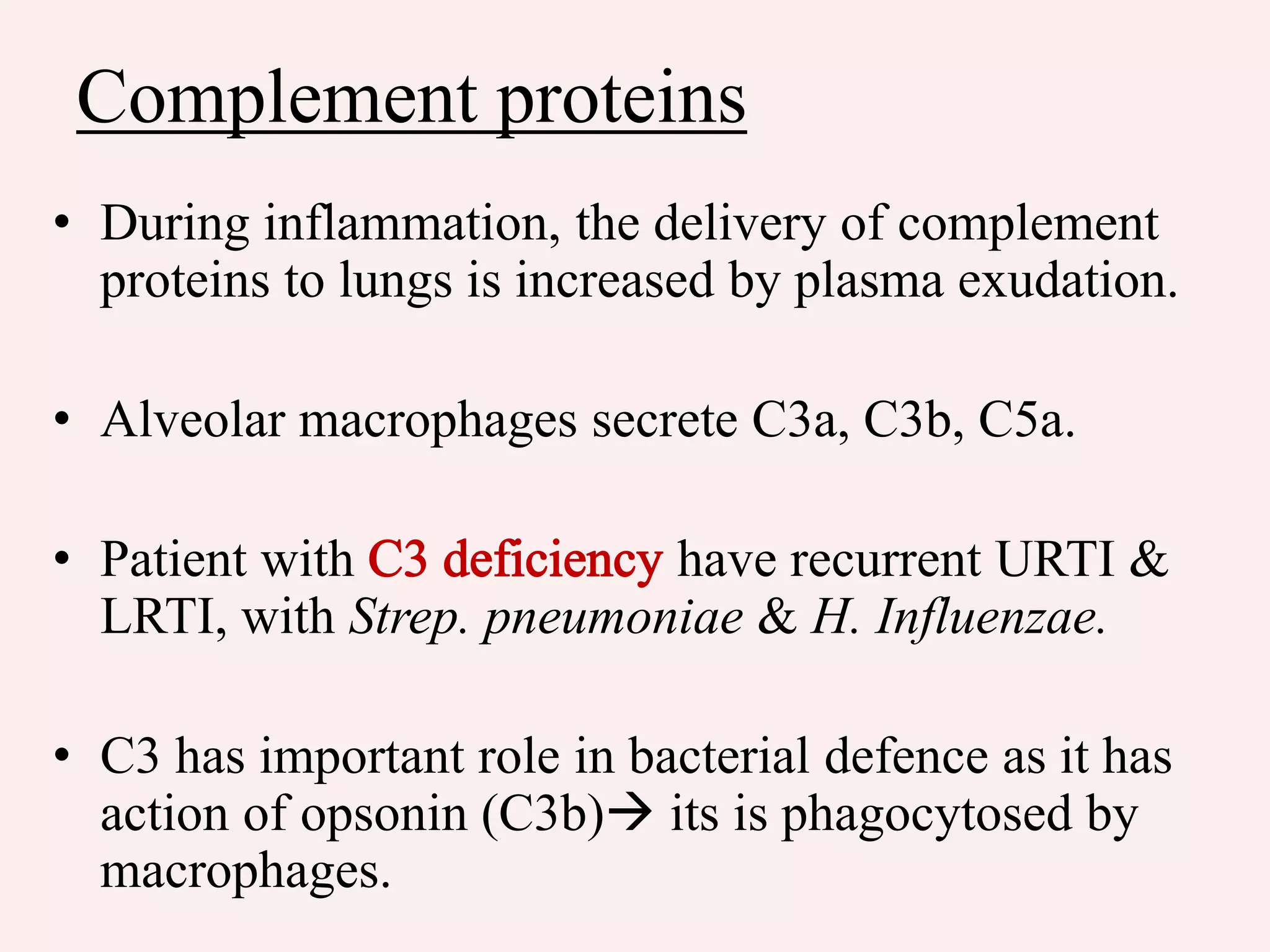
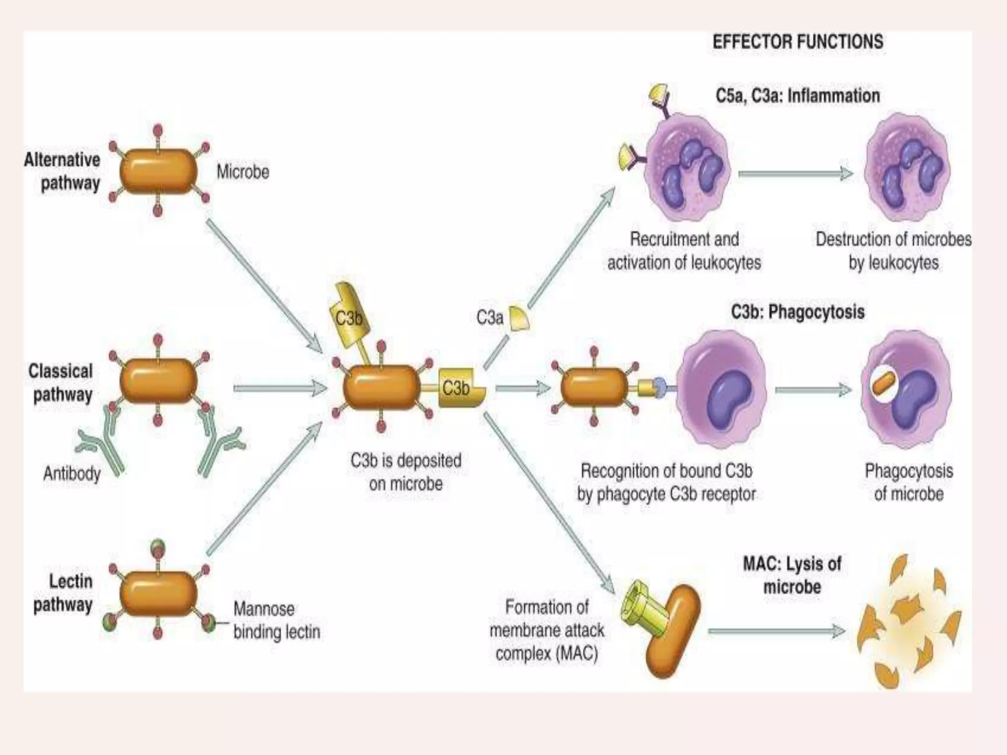
Complement proteins
• Duringinflammation, the delivery of complement
proteins to lungs is increased by plasma exudation.
• Alveolar macrophages secrete C3a, C3b, C5a.
• Patient with have recurrent URTI &
LRTI, with Strep. pneumoniae & H. Influenzae.
• C3 has important role in bacterial defence as it has
action of opsonin (C3b) its is phagocytosed by
macrophages.
29.
• Many ofthese agents may be derived from
alveolar macrophages & airway epithelial
cells.
• During inflammatory & injurious processes
the rate of secretion of these important
protective agents is likely to be greatly
enhanced
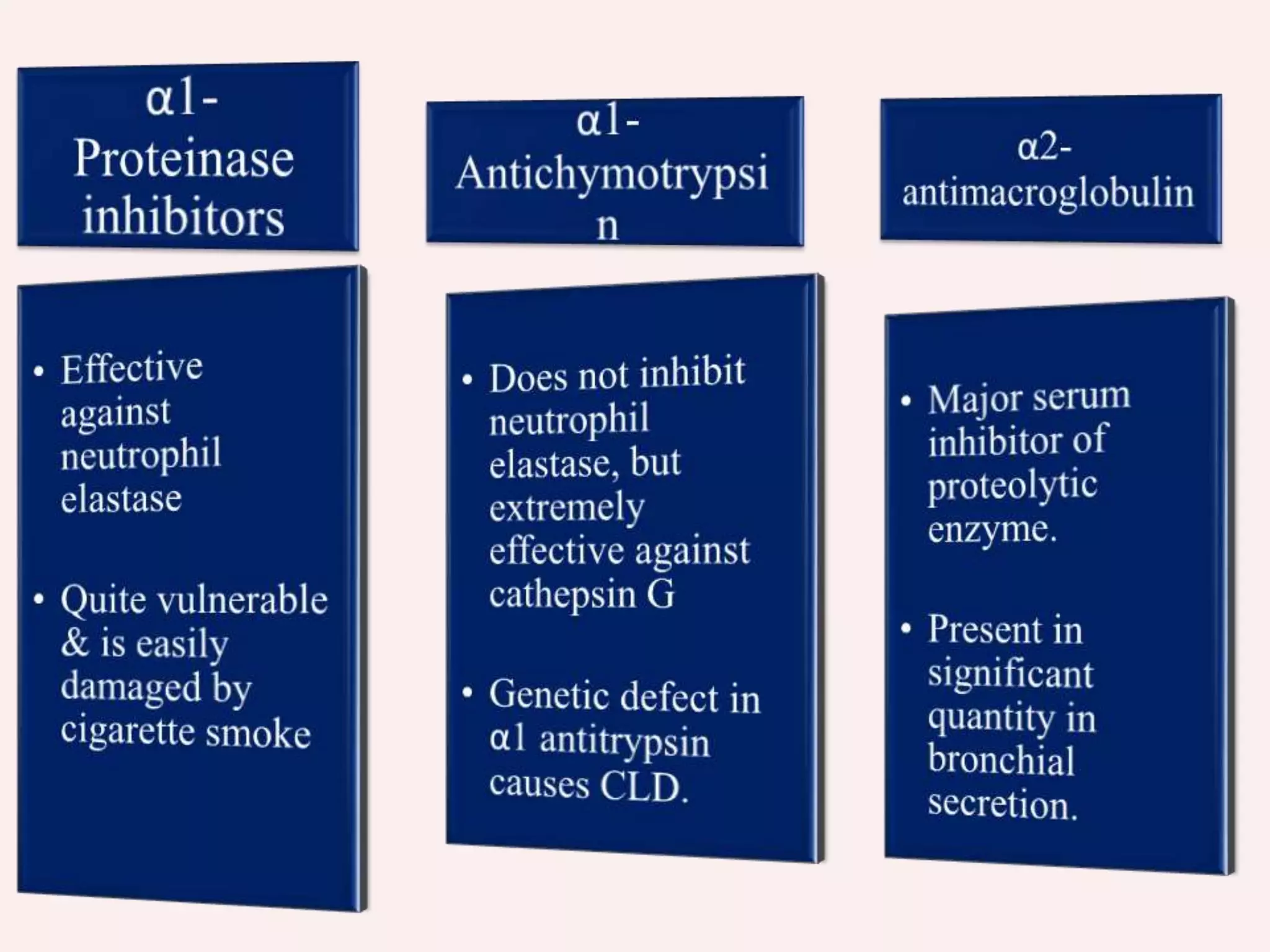
31.
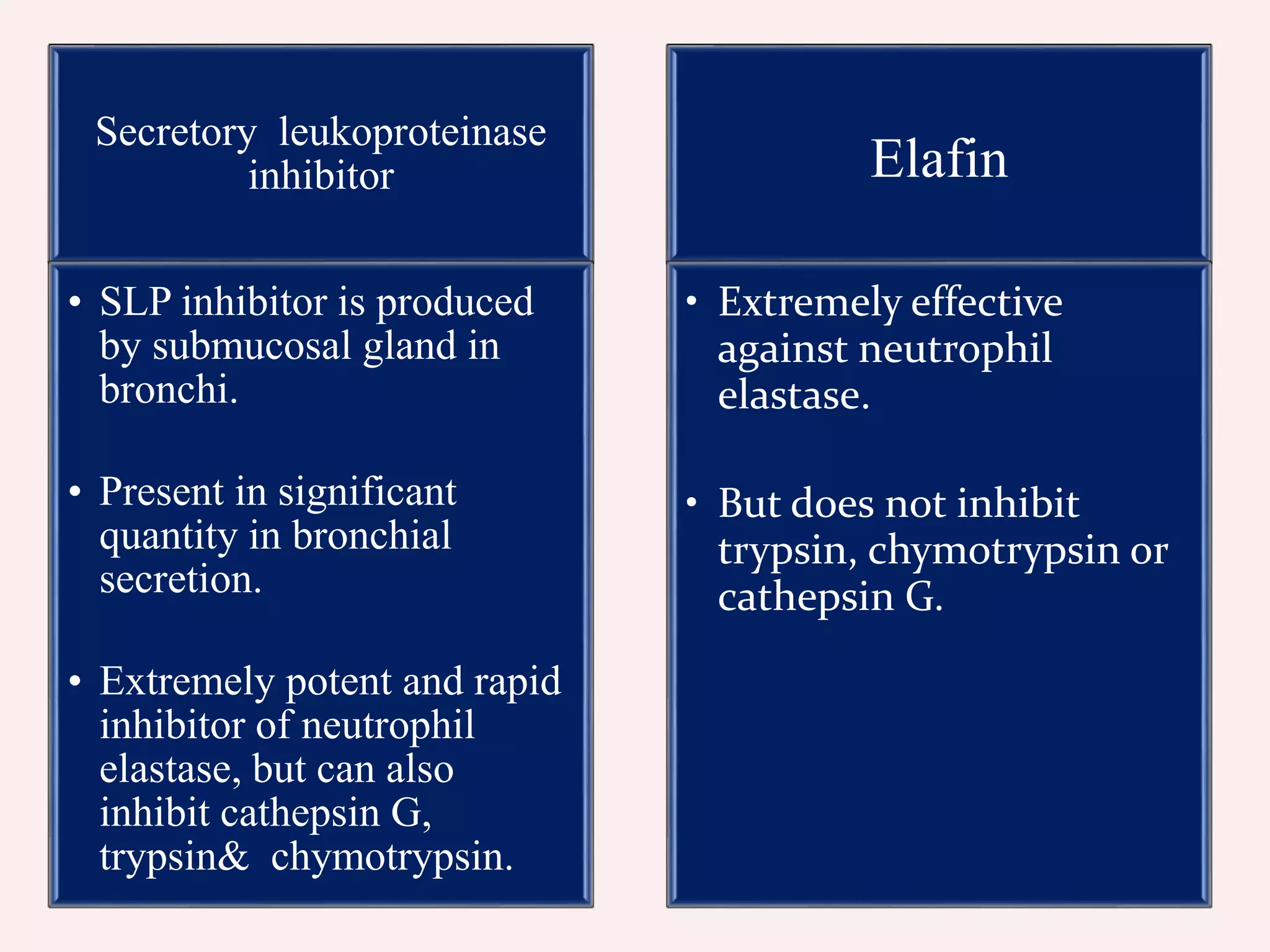
Secretory leukoproteinase
inhibitor
• SLPinhibitor is produced
by submucosal gland in
bronchi.
• Present in significant
quantity in bronchial
secretion.
• Extremely potent and rapid
inhibitor of neutrophil
elastase, but can also
inhibit cathepsin G,
trypsin& chymotrypsin.
Elafin
• Extremely effective
against neutrophil
elastase.
• But does not inhibit
trypsin, chymotrypsin or
cathepsin G.
32.
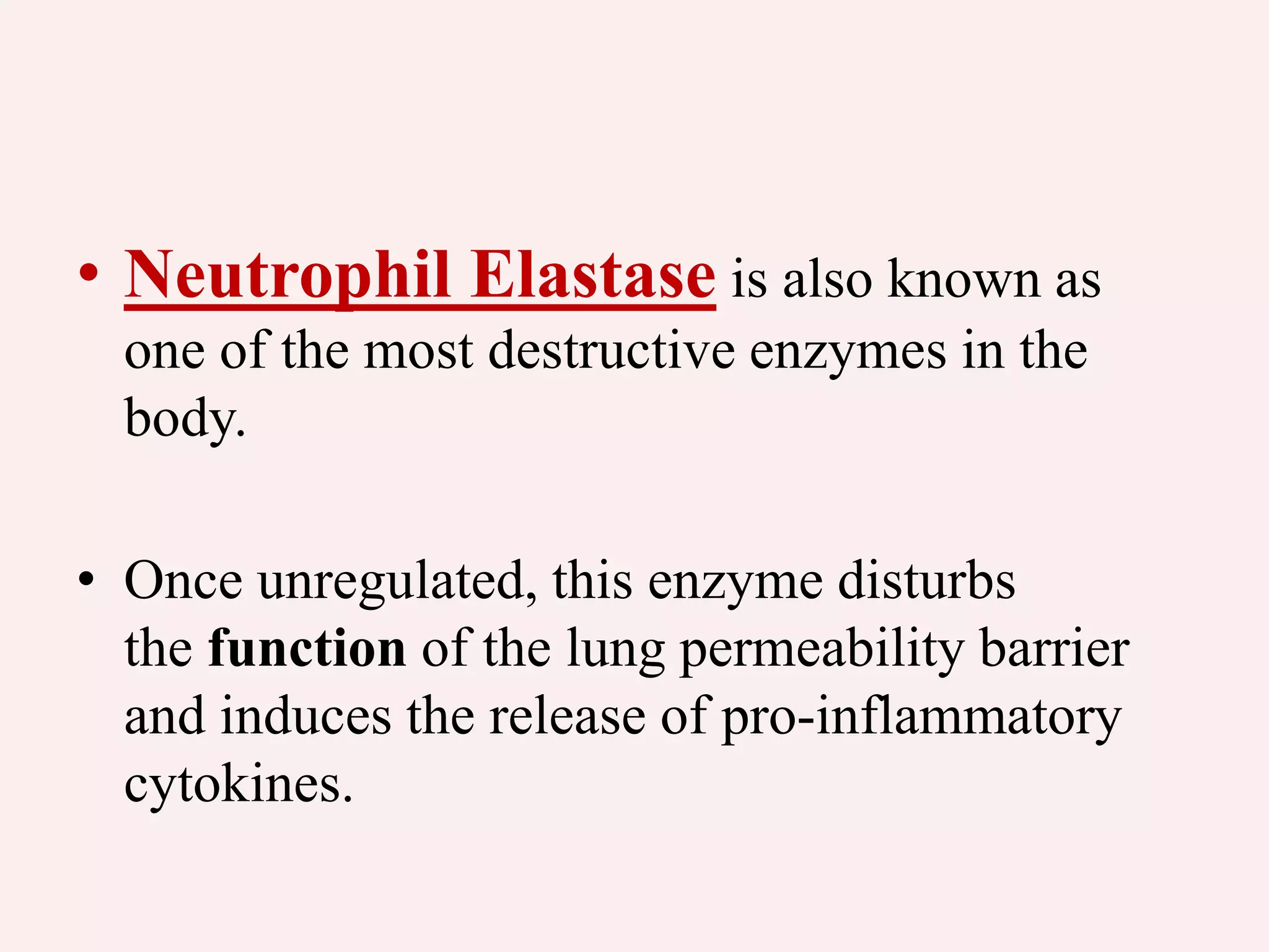
• Neutrophil Elastaseis also known as
one of the most destructive enzymes in the
body.
• Once unregulated, this enzyme disturbs
the function of the lung permeability barrier
and induces the release of pro-inflammatory
cytokines.
33.
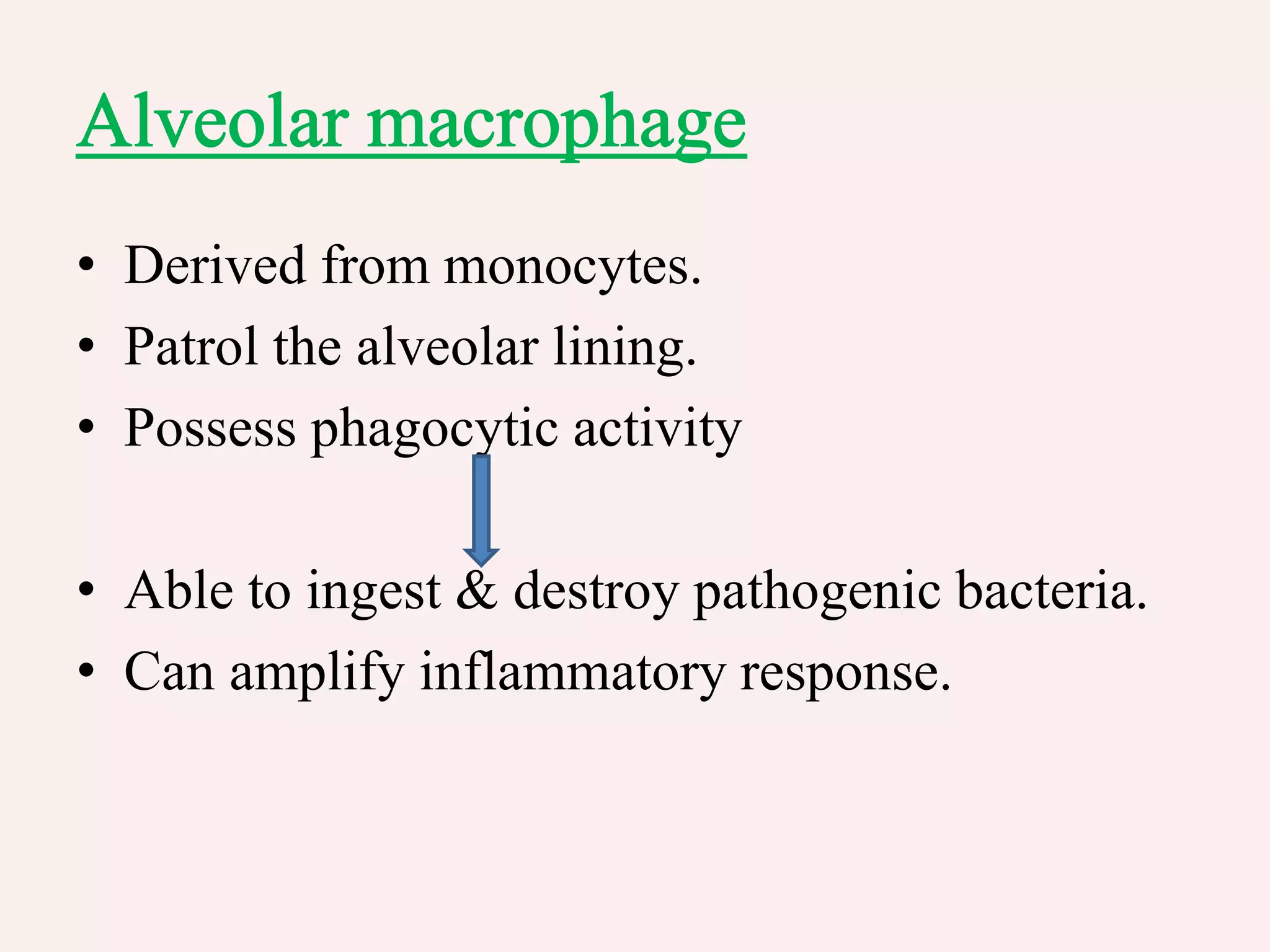
• Derived frommonocytes.
• Patrol the alveolar lining.
• Possess phagocytic activity
• Able to ingest & destroy pathogenic bacteria.
• Can amplify inflammatory response.
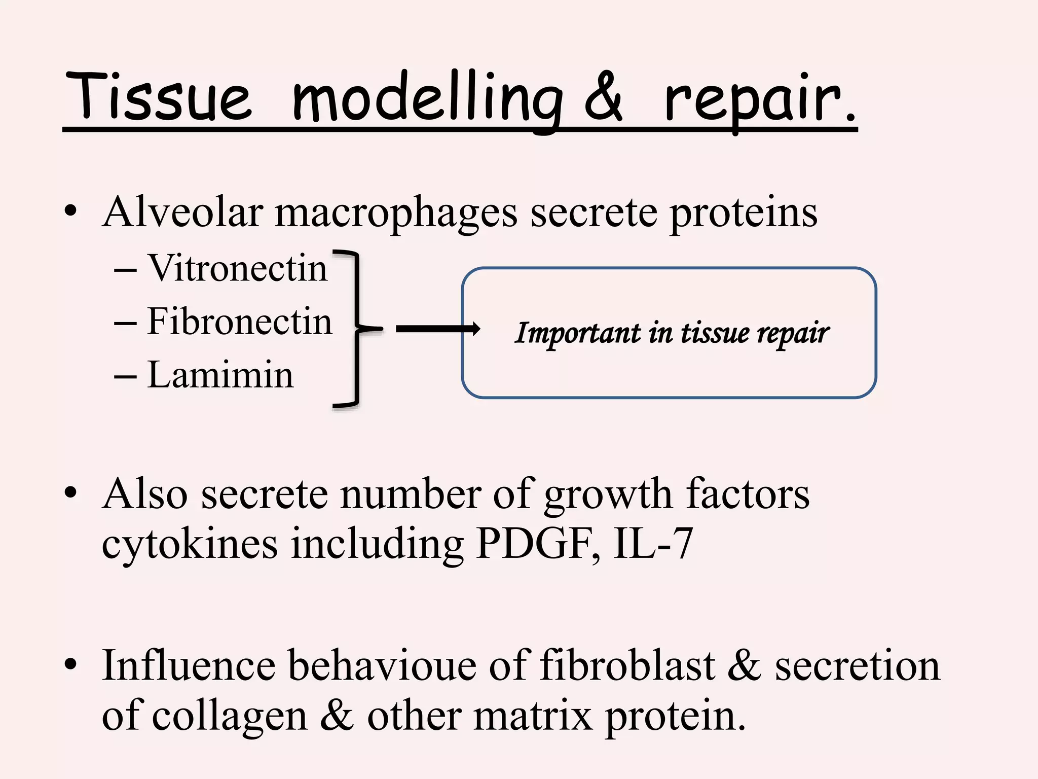
34.
• Role inrepairing inflammatory tissue.
• Have a wide range of degradative enzymes.
• Have capacity to digest proteins, lipids,
carbohydrates.
35.
• Activated macrophagesform nitrite & nitrate,
which contribute to antifungal, antiparasitic, &
tumorocidal activities.
• Macrophages also call in a number of other
phagocytic cells e.g neutrophil, eosinophil, by
specific generation of chemokines.
• Despite such powerful mechanism, not all
phagocytosed particles are destroyed.
– Minerals such as Quartz & Abestos.
– Number of microorganisms including MTB.
36.
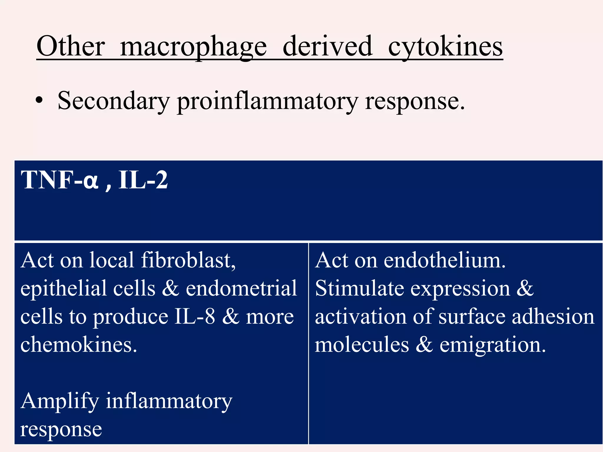
Initiation & controlof inflammatory response.
• Macrophages secrete a number of chemotactic
proteins including members of 5-LOX & COX
pathway which exert important proinflammatory
effects.
• Leukotriene B4 which is a specific neutrophil
chemotoxin.