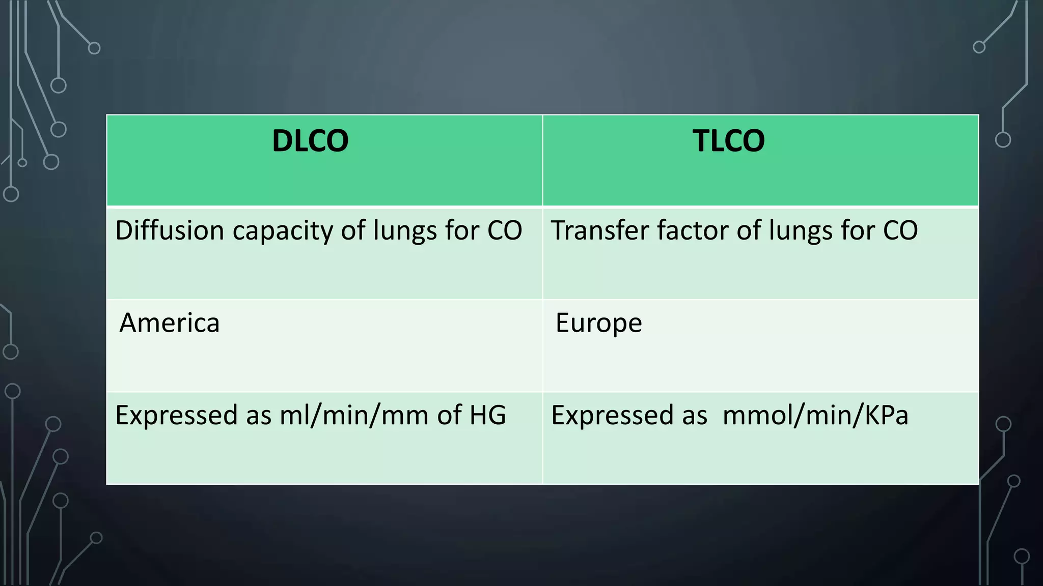
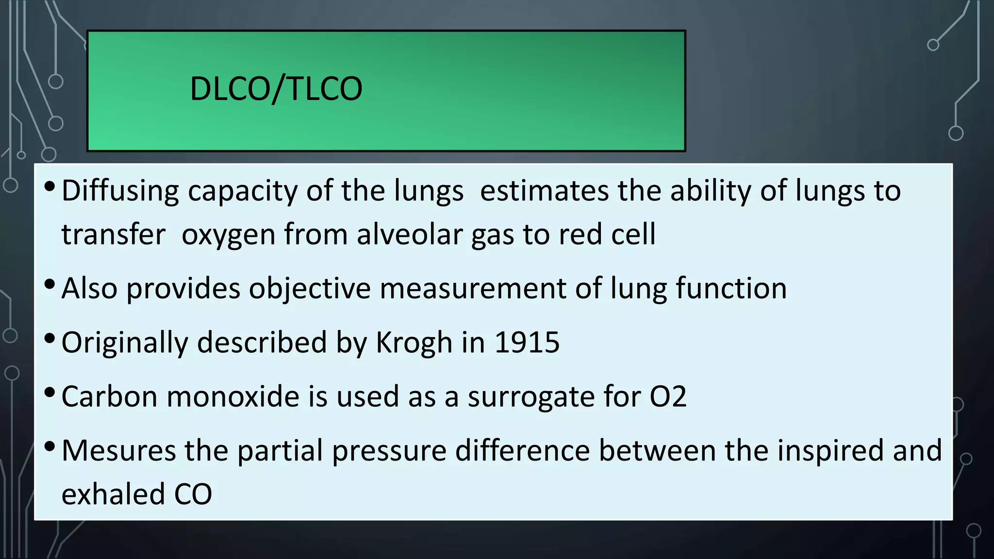
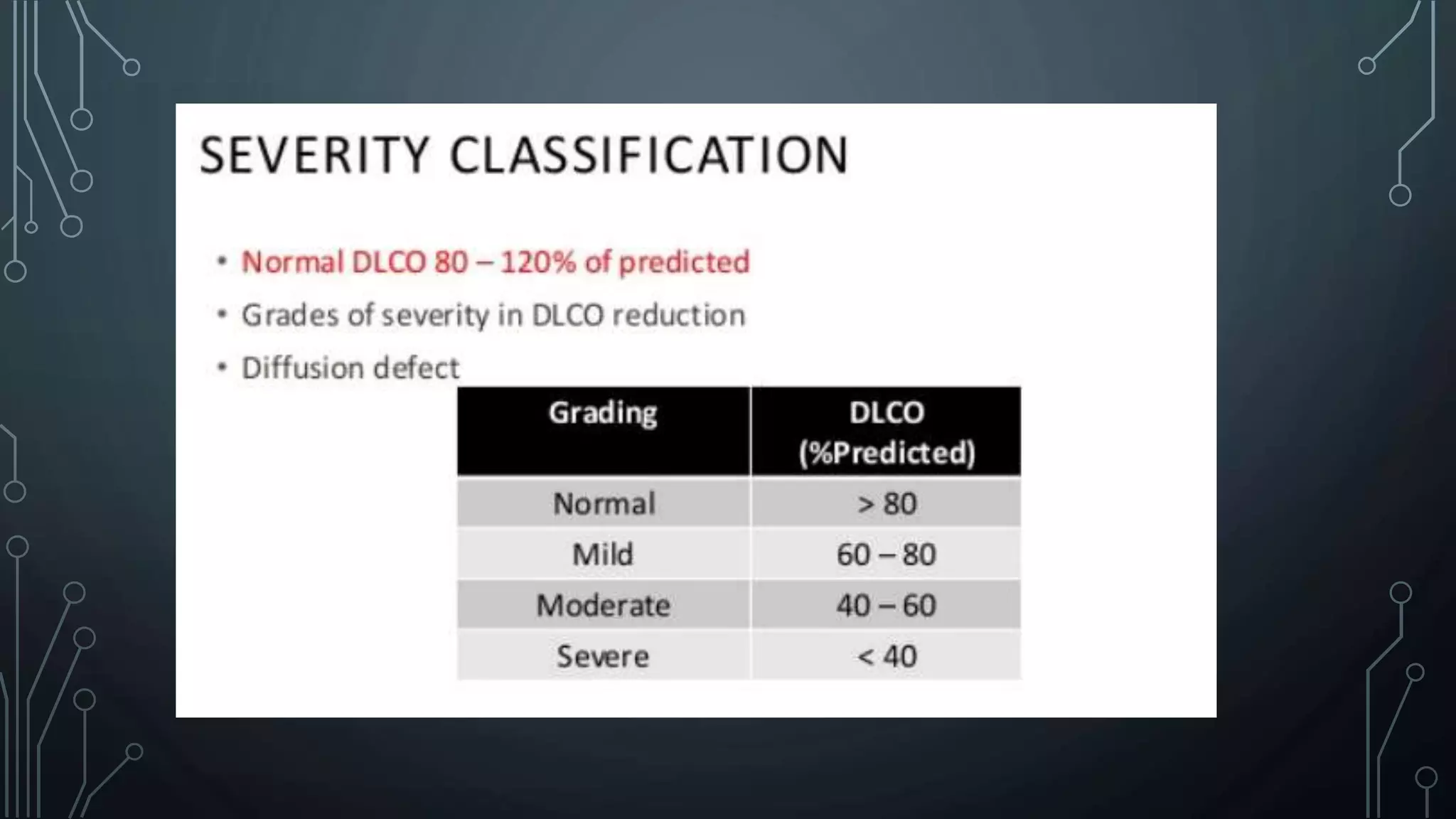
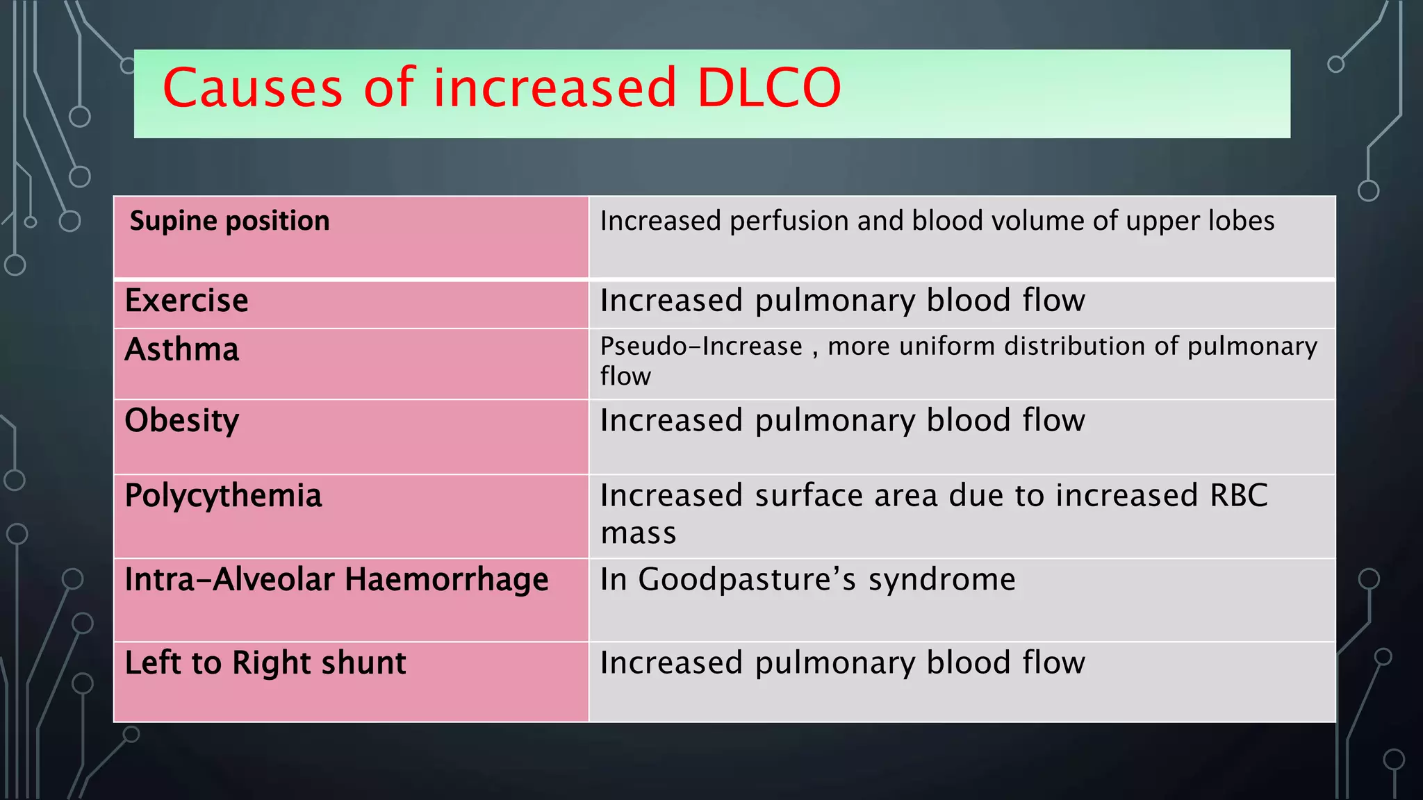
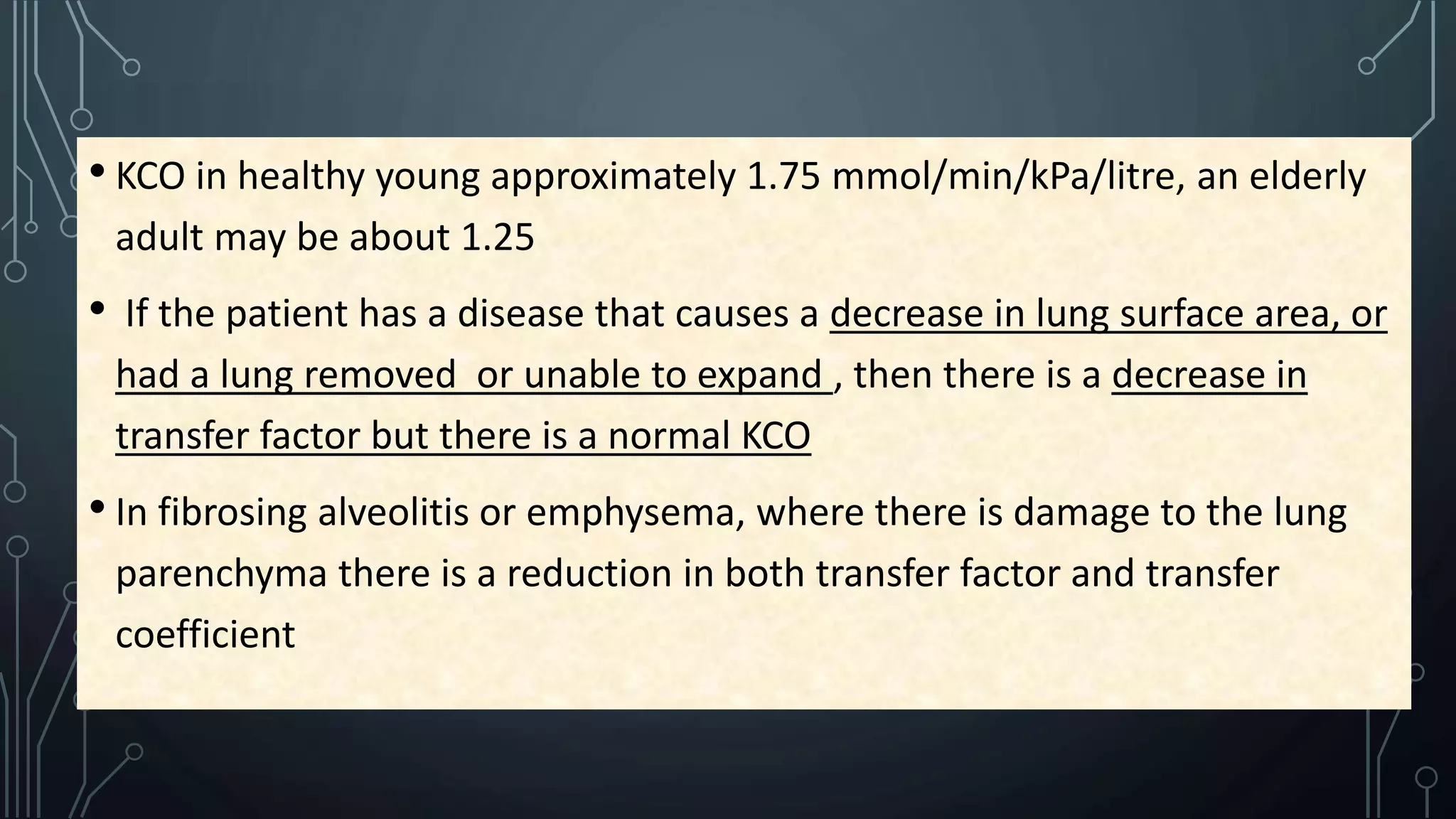
DLCO/TLCO
DIFFUSION CAPACITY OF LUNGS for CO
( DLCO ) or
TRANSFER FACTOR of LUNGS for CO
( TLCO )
Dr Tanveer Kamal Fahim
Phase B Resident , MD ( Pulmonology)
Medicine Unit VII , NIDCH
DLCO TLCO
Diffusion capacity of lungs for CO Transfer factor of lungs for CO
America Europe
Expressed as ml/min/mm of HG Expressed as mmol/min/KPa
DLCO/TLCO
•Diffusing capacity of the lungs estimates the ability of lungs to
transfer oxygen from alveolar gas to red cell
•Also provides objective measurement of lung function
•Originally described by Krogh in 1915
•Carbon monoxide is used as a surrogate for O2
•Mesures the partial pressure difference between the inspired and
exhaled CO
The amount of oxygen transferred is largely determined by three
factors :
• SURFACE AREA (A) of the ALVEOLAR-CAPILLARY MEMBRANE, which consists of the
alveolar and capillary walls
• THICKNESS (T) of the membrane
• DRIVING PRESSURE , that is , the difference in oxygen tension between the alveolar
gas and the venous blood (Δ PO2)
A*Δ PO2
T
Diffusion of Lung =
 Why use of CO ?
•Not normally present in alveoli/blood
•Transfer is diffusion limited rather than perfusion limited
•Avidly binds to Hb (210 times of Oxygen)
• As capillary PCO is very low normally , can be assumed to be negligible
and DlCO is calculated by dividing CO uptake (˙VCO) by alveolar PCO
•Harmless at low concentrations(<0.3%)
•Less affected by other factors
Oxygen :
• Perfusion limited
• Limited by ventilation perfusion mismatch , shunt etc
• Accurate measurement of PO2 during capillary transfer is very difficult
NO :
• Highly reactive with oxygen so requires special equipment
• Potential cardiovascular side effect
• Under research
Indications of DLCO
1. Categorize patients with Restrictive disease or Extrathoracic restriction
(obesity,neuromascular disease)
2. Identify early ILD in high risk patients ( H/O chest radiation,chronic Amiodarone etc)
3. To quantify anatomic emphysema
4. Before lung surgery
5. Pulmonary Vascular disease , chemotherapeutic agents
6. Response to treatment
7. Assess severity and progression
8. Disability documentation for legal purpose
Single – Breath Holding method most widely used and
standard
Procedure
1. Normal breathing ( upto RV)
2. Take deep breathing and blow out all in the air (upto TLC , panting )
3. Deep inspiration of supplied gas mixture and hold it for 10 s ( contains
Nitrogen , 0.3% CO , inert tracer gas Heliium , oxygen 18-21%)
4. Blow all the air out
5. Compute all
Contraindications
• Patient unable to perform maneuver, breath hold
•Smoking within 24 hr
•Vigorous exercise before test
•Significant desaturation
Normal value
Causes of increased DLCO
Supine position Increased perfusion and blood volume of upper lobes
Exercise Increased pulmonary blood flow
Asthma Pseudo-Increase , more uniform distribution of pulmonary
flow
Obesity Increased pulmonary blood flow
Polycythemia Increased surface area due to increased RBC
mass
Intra-Alveolar Haemorrhage In Goodpasture’s syndrome
Left to Right shunt Increased pulmonary blood flow
Causes of decreased DLCO
Reduced surface area-
• Anaemia
• Emphysema
• Lung/Lobe resection
• Bronchial obstruction by tumor
• Multiple emboli
 Other –Elderly, female , Smoking , Pregnancy ,
ventilation perfusion mismatch
Increased thickness of membrane
• IPF , Sarcoidosis , Asbestosis ,
Hypersensivity pneumonitis
• Heart Failure , Pulmonary HTN
• Collagen Vascular Disease-SLE ,
Scleroderma
• Drug induced fibrosis
Reduced DLCO
Anaemia Reduced Hb , reduced surface area
Emphysema Alveolar walls and capillaries are destroyed ,
reducing surface area
Emboli By blocking perfusion , reduces surface are
Bronchial obstruction Reduces lung area and volume
Heart failure lengthens the pathway for diffusion due to
fluid
Smoker high CO tension in blood educes
driving pressure
Isolated unexplained reduction of
DLCO with normal spirometry and
lung volume
Primary Pulmonary HTN, Emboli , Obliterative
Vasculopathy
•Adjusting DLCO
•For anaemia : adjusted downwards
•For polycythemia : adjusted upwards
• For low lung volumes : adjusted downwards
•For high lung volumes : adjusted upwards
CRITERIA for ACCEPTABILITY
•Inspired volume ⩾90% of the largest VC in the same test
session
•85% of test gas Inspired volume inhaled in < 4 second
•A stable calculated breath-hold for 10±2 seconds
•Sample collection completed within 4 second of the start of
exhalation
KCO- TRANSFER CO-EFFICIENT for CO
• KCO in healthy young approximately 1.75 mmol/min/kPa/litre, an elderly
adult may be about 1.25
• If the patient has a disease that causes a decrease in lung surface area, or
had a lung removed or unable to expand , then there is a decrease in
transfer factor but there is a normal KCO
• In fibrosing alveolitis or emphysema, where there is damage to the lung
parenchyma there is a reduction in both transfer factor and transfer
coefficient
•KCO increases with age
•Raised/normal KCO but normal/reduced DLCO :
kypho-scoliosis
Pneumonectomy,lobectomy
Neuromascular disaeses
Ankylosing spondylitis
Dlco/tlco

Dlco/tlco

  • 1.
  • 2.
    DIFFUSION CAPACITY OFLUNGS for CO ( DLCO ) or TRANSFER FACTOR of LUNGS for CO ( TLCO ) Dr Tanveer Kamal Fahim Phase B Resident , MD ( Pulmonology) Medicine Unit VII , NIDCH
  • 3.
    DLCO TLCO Diffusion capacityof lungs for CO Transfer factor of lungs for CO America Europe Expressed as ml/min/mm of HG Expressed as mmol/min/KPa
  • 4.
    DLCO/TLCO •Diffusing capacity ofthe lungs estimates the ability of lungs to transfer oxygen from alveolar gas to red cell •Also provides objective measurement of lung function •Originally described by Krogh in 1915 •Carbon monoxide is used as a surrogate for O2 •Mesures the partial pressure difference between the inspired and exhaled CO
  • 6.
    The amount ofoxygen transferred is largely determined by three factors : • SURFACE AREA (A) of the ALVEOLAR-CAPILLARY MEMBRANE, which consists of the alveolar and capillary walls • THICKNESS (T) of the membrane • DRIVING PRESSURE , that is , the difference in oxygen tension between the alveolar gas and the venous blood (Δ PO2) A*Δ PO2 T Diffusion of Lung =
  • 7.
     Why useof CO ? •Not normally present in alveoli/blood •Transfer is diffusion limited rather than perfusion limited •Avidly binds to Hb (210 times of Oxygen) • As capillary PCO is very low normally , can be assumed to be negligible and DlCO is calculated by dividing CO uptake (˙VCO) by alveolar PCO •Harmless at low concentrations(<0.3%) •Less affected by other factors
  • 8.
    Oxygen : • Perfusionlimited • Limited by ventilation perfusion mismatch , shunt etc • Accurate measurement of PO2 during capillary transfer is very difficult NO : • Highly reactive with oxygen so requires special equipment • Potential cardiovascular side effect • Under research
  • 9.
    Indications of DLCO 1.Categorize patients with Restrictive disease or Extrathoracic restriction (obesity,neuromascular disease) 2. Identify early ILD in high risk patients ( H/O chest radiation,chronic Amiodarone etc) 3. To quantify anatomic emphysema 4. Before lung surgery 5. Pulmonary Vascular disease , chemotherapeutic agents 6. Response to treatment 7. Assess severity and progression 8. Disability documentation for legal purpose
  • 11.
    Single – BreathHolding method most widely used and standard
  • 12.
    Procedure 1. Normal breathing( upto RV) 2. Take deep breathing and blow out all in the air (upto TLC , panting ) 3. Deep inspiration of supplied gas mixture and hold it for 10 s ( contains Nitrogen , 0.3% CO , inert tracer gas Heliium , oxygen 18-21%) 4. Blow all the air out 5. Compute all
  • 15.
    Contraindications • Patient unableto perform maneuver, breath hold •Smoking within 24 hr •Vigorous exercise before test •Significant desaturation
  • 16.
  • 20.
    Causes of increasedDLCO Supine position Increased perfusion and blood volume of upper lobes Exercise Increased pulmonary blood flow Asthma Pseudo-Increase , more uniform distribution of pulmonary flow Obesity Increased pulmonary blood flow Polycythemia Increased surface area due to increased RBC mass Intra-Alveolar Haemorrhage In Goodpasture’s syndrome Left to Right shunt Increased pulmonary blood flow
  • 21.
    Causes of decreasedDLCO Reduced surface area- • Anaemia • Emphysema • Lung/Lobe resection • Bronchial obstruction by tumor • Multiple emboli  Other –Elderly, female , Smoking , Pregnancy , ventilation perfusion mismatch Increased thickness of membrane • IPF , Sarcoidosis , Asbestosis , Hypersensivity pneumonitis • Heart Failure , Pulmonary HTN • Collagen Vascular Disease-SLE , Scleroderma • Drug induced fibrosis
  • 22.
    Reduced DLCO Anaemia ReducedHb , reduced surface area Emphysema Alveolar walls and capillaries are destroyed , reducing surface area Emboli By blocking perfusion , reduces surface are Bronchial obstruction Reduces lung area and volume Heart failure lengthens the pathway for diffusion due to fluid Smoker high CO tension in blood educes driving pressure Isolated unexplained reduction of DLCO with normal spirometry and lung volume Primary Pulmonary HTN, Emboli , Obliterative Vasculopathy
  • 24.
    •Adjusting DLCO •For anaemia: adjusted downwards •For polycythemia : adjusted upwards • For low lung volumes : adjusted downwards •For high lung volumes : adjusted upwards
  • 25.
    CRITERIA for ACCEPTABILITY •Inspiredvolume ⩾90% of the largest VC in the same test session •85% of test gas Inspired volume inhaled in < 4 second •A stable calculated breath-hold for 10±2 seconds •Sample collection completed within 4 second of the start of exhalation
  • 26.
  • 27.
    • KCO inhealthy young approximately 1.75 mmol/min/kPa/litre, an elderly adult may be about 1.25 • If the patient has a disease that causes a decrease in lung surface area, or had a lung removed or unable to expand , then there is a decrease in transfer factor but there is a normal KCO • In fibrosing alveolitis or emphysema, where there is damage to the lung parenchyma there is a reduction in both transfer factor and transfer coefficient
  • 28.
    •KCO increases withage •Raised/normal KCO but normal/reduced DLCO : kypho-scoliosis Pneumonectomy,lobectomy Neuromascular disaeses Ankylosing spondylitis