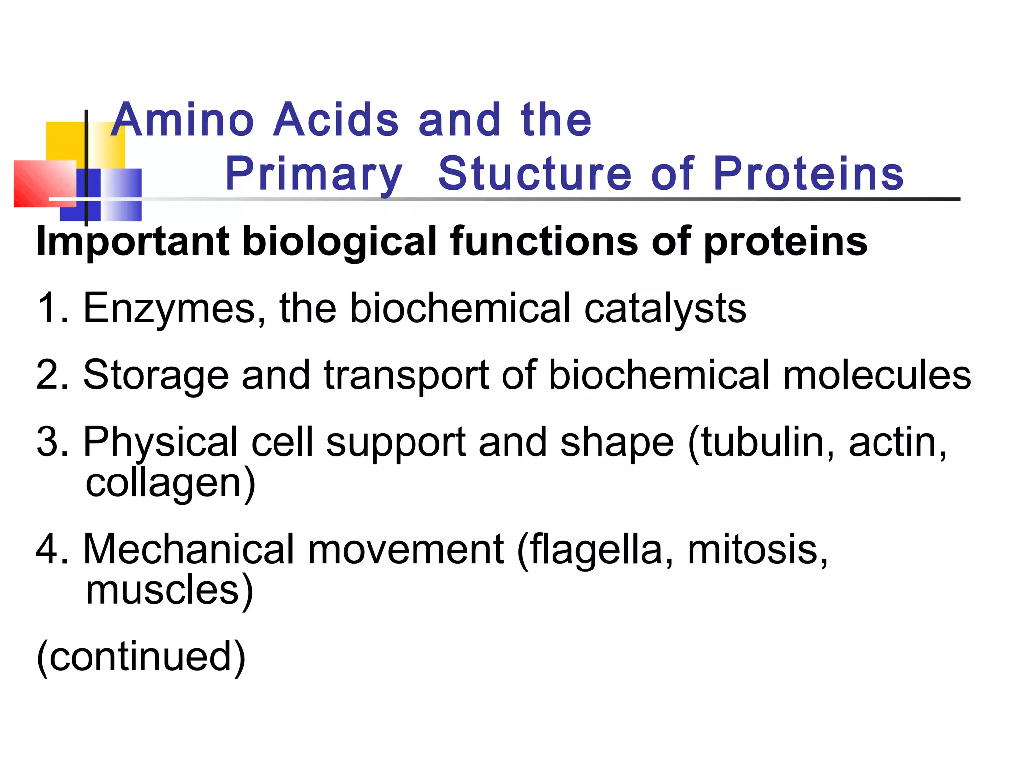
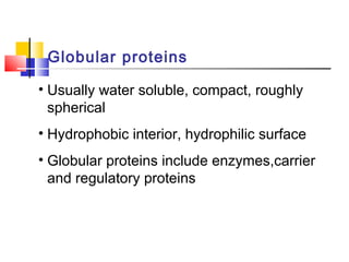
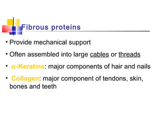
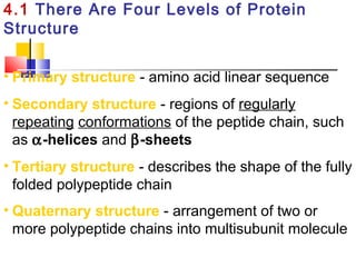
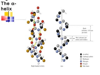
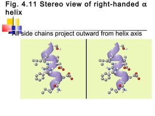
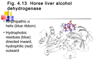
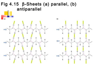
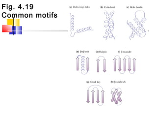
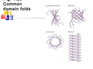
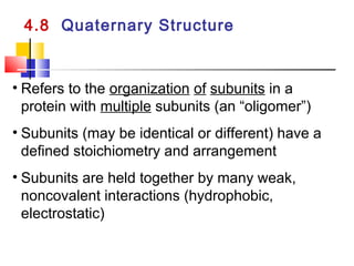
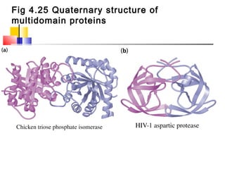
Amino Acids and the
Primary Stucture of Proteins
Important biological functions of proteins
1. Enzymes, the biochemical catalysts
2. Storage and transport of biochemical molecules
3. Physical cell support and shape (tubulin, actin,
collagen)
4. Mechanical movement (flagella, mitosis,
muscles)
(continued)
Globular proteins
• Usually water soluble, compact, roughly
spherical
• Hydrophobic interior, hydrophilic surface
• Globular proteins include enzymes,carrier
and regulatory proteins
Fibrous proteins
• Provide mechanical support
• Often assembled into large cables or threads
• α-Keratins: major components of hair and nails
• Collagen: major component of tendons, skin,
bones and teeth
General Structure of Amino Acids
• Twenty common α-amino acids have carboxyl
and amino groups bonded to the α-carbon atom
• A hydrogen atom and a side chain (R) are also
attached to the α-carbon atom
Zwitterionic form of amino acids
• Under normal cellular conditions amino
acids are zwitterions (dipolar ions):
Amino group = -NH3
+
Carboxyl group = -COO-
Stereochemistry of amino acids
• 19 of the 20 common amino acids have a
chiral α-carbon atom (Gly does not)
• Threonine and isoleucine have 2 chiral
carbons each (4 possible stereoisomers each)
• Mirror image pairs of amino acids are
designated L (levo) and D (dextro)
• Proteins are assembled from L-amino acids
(a few D-amino acids occur in nature)
Four aliphatic amino acid
structures
Proline has a nitrogen in the
aliphatic ring system
• Proline (Pro, P) - has a three
carbon side chain bonded to
the α-amino nitrogen
• The heterocyclic pyrrolidine
ring restricts the geometry of
polypeptides
Aromatic amino acid structures
Methionine and cysteine
Fig 3.4 Formation of cystine
D. Side Chains with Alcohol
Groups
• Serine (Ser, S) and Threonine (Thr, T) have
uncharged polar side chains
Structures of histidine, lysine and
arginine
Structures of aspartate,
glutamate, asparagine and
glutamine
G. The Hydrophobicity of
Amino Acid Side Chains
• Hydropathy: the relative hydrophobicity of each
amino acid
• The larger the hydropathy, the greater the
tendency of an amino acid to prefer a
hydrophobic environment
• Hydropathy affects protein folding:
hydrophobic side chains tend to be in the interior
hydrophilic residues tend to be on the surface
Table
3.1
• Hydropathy scale for
amino acid residues
(Free-energy change for
transfer of an amino acid
from interior of a lipid
bilayer to water)
Free-energy change
for transfer (kjmol-1
)
Amino
acid
Fig 3.5 Compounds derived from
common amino acids
Fig 3.6 Titration curve for alanine
• Titration curves
are used to
determine pKa
values
• pK1 = 2.4
• pK2 = 9.9
• pIAla = isoelectric
point
Fig 3.7 Ionization of Histidine
(a) Titration curve
of histidine
pK1 = 1.8
pK2 = 6.0
pK3 = 9.3
Fig 3.7 (b) Deprotonation of imidazolium
ring
Table
3.2
pKa values of
amino acid
ionizable groups
3.5 Peptide Bonds Link Amino Acids
in Proteins
• Peptide bond - linkage between amino acids
is a secondary amide bond
• Formed by condensation of the α-carboxyl of
one amino acid with the α-amino of another
amino acid (loss of H2O molecule)
• Primary structure - linear sequence of
amino acids in a polypeptide or protein
Fig 3.9 Peptide bond between
two amino acids
Polypeptide chain nomenclature
• Amino acid “residues” compose peptide chains
• Peptide chains are numbered from the N (amino)
terminus to the C (carboxyl) terminus
• Example: (N) Gly-Arg-Phe-Ala-Lys (C)
(or GRFAK)
• Formation of peptide bonds eliminates the
ionizable α-carboxyl and α-amino groups of the
free amino acids
Fig 3.10 Aspartame, an artificial
sweetener
• Aspartame is a
dipeptide methyl ester
(aspartylphenylalanine
methyl ester)
• About 200 times
sweeter than table
sugar
• Used in diet drinks
3.7 Amino Acid Composition of Proteins
• Amino acid analysis - determination of the
amino acid composition of a protein
• Peptide bonds are cleaved by acid hydrolysis
(6M HCl, 110o
, 16-72 hours)
• Amino acids are separated
chromatographically and quantitated
• Phenylisothiocyanate (PITC) used to derivatize
the amino acids prior to HPLC analysis
Fig 3.13 Acid-catalyzed hydrolysis of a
peptide
of the peptide
bond
(a) Peptide bond shown as a
C-N single bond
(b) Peptide bond shown as a
double bond
(c) Actual structure is a hybrid
of the two resonance
forms. Electrons are
delocalized over three
atoms: O, C, N
Fig. 4.6 Planar peptide groups in a
polypeptide chain
• Rotation around C-N bond is restricted due to the
double-bond nature of the resonance hybrid form
• Peptide groups (blue planes) are therefore planar
Fig. 4.7 Trans and cis conformations
of a peptide group
• Nearly all peptide groups in proteins are
in the trans conformation
4.1 There Are Four Levels of Protein
Structure
• Primary structure - amino acid linear sequence
• Secondary structure - regions of regularly
repeating conformations of the peptide chain, such
as α-helices and β-sheets
• Tertiary structure - describes the shape of the fully
folded polypeptide chain
• Quaternary structure - arrangement of two or
more polypeptide chains into multisubunit molecule
The α-
helix
Fig. 4.11 Stereo view of right-handed α
helix
• All side chains project outward from helix axis
Fig. 4.13 Horse liver alcohol
dehydrogenase
• Amphipathic α
helix (blue ribbon)
• Hydrophobic
residues (blue)
directed inward,
hydrophilic (red)
outward
Fig 4.15 β-Sheets (a) parallel, (b)
antiparallel
Fig. 4.19
Common motifs
Fig. 4.23
Common
domain folds
4.8 Quaternary Structure
• Refers to the organization of subunits in a
protein with multiple subunits (an “oligomer”)
• Subunits (may be identical or different) have a
defined stoichiometry and arrangement
• Subunits are held together by many weak,
noncovalent interactions (hydrophobic,
electrostatic)
Fig 4.25 Quaternary structure of
multidomain proteins
Fig. 4.42 Hemoglobin tetramer
(a) Human oxyhemoglobin (b) Tetramer schematic

Proteins basics

  • 1.
    Amino Acids andthe Primary Stucture of Proteins Important biological functions of proteins 1. Enzymes, the biochemical catalysts 2. Storage and transport of biochemical molecules 3. Physical cell support and shape (tubulin, actin, collagen) 4. Mechanical movement (flagella, mitosis, muscles) (continued)
  • 2.
    Globular proteins • Usuallywater soluble, compact, roughly spherical • Hydrophobic interior, hydrophilic surface • Globular proteins include enzymes,carrier and regulatory proteins
  • 3.
    Fibrous proteins • Providemechanical support • Often assembled into large cables or threads • α-Keratins: major components of hair and nails • Collagen: major component of tendons, skin, bones and teeth
  • 4.
    General Structure ofAmino Acids • Twenty common α-amino acids have carboxyl and amino groups bonded to the α-carbon atom • A hydrogen atom and a side chain (R) are also attached to the α-carbon atom
  • 5.
    Zwitterionic form ofamino acids • Under normal cellular conditions amino acids are zwitterions (dipolar ions): Amino group = -NH3 + Carboxyl group = -COO-
  • 6.
    Stereochemistry of aminoacids • 19 of the 20 common amino acids have a chiral α-carbon atom (Gly does not) • Threonine and isoleucine have 2 chiral carbons each (4 possible stereoisomers each) • Mirror image pairs of amino acids are designated L (levo) and D (dextro) • Proteins are assembled from L-amino acids (a few D-amino acids occur in nature)
  • 7.
    Four aliphatic aminoacid structures
  • 8.
    Proline has anitrogen in the aliphatic ring system • Proline (Pro, P) - has a three carbon side chain bonded to the α-amino nitrogen • The heterocyclic pyrrolidine ring restricts the geometry of polypeptides
  • 9.
  • 10.
  • 11.
  • 12.
    D. Side Chainswith Alcohol Groups • Serine (Ser, S) and Threonine (Thr, T) have uncharged polar side chains
  • 13.
    Structures of histidine,lysine and arginine
  • 14.
    Structures of aspartate, glutamate,asparagine and glutamine
  • 15.
    G. The Hydrophobicityof Amino Acid Side Chains • Hydropathy: the relative hydrophobicity of each amino acid • The larger the hydropathy, the greater the tendency of an amino acid to prefer a hydrophobic environment • Hydropathy affects protein folding: hydrophobic side chains tend to be in the interior hydrophilic residues tend to be on the surface
  • 16.
    Table 3.1 • Hydropathy scalefor amino acid residues (Free-energy change for transfer of an amino acid from interior of a lipid bilayer to water) Free-energy change for transfer (kjmol-1 ) Amino acid
  • 17.
    Fig 3.5 Compoundsderived from common amino acids
  • 18.
    Fig 3.6 Titrationcurve for alanine • Titration curves are used to determine pKa values • pK1 = 2.4 • pK2 = 9.9 • pIAla = isoelectric point
  • 19.
    Fig 3.7 Ionizationof Histidine (a) Titration curve of histidine pK1 = 1.8 pK2 = 6.0 pK3 = 9.3
  • 20.
    Fig 3.7 (b)Deprotonation of imidazolium ring
  • 21.
    Table 3.2 pKa values of aminoacid ionizable groups
  • 22.
    3.5 Peptide BondsLink Amino Acids in Proteins • Peptide bond - linkage between amino acids is a secondary amide bond • Formed by condensation of the α-carboxyl of one amino acid with the α-amino of another amino acid (loss of H2O molecule) • Primary structure - linear sequence of amino acids in a polypeptide or protein
  • 23.
    Fig 3.9 Peptidebond between two amino acids
  • 24.
    Polypeptide chain nomenclature •Amino acid “residues” compose peptide chains • Peptide chains are numbered from the N (amino) terminus to the C (carboxyl) terminus • Example: (N) Gly-Arg-Phe-Ala-Lys (C) (or GRFAK) • Formation of peptide bonds eliminates the ionizable α-carboxyl and α-amino groups of the free amino acids
  • 25.
    Fig 3.10 Aspartame,an artificial sweetener • Aspartame is a dipeptide methyl ester (aspartylphenylalanine methyl ester) • About 200 times sweeter than table sugar • Used in diet drinks
  • 26.
    3.7 Amino AcidComposition of Proteins • Amino acid analysis - determination of the amino acid composition of a protein • Peptide bonds are cleaved by acid hydrolysis (6M HCl, 110o , 16-72 hours) • Amino acids are separated chromatographically and quantitated • Phenylisothiocyanate (PITC) used to derivatize the amino acids prior to HPLC analysis
  • 27.
    Fig 3.13 Acid-catalyzedhydrolysis of a peptide
  • 28.
    of the peptide bond (a)Peptide bond shown as a C-N single bond (b) Peptide bond shown as a double bond (c) Actual structure is a hybrid of the two resonance forms. Electrons are delocalized over three atoms: O, C, N
  • 29.
    Fig. 4.6 Planarpeptide groups in a polypeptide chain • Rotation around C-N bond is restricted due to the double-bond nature of the resonance hybrid form • Peptide groups (blue planes) are therefore planar
  • 30.
    Fig. 4.7 Transand cis conformations of a peptide group • Nearly all peptide groups in proteins are in the trans conformation
  • 31.
    4.1 There AreFour Levels of Protein Structure • Primary structure - amino acid linear sequence • Secondary structure - regions of regularly repeating conformations of the peptide chain, such as α-helices and β-sheets • Tertiary structure - describes the shape of the fully folded polypeptide chain • Quaternary structure - arrangement of two or more polypeptide chains into multisubunit molecule
  • 32.
  • 33.
    Fig. 4.11 Stereoview of right-handed α helix • All side chains project outward from helix axis
  • 34.
    Fig. 4.13 Horseliver alcohol dehydrogenase • Amphipathic α helix (blue ribbon) • Hydrophobic residues (blue) directed inward, hydrophilic (red) outward
  • 35.
    Fig 4.15 β-Sheets(a) parallel, (b) antiparallel
  • 36.
  • 37.
  • 38.
    4.8 Quaternary Structure •Refers to the organization of subunits in a protein with multiple subunits (an “oligomer”) • Subunits (may be identical or different) have a defined stoichiometry and arrangement • Subunits are held together by many weak, noncovalent interactions (hydrophobic, electrostatic)
  • 39.
    Fig 4.25 Quaternarystructure of multidomain proteins
  • 40.
    Fig. 4.42 Hemoglobintetramer (a) Human oxyhemoglobin (b) Tetramer schematic