
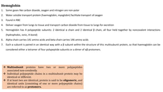
![Physical Properties
1. Most aa are soluble in water
2. Most of them melt above 200 degree
Centigrade
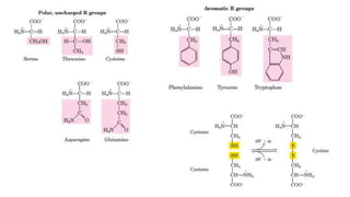
3. Sweet (Gly, Ala, Val), tasteless (Leu),
bitter (Arg, Ile) ..[Monosodium
glutamate in food]
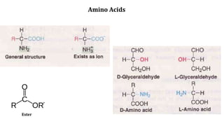
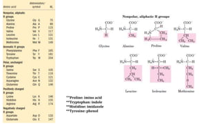
4. All aa except glycine possesses optical
isomer due to presence of asymmetric
Carbon
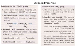
5. Aa are ampholytes – contain both –
COOH and –NH2, ie, can donate a
proton or can accept a proton. They
can exist as dipolar or Zwitterion
Isoelectric pH (pI): pH at which molecule
exist in Zwitterionic form.](https://image.slidesharecdn.com/aminoacidprotein-191018092150/85/Amino-Acids-and-Protein-8-320.jpg)
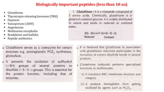
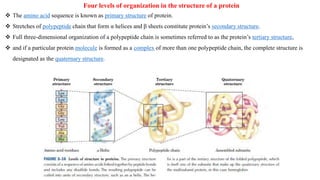
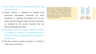
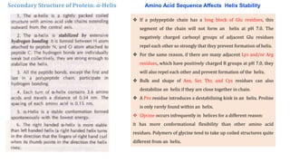
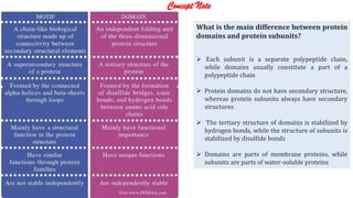
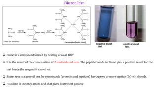
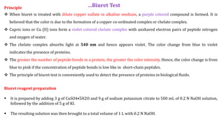
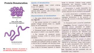


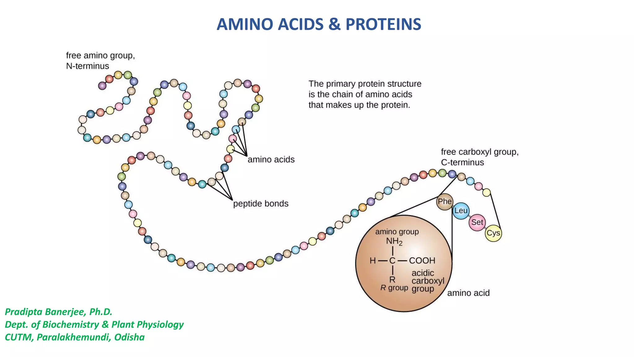
The document discusses the structure and function of amino acids and proteins, including their classification, absorption, and metabolic fate. It details the different types of proteins, their structural organization (primary, secondary, tertiary, and quaternary), and the bonds that stabilize these structures. Additionally, it covers biochemical tests like the Biuret test for detecting proteins and references various sources for further reading.


![Complete E material on Fundamentals of Biochemistry [2+1]; (32 Lectures)](https://cdn.slidesharecdn.com/ss_thumbnails/e-materialofbiochemistry-200405111750-thumbnail.jpg?width=640&height=640&fit=bounds)


