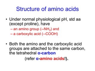
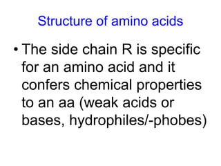
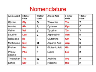
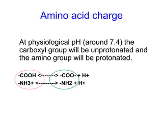
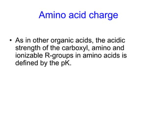
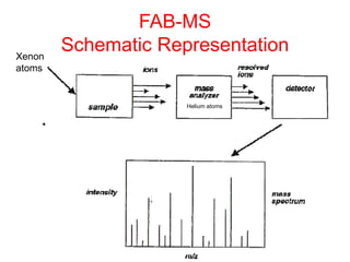
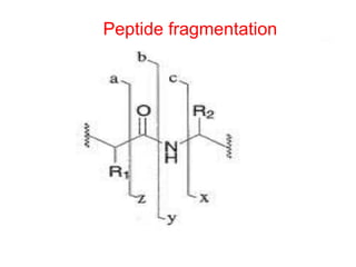
Amino acids
Introduction 
• All poly/peptides are polymers of amino 
acids 
• Naturally, there are more than 300 known 
amino acids 
• Only 20 amino acids occur in (mammalian) 
proteins through protein synthesis. They are 
also called 
– basic/primary/standard/proteinogenic 
– They are almost all L-α-amino acids
Introduction 
• In addition to the 20 standard amino acids 
coded by DNA, 2 additional amino acids 
are also DNA coded  in a non-standard 
manner. 
– Selenocysteine: in some proteins is coded by 
the stop codon UGA 
– Pyrrolysine: used by some methanogenic 
bacteria in methane producing enzyme 
systems. Coded for by UAG.
Introduction 
• The standard amino acids have a wide 
range of biological functions 
–Monomers of proteins through the 
standard genetic code-protein synthesis 
– Glycine and glutamate are also 
neurotransmitters. 
–Tyrosine is involved in the formation of 
thyroid hormone etc
Introduction 
– Standard amino acids also serve as 
precursors of other molecules 
• Tryptophan: precursor of the 
neurotransmitter serotonin 
• Glycine: is one of the reactants in the 
synthesis of porphyrins e.g. heme 
• Arginine: used in the synthesis of (the 
hormone) nitric oxide
Introduction 
• Other amino acids in proteins are usually 
formed by post-transcriptional 
modification of proteins, i.e. after 
translation. 
• Such modifications are often important for 
protein function
Non std aa: functions and 
occurrence 
• Occur as free or in combined states. 
• Commonly formed as modifications of std aa 
– E.g taurine is formed by decarboxylation of 
cysteine 
– Dopamine is made from tyrosine 
– Hydroxyproline (in collagen) is made from 
proline (post transcriptionally)
Non std aa: functions and 
occurrence 
• They serve specialized functions, 
– e.g. dopamine and γ-amino butyric acid 
(GABA) are neurotransmitters 
• They usually occur in the metabolic pathways for 
standard amino acids, e.g. 
– Ornithine and citruline: occur in the urea cycle 
(part of aa breakdown). 
– Carnitine is used in lipid transport within a cell 
etc.
Non std aa: functions and 
occurrence 
• Hundreds of other non proteinogenic 
α- amino acids have been identified in 
nature 
–E.g in carbonaceous chondrites 
(the primitive Murchison meteorite 
(>79 aa)), 
–in plants (nisin or alamethicin) etc
Structure of amino acids
Structure of amino acids 
• Under normal physiological pH, std aa 
(except proline), have 
– an amino group (--NH2) and 
– a carboxylic acid (--COOH) 
• Both the amino and the carboxylic acid 
groups are attached to the same carbon, 
the tetrahedral α-carbon 
(refer α-amino acids!).
Structure of amino acids 
• Amino acids are distinguished from one 
another by their R groups, attached to the α- 
carbon 
– One exception is glycine (gly) where the R is 
hydrogen 
• The 4th substitution is usually hydrogen 
• The α-carbon is chiral (except gly), ie is 
asymmetrical due to having 4 different groups 
attached (-NH2, -COOH, -R and -H)
Structure of amino acids 
• The side chain R is specific 
for an amino acid and it 
confers chemical properties 
to an aa (weak acids or 
bases, hydrophiles/-phobes)
Structure of amino acids 
• Understanding the R groups of aa is 
important in order to devise: 
– Methods of aa analysis 
– Methods of aa purification 
– Characterization and Identification of proteins 
• e.g. aromatic aa absorb UV light at 275-280nm. Trp has a maximal 
peak at 280nm etc. proteins can be sequenced and distinguished as 
in Functional Genomics.
Nomenclature of amino acids
Nomenclature 
Amino Acid 3 letter 
code 
1 letter 
code 
Amino Acid 3 letter 
code 
1 letter 
code 
Glycine Gly G Threonine Thr T 
Alanine Ala A Cysteine Cys C 
Valine Val V Tyrosine Tyr Y 
Leucine Leu L Asparagine Asn N 
Isoleucine Ile I Glutamine Gln Q 
Methionine Met M Aspartic Acid Asp D 
Proline Pro P Glutamic Acid Glu E 
Phenyl 
Phe F Lysine Lys K 
alanine 
Tryptophan Trp W Arginine Arg R 
Serine Ser S Histidine His H
Classification
Classification 
• R polarity: 
1. hydrophilic (if R is polar) 
Interact with aqueous 
environment, Involved in H-bond 
formation. Found on exterior 
parts of proteins & reactive sites 
of enzymes 
2. hydrophobic (if R is non-polar) 
Reside in the interior of 
proteins,They do not ionizedo 
not participate in H-bond 
formation
Hydrophobic 
and Neutral 
at pH=7 
Hydrophilic 
and Neutral 
at pH=7
Classification 
• Acid base reaction: Depends on 
ionizability of 
1. # of Amino groups 
2. # of Carboxylic groups 
3. R group properties
Classification 
Acidic 
(Two Carboxylic Groups) 
Basic 
(more than one basic group)
Amino acid charge 
At physiological pH (around 7.4) the 
carboxyl group will be unprotonated and 
the amino group will be protonated. 
-COOH <--------> -COO- + H+ 
-NH3+ <---------> -NH2 + H+
Amino acid charge 
• An amino acid with 
no ionizable R-group 
would be 
electrically neutral 
at this pH (ie + and 
– charge balanced). 
• This species is 
called a zwitterion.
Amino acid charge 
• As in other organic acids, the acidic 
strength of the carboxyl, amino and 
ionizable R-groups in amino acids is 
defined by the pK.
Amino acid charge 
• The net charge of an amino acid will thus be the 
summation of all the charges in the amino acid at a 
specified pH 
• Amino acids have different charges at different pH 
value of the aqueous medium around 
• When the net charge of an amino acid or protein is 
zero the pH will be equivalent to the isoelectric 
point: pI.
Amino acid charge 
NOTE: 
• At its isoelectric pH (pI) an amino acid 
bears no net charge (The net – and + 
charge balances)
Titration curve of leucine 
pI = pK1 + pK2 
2
pI = pK2 + pK3 
2 
Titration of Histidine
NOTE THAT, YOU 
• Know the structure of an amino acid. 
• Know why amino acids are optically active. 
• Know why some amino acids are called 
neutral. 
• Know why some amino acids are called 
acidic and others basic, hydrophobic/phillic. 
• Know a zwitterion 
• Know the definition of an isoelectric point and 
how this point is calculated for amino acids.
Peptides 
and 
Peptide Bonds
Definitions 
– Peptide bond=the bond between 2 amino acids 
– Peptide=a molecule made of the union of aa joined by 
peptide bonds 
• Di-ppd= 2 amino acid residues/1 peptide bond 
• Tri-ppd= 3 amino acid residues/2 peptide bonds 
• Tetra--=? Etc 
– Residue=an amino acid in a peptide whose carboxyl 
group has been used in a peptide bond 
• Named by replacing the –ate or ine end of aa with –yl 
• Eg alanine=alanyl, cycteine=cyctenyl etc 
aspartate= ? 
Also called aminoacyls
Peptides 
IMPORTANCE 
• Hormones eg insulin 
• Neurotransmitters/modulators eg 
• Drugs (antibiotics) eg valinomycin 
• Beverage sweeterners eg aspartame 
• Some mediate tumor development
• Naming of peptides 
• Peptides are named as derivatives of the amino 
acid with intact –COOH 
– Eg: Alanyl-aspartyl-histidyl-valine 
– It means the whole residue sequence is a derivative 
of valine (valine has an intact –COOH in the above 
sequence) 
– Conventinal presentation of peptides: N---------C, 
– Ie N terminus on the left and the C terminus on the 
right
Peptide bond formation 
• Condensation reaction that result in 
polymerization of aapeptideprotein 
• It involves the alfa amino group of one aa 
and the alfa carboxyl group of the other, 
with elimination of a water molecule
H2O
Electron Resonance Stability of 
peptide bonds 
• The presence of a 
carbonyl group in the 
peptide bond allows 
for ERS 
• The peptide bond 
shows rigidity, ie has 
a partial double bond 
character
Characteristics of the peptide 
bond 
• Due to partial double bond, there is no 
freedom of rotation around C—N 
• All 4 atoms of the bond lie on the same 
plane- it is coplanar 
– There is free rotation in the rest of the bonds 
in the peptide backbone 
– Thus 2/3 of atoms in a peptide are co-planar 
while 1/3 have free rotation 
• Peptides are therefore semi-rigid
Abnormal peptide bonds 
• Non alfa peptidyl bonds may occur in 
peptides 
– Eg in thyrotropin releasing hormone (TRH) 
where the glutamate is made into a cycle 
(pyroglutamate) and the carboxyl end is 
‘amidated’ (pyrolyl carboxyl) 
– In glutathione
Some peptides of biological 
importance 
• Glutathione 
– A tripeptide containing glutamic acid, cystein 
and glycine 
– Present in RBC and other cells to maintain 
optimal redox potentials 
– Anhances/optimizes the action of enzymes 
(eg glutathione reductase) by preventing 
oxidation of their –SH to –SS
Some peptides of biological 
importance 
• Bradykinin and Kallidin 
– 9 and 10 residues respectively 
– From partial hydrolysis of plasma proteins by 
snake venoms (hemolytic venoms) 
– Powerful vasodepressors and cardiac 
inhibitors
Some peptides of biological 
importance 
• Tyrocidin and Gramicidin 
– Cyclic peptides, 10 aa each 
– They form rings their terminal 2 amino acid 
residues 
– They are useful antibiotics
Some peptides of biological 
importance 
• Peptide hormones and factors 
– Posterior Pituitary-oxytocin and vassopressin 
– Hypothalamic hormones-somatostatin 
– Intestinal hormones-gastrin 
– Kidney and brain factors-hypertensin and 
enkephalins
• PLEASE NOTE: 
– Do you know what is a peptide and peptide 
bond? 
– Dou you know an amino acid and an amino 
acid residue? 
– Dou you know how to name residues and 
peptides? 
– Peptide bond formation? 
– Do you know the features of a peptide bond? 
– Do you know why peptides are semi-rigid?
The primary structure of 
proteins
Primary structure 
• Refers to 
– the number and the order of amino acid 
residues in the peptide/polypeptide 
– This is the linear sequence of residues
Primary structure and biological 
• Mutations 
activity 
– SNPs: DNA changechange in the order & 
sometimes the number of amino acid 
residues. 
eg 
1. Alcohol intolerance in many Asians 
2. Susceptibility to malaria (K189M in the 
ICAM-1 gene susceptibility to cerebral 
malaria)
Primary structure Nomenclature 
• Both 3 letter and single letter systems 
used to determine primary structures of 
ppds (remember aa nomenclature) 
• In the 3 letter system, residues are joined 
by a dash/line: Gly-Tyr-Ala-Glu-Lys 
• In the 1 letter system the same ppd 
becomes: GYAEK, ie no lines
Primary structure Nomenclature 
• If uncertain about a residue, it is enclosed 
in brackets and the possible residues 
separated by a comma 
– Thus Gly- (Tyr,Ala)-Glu-Lys
Charge on peptides 
• Formation of the peptide bond involves the 
loss of +1 and -1 =0 charge. 
• Peptide bonds are neutral at any pH 
• At pH 7.4, peptides are charged due to 
– N-terminal group 
– C-terminal group 
– Basic or acidic R groups
Determination of primary structure 
• Sanger worked with insulin in the 60s. 
–Separated the 2 chains, A and B, 
cleaved the peptides into smaller 
peptides with overlapping sequences 
– Using 1-fluoro-2,4-dinitrobenzene, he 
could remove and identify each aa at a 
time
Protein 
mixture 
Stages of sequencing 
Proteins 
Peptide 
mixture 
Peptides MS analysis 
MS data 
Identification 
separation 
digestion digestion 
separation 
Database search 
algorithms
Stages of sequencing 
• Purify peptides and asses and purify by 
any suitable technique e.g. SDS-PAGE, 
chromatography 
• Homogeneity should be 90-95%
FAB-MS 
Schematic Representation 
Xenon 
atoms 
Helium atoms
Peptide fragmentation
From public online data banks
Applications of sequencing 
To study 
Protein functions, localization, modifications 
and protein interactions 
Also therapeutics and diagnostics
• DO YOU KNOW 
– The biological implications of the primary 
structure of proteins (peptides) 
– Nomenclature of peptides? 
– Basic steps of sequencing a peptide? 
– Importance of peptide sequencing? 
– The principle of FAB MS in peptide 
sequencing? 
• Then GOOD DAY!

Amino acid

  • 1.
  • 2.
    Introduction • Allpoly/peptides are polymers of amino acids • Naturally, there are more than 300 known amino acids • Only 20 amino acids occur in (mammalian) proteins through protein synthesis. They are also called – basic/primary/standard/proteinogenic – They are almost all L-α-amino acids
  • 3.
    Introduction • Inaddition to the 20 standard amino acids coded by DNA, 2 additional amino acids are also DNA coded  in a non-standard manner. – Selenocysteine: in some proteins is coded by the stop codon UGA – Pyrrolysine: used by some methanogenic bacteria in methane producing enzyme systems. Coded for by UAG.
  • 4.
    Introduction • Thestandard amino acids have a wide range of biological functions –Monomers of proteins through the standard genetic code-protein synthesis – Glycine and glutamate are also neurotransmitters. –Tyrosine is involved in the formation of thyroid hormone etc
  • 5.
    Introduction – Standardamino acids also serve as precursors of other molecules • Tryptophan: precursor of the neurotransmitter serotonin • Glycine: is one of the reactants in the synthesis of porphyrins e.g. heme • Arginine: used in the synthesis of (the hormone) nitric oxide
  • 6.
    Introduction • Otheramino acids in proteins are usually formed by post-transcriptional modification of proteins, i.e. after translation. • Such modifications are often important for protein function
  • 7.
    Non std aa:functions and occurrence • Occur as free or in combined states. • Commonly formed as modifications of std aa – E.g taurine is formed by decarboxylation of cysteine – Dopamine is made from tyrosine – Hydroxyproline (in collagen) is made from proline (post transcriptionally)
  • 8.
    Non std aa:functions and occurrence • They serve specialized functions, – e.g. dopamine and γ-amino butyric acid (GABA) are neurotransmitters • They usually occur in the metabolic pathways for standard amino acids, e.g. – Ornithine and citruline: occur in the urea cycle (part of aa breakdown). – Carnitine is used in lipid transport within a cell etc.
  • 9.
    Non std aa:functions and occurrence • Hundreds of other non proteinogenic α- amino acids have been identified in nature –E.g in carbonaceous chondrites (the primitive Murchison meteorite (>79 aa)), –in plants (nisin or alamethicin) etc
  • 11.
  • 12.
    Structure of aminoacids • Under normal physiological pH, std aa (except proline), have – an amino group (--NH2) and – a carboxylic acid (--COOH) • Both the amino and the carboxylic acid groups are attached to the same carbon, the tetrahedral α-carbon (refer α-amino acids!).
  • 13.
    Structure of aminoacids • Amino acids are distinguished from one another by their R groups, attached to the α- carbon – One exception is glycine (gly) where the R is hydrogen • The 4th substitution is usually hydrogen • The α-carbon is chiral (except gly), ie is asymmetrical due to having 4 different groups attached (-NH2, -COOH, -R and -H)
  • 14.
    Structure of aminoacids • The side chain R is specific for an amino acid and it confers chemical properties to an aa (weak acids or bases, hydrophiles/-phobes)
  • 15.
    Structure of aminoacids • Understanding the R groups of aa is important in order to devise: – Methods of aa analysis – Methods of aa purification – Characterization and Identification of proteins • e.g. aromatic aa absorb UV light at 275-280nm. Trp has a maximal peak at 280nm etc. proteins can be sequenced and distinguished as in Functional Genomics.
  • 16.
  • 17.
    Nomenclature Amino Acid3 letter code 1 letter code Amino Acid 3 letter code 1 letter code Glycine Gly G Threonine Thr T Alanine Ala A Cysteine Cys C Valine Val V Tyrosine Tyr Y Leucine Leu L Asparagine Asn N Isoleucine Ile I Glutamine Gln Q Methionine Met M Aspartic Acid Asp D Proline Pro P Glutamic Acid Glu E Phenyl Phe F Lysine Lys K alanine Tryptophan Trp W Arginine Arg R Serine Ser S Histidine His H
  • 18.
  • 19.
    Classification • Rpolarity: 1. hydrophilic (if R is polar) Interact with aqueous environment, Involved in H-bond formation. Found on exterior parts of proteins & reactive sites of enzymes 2. hydrophobic (if R is non-polar) Reside in the interior of proteins,They do not ionizedo not participate in H-bond formation
  • 20.
    Hydrophobic and Neutral at pH=7 Hydrophilic and Neutral at pH=7
  • 21.
    Classification • Acidbase reaction: Depends on ionizability of 1. # of Amino groups 2. # of Carboxylic groups 3. R group properties
  • 22.
    Classification Acidic (TwoCarboxylic Groups) Basic (more than one basic group)
  • 23.
    Amino acid charge At physiological pH (around 7.4) the carboxyl group will be unprotonated and the amino group will be protonated. -COOH <--------> -COO- + H+ -NH3+ <---------> -NH2 + H+
  • 24.
    Amino acid charge • An amino acid with no ionizable R-group would be electrically neutral at this pH (ie + and – charge balanced). • This species is called a zwitterion.
  • 25.
    Amino acid charge • As in other organic acids, the acidic strength of the carboxyl, amino and ionizable R-groups in amino acids is defined by the pK.
  • 26.
    Amino acid charge • The net charge of an amino acid will thus be the summation of all the charges in the amino acid at a specified pH • Amino acids have different charges at different pH value of the aqueous medium around • When the net charge of an amino acid or protein is zero the pH will be equivalent to the isoelectric point: pI.
  • 27.
    Amino acid charge NOTE: • At its isoelectric pH (pI) an amino acid bears no net charge (The net – and + charge balances)
  • 28.
    Titration curve ofleucine pI = pK1 + pK2 2
  • 29.
    pI = pK2+ pK3 2 Titration of Histidine
  • 30.
    NOTE THAT, YOU • Know the structure of an amino acid. • Know why amino acids are optically active. • Know why some amino acids are called neutral. • Know why some amino acids are called acidic and others basic, hydrophobic/phillic. • Know a zwitterion • Know the definition of an isoelectric point and how this point is calculated for amino acids.
  • 31.
  • 32.
    Definitions – Peptidebond=the bond between 2 amino acids – Peptide=a molecule made of the union of aa joined by peptide bonds • Di-ppd= 2 amino acid residues/1 peptide bond • Tri-ppd= 3 amino acid residues/2 peptide bonds • Tetra--=? Etc – Residue=an amino acid in a peptide whose carboxyl group has been used in a peptide bond • Named by replacing the –ate or ine end of aa with –yl • Eg alanine=alanyl, cycteine=cyctenyl etc aspartate= ? Also called aminoacyls
  • 33.
    Peptides IMPORTANCE •Hormones eg insulin • Neurotransmitters/modulators eg • Drugs (antibiotics) eg valinomycin • Beverage sweeterners eg aspartame • Some mediate tumor development
  • 34.
    • Naming ofpeptides • Peptides are named as derivatives of the amino acid with intact –COOH – Eg: Alanyl-aspartyl-histidyl-valine – It means the whole residue sequence is a derivative of valine (valine has an intact –COOH in the above sequence) – Conventinal presentation of peptides: N---------C, – Ie N terminus on the left and the C terminus on the right
  • 35.
    Peptide bond formation • Condensation reaction that result in polymerization of aapeptideprotein • It involves the alfa amino group of one aa and the alfa carboxyl group of the other, with elimination of a water molecule
  • 36.
  • 37.
    Electron Resonance Stabilityof peptide bonds • The presence of a carbonyl group in the peptide bond allows for ERS • The peptide bond shows rigidity, ie has a partial double bond character
  • 39.
    Characteristics of thepeptide bond • Due to partial double bond, there is no freedom of rotation around C—N • All 4 atoms of the bond lie on the same plane- it is coplanar – There is free rotation in the rest of the bonds in the peptide backbone – Thus 2/3 of atoms in a peptide are co-planar while 1/3 have free rotation • Peptides are therefore semi-rigid
  • 40.
    Abnormal peptide bonds • Non alfa peptidyl bonds may occur in peptides – Eg in thyrotropin releasing hormone (TRH) where the glutamate is made into a cycle (pyroglutamate) and the carboxyl end is ‘amidated’ (pyrolyl carboxyl) – In glutathione
  • 41.
    Some peptides ofbiological importance • Glutathione – A tripeptide containing glutamic acid, cystein and glycine – Present in RBC and other cells to maintain optimal redox potentials – Anhances/optimizes the action of enzymes (eg glutathione reductase) by preventing oxidation of their –SH to –SS
  • 42.
    Some peptides ofbiological importance • Bradykinin and Kallidin – 9 and 10 residues respectively – From partial hydrolysis of plasma proteins by snake venoms (hemolytic venoms) – Powerful vasodepressors and cardiac inhibitors
  • 43.
    Some peptides ofbiological importance • Tyrocidin and Gramicidin – Cyclic peptides, 10 aa each – They form rings their terminal 2 amino acid residues – They are useful antibiotics
  • 44.
    Some peptides ofbiological importance • Peptide hormones and factors – Posterior Pituitary-oxytocin and vassopressin – Hypothalamic hormones-somatostatin – Intestinal hormones-gastrin – Kidney and brain factors-hypertensin and enkephalins
  • 45.
    • PLEASE NOTE: – Do you know what is a peptide and peptide bond? – Dou you know an amino acid and an amino acid residue? – Dou you know how to name residues and peptides? – Peptide bond formation? – Do you know the features of a peptide bond? – Do you know why peptides are semi-rigid?
  • 46.
  • 47.
    Primary structure •Refers to – the number and the order of amino acid residues in the peptide/polypeptide – This is the linear sequence of residues
  • 48.
    Primary structure andbiological • Mutations activity – SNPs: DNA changechange in the order & sometimes the number of amino acid residues. eg 1. Alcohol intolerance in many Asians 2. Susceptibility to malaria (K189M in the ICAM-1 gene susceptibility to cerebral malaria)
  • 49.
    Primary structure Nomenclature • Both 3 letter and single letter systems used to determine primary structures of ppds (remember aa nomenclature) • In the 3 letter system, residues are joined by a dash/line: Gly-Tyr-Ala-Glu-Lys • In the 1 letter system the same ppd becomes: GYAEK, ie no lines
  • 50.
    Primary structure Nomenclature • If uncertain about a residue, it is enclosed in brackets and the possible residues separated by a comma – Thus Gly- (Tyr,Ala)-Glu-Lys
  • 51.
    Charge on peptides • Formation of the peptide bond involves the loss of +1 and -1 =0 charge. • Peptide bonds are neutral at any pH • At pH 7.4, peptides are charged due to – N-terminal group – C-terminal group – Basic or acidic R groups
  • 52.
    Determination of primarystructure • Sanger worked with insulin in the 60s. –Separated the 2 chains, A and B, cleaved the peptides into smaller peptides with overlapping sequences – Using 1-fluoro-2,4-dinitrobenzene, he could remove and identify each aa at a time
  • 53.
    Protein mixture Stagesof sequencing Proteins Peptide mixture Peptides MS analysis MS data Identification separation digestion digestion separation Database search algorithms
  • 54.
    Stages of sequencing • Purify peptides and asses and purify by any suitable technique e.g. SDS-PAGE, chromatography • Homogeneity should be 90-95%
  • 56.
    FAB-MS Schematic Representation Xenon atoms Helium atoms
  • 57.
  • 58.
  • 59.
    Applications of sequencing To study Protein functions, localization, modifications and protein interactions Also therapeutics and diagnostics
  • 60.
    • DO YOUKNOW – The biological implications of the primary structure of proteins (peptides) – Nomenclature of peptides? – Basic steps of sequencing a peptide? – Importance of peptide sequencing? – The principle of FAB MS in peptide sequencing? • Then GOOD DAY!