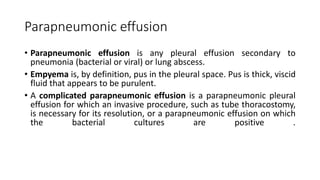
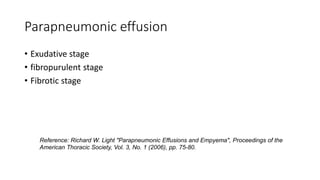
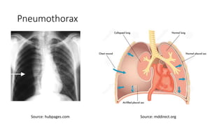
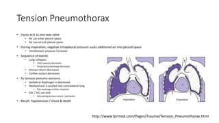
Parapneumonic effusion and
Pneumothorax
Pratap Sagar Tiwari, MD, Internal Medicine
Note: This is lecture class slide for MBBS students
Parapneumonic effusion
• Parapneumonic effusion is any pleural effusion secondary to
pneumonia (bacterial or viral) or lung abscess.
• Empyema is, by definition, pus in the pleural space. Pus is thick, viscid
fluid that appears to be purulent.
• A complicated parapneumonic effusion is a parapneumonic pleural
effusion for which an invasive procedure, such as tube thoracostomy,
is necessary for its resolution, or a parapneumonic effusion on which
the bacterial cultures are positive .
Complicated Pleural effusion
• Positive bacterial studies
• a glucose level < 60 mg/dl
• a pH < 7.20.
• a lactic acid dehydrogenase (LDH) level of >three times the upper
normal limit of serum.
Parapneumonic effusion
• Exudative stage
• fibropurulent stage
• Fibrotic stage
Reference: Richard W. Light "Parapneumonic Effusions and Empyema", Proceedings of the
American Thoracic Society, Vol. 3, No. 1 (2006), pp. 75-80.
Parapneumonic effusion: exudative stage
• The first stage is the exudative stage in which there is rapid
outpouring of fluid into the pleural space.
• Most of the fluid is due to increased pulmonary interstitial fluid
traversing the pleura to enter the pleural space but some of this is
due to increased permeability of the capillaries in the pleural space.
• The pleural fluid in this stage is characterized by negative bacterial
studies, a glucose level >60 mg/dl, a pH >7.20, and LDH level of < 3X
the upper normal limit of serum.
Parapneumonic effusion: fibropurulent stage
• If untreated, the effusion may proceed to the second stage, which is
the fibropurulent stage.
• The pleural fluid in this stage is characterized by positive bacterial
studies, a glucose level <60 mg/dl, a pH <7.20, and a pleural fluid LDH
>3 times the upper normal limit for serum.
• In this stage, the pleural fluid becomes infected and progressively
loculated.
• The pleural fluid needs to be drained in this stage and drainage
becomes progressively difficult as more loculations form.
Parapneumonic effusion: fibrotic stage
• If a stage 2 effusion is not drained, the effusion may progress to the
third stage in which fibroblasts grow into the pleural fluid from both
the visceral and parietal pleurae, producing a thick pleural peel. The
peel over the visceral pleura prevents the lung from expanding.
• Because the pleural space must be eradicated if a pleural infection is
going to be eliminated, this peel must be removed if the infection is
going to be cured.
Pneumothorax
Pneumothorax
• The term ‘pneumothorax’ was first coined by Itard and then Laennec in
1803 and 1819 respectively, (1) and refers to air in the pleural cavity (ie,
interspersed between the lung and the chest wall).
• Primary spontaneous pneumothorax (PSP): occurring in absence of known
lung disease.
• Secondary pneumothorax (SSP) is associated with underlying lung disease
most commonly COPD.
• Subpleural blebs and bullae are found at the lung apices at thoracoscopy
and on CT scanning in up to 90% of cases of PSP,(5) and are thought to
play a role.
1. Laennec RTH. Traite´ du diagnostic des maladies des poumons et du coeur. Tome Second, Paris: Brosson and Chaude´, 1819.
2. Donahue DM, Wright CD, Viale G, et al. Resection of pulmonary blebs and pleurodesis for spontaneous pneumothorax. Chest 1993;104:1767e9.
Smoking: a risk factor for pneumothorax
• Smoking has been implicated in this aetiological pathway, the
smoking habit being associated with a 12% risk of developing
pneumothorax in healthy smoking men compared with 0.1% in
nonsmokers. 1
• Ref: 1. Bense L, Eklund G, Odont D, et al. Smoking and the increased risk of contracting pneumothorax. Chest 1987;92:1009e12.
Risks factors for PSP include the following:
1. Smoking
2. Tall, thin stature in a healthy person
3. Marfan syndrome
4. Pregnancy
5. Familial pneumothorax
6. Blebs and bullae
• Typical PSP patients also tend to have a tall and thin body habitus. Whether
height affects development of subpleural blebs or whether more negative apical
pleural pressures cause preexisting blebs to rupture is unclear.
• Pregnancy is an unrecognized risk factor, as suggested by a 10-year retrospective
series in which 5 of 250 spontaneous pneumothorax cases were in pregnant
women.
Signs and symptoms
• The presentation of patients with pneumothorax varies depending on the
following types of pneumothorax and ranges from completely asymptomatic to
life-threatening respiratory distress:
• Spontaneous pneumothorax: No clinical signs or symptoms in primary
spontaneous pneumothorax until a bleb ruptures and causes pneumothorax;
typically, the result is acute onset of chest pain and SOB, particularly with SSP
• Iatrogenic pneumothorax: Symptoms similar to those of spontaneous
pneumothorax, depending on patient’s age, presence of underlying lung disease,
and extent of pneumothorax
• Tension pneumothorax: Hypotension, hypoxia, chest pain, dyspnea
• Catamenial pneumothorax: Women aged 30-40 years with onset of symptoms
within 48 hours of menstruation, right-sided pneumothorax, and recurrence.
Management : SP
• Breathlessness indicates the need for active intervention(needle aspiration or chest tube
drainage) as well as supportive treatment (including oxygen).
• Patients with PSP or SSP and significant breathlessness associated with any size of pneumothorax
should undergo active intervention.
• The size of the pneumothorax determines the rate of resolution and is a relative indication for
active intervention.
• For small PSP: Observation is the treatment of choice if without significant breathlessness.
• All patients with SSP should be admitted to hospital for at least 24 h and receive supplemental
oxygen and most will require chest tube drainage.
• For recurrent or persistent air leak : Surgical treatment
• The surgical treatments usually involve pleurodesis (in which the layers of pleura are induced to
stick together) or pleurectomy (the surgical removal of pleural membranes).
• Tetracycline used to be recommended as the first-line sclerosant therapy for both PSP and SSP.
Pneumothorax : Chest tube drainage
Source:www.aic.cuhk.edu.hk
Tension Pneumothorax
• This is a medical emergency that can arise in a variety of clinical situations
like Ventilated patients on ICU, Trauma patients, Resuscitation patients
(CPR), Lung disease, especially acute presentations of asthma and COPD.
• It arises as a result of the development of a one-way valve system at the
site of the breach in the pleural membrane, permitting air to enter the
pleural cavity during inspiration but preventing egress of air during
expiration, with consequent increase in the intrapleural pressure such that
it exceeds atmospheric pressure for much of the respiratory cycle.
• As a result, impaired venous return and reduced cardiac output results in
the typical features of hypoxaemia and haemodynamic compromise.
Pneumothorax
Source: hubpages.com Source: mddirect.org
Tension Pneumothorax
• Injury acts as one-way valve
• Air can enter pleural space
• Air cannot exit pleural space
• During inspiration, negative intrapleural pressure sucks additional air into pleural space
• Intrathoracic pressure increases
• Sequence of events
• Lung collapses
• Vital capacity decreases
• Respiratory exchange decreases
• Venous return decreases
• Cardiac output decreases
• As tension pneumo worsens:
• Ipsilateral diaphragm is depressed
• Mediastinum is pushed into contralateral lung
• Gas exchange further impaired
• SVC / IVC can kink
• Worsening venous return / perfusion
• Result: hypotension / shock & death
http://www.fprmed.com/Pages/Trauma/Tension_Pneumothorax.html
Treatment
• Immediate placement of a 14-g catheter into the second intercostal space at the
midclavicular line should yield a rush of air and decompression of the
pneumothorax.
• All patients require subsequent chest tube placement.
• Immediate Needle decompression
• Enter chest
• 2nd or 3rd intercostal space
• Mid-clavicular line
• Leave plastic sheath on needle
• Several needles may need to be placed
• Should hear a rush of air through needle
• Usually very obvious
• This is initially diagnostic AND therapeutic
• Patient MUST have definitive chest tube place after this
• Regardless of air rush or not
http://www.fprmed.com/Pages/Trauma/Tension_Pneumothorax.html
Chest tube placement
• Can be done initially (before CXR) or after needle thoracostomy
• If done before CXR
• Weigh the benefit of a chest tube without CXR against the risk of respiratory distress and hemodynamic compromise
• If uncertain of diagnosis, begin with needle decompression then convert to a chest tube
• Definitive treatment for tension pneumothorax
• Tube size selection
• For most trauma cases, use 36-40F tube
• May consider smaller thoracostomy tube (24-28F) if non-trauma situation
• Insertion point
• Adult:
• 4th-6th intercostal space at mid/ ant axillary line
• Monitor vital signs and ABG
• Tetanus prophylaxis, if penetrating injury
• Prophylaxis :Td 0.5 cc IM
• If hypotension persists
• Persistent hypotension frequently suggests hypovolemia
End of slides
• Next class: Mesothelioma and Ca Lung

Parapneumonic effusion and Pneumothorax

  • 1.
    Parapneumonic effusion and Pneumothorax PratapSagar Tiwari, MD, Internal Medicine Note: This is lecture class slide for MBBS students
  • 2.
    Parapneumonic effusion • Parapneumoniceffusion is any pleural effusion secondary to pneumonia (bacterial or viral) or lung abscess. • Empyema is, by definition, pus in the pleural space. Pus is thick, viscid fluid that appears to be purulent. • A complicated parapneumonic effusion is a parapneumonic pleural effusion for which an invasive procedure, such as tube thoracostomy, is necessary for its resolution, or a parapneumonic effusion on which the bacterial cultures are positive .
  • 3.
    Complicated Pleural effusion •Positive bacterial studies • a glucose level < 60 mg/dl • a pH < 7.20. • a lactic acid dehydrogenase (LDH) level of >three times the upper normal limit of serum.
  • 4.
    Parapneumonic effusion • Exudativestage • fibropurulent stage • Fibrotic stage Reference: Richard W. Light "Parapneumonic Effusions and Empyema", Proceedings of the American Thoracic Society, Vol. 3, No. 1 (2006), pp. 75-80.
  • 5.
    Parapneumonic effusion: exudativestage • The first stage is the exudative stage in which there is rapid outpouring of fluid into the pleural space. • Most of the fluid is due to increased pulmonary interstitial fluid traversing the pleura to enter the pleural space but some of this is due to increased permeability of the capillaries in the pleural space. • The pleural fluid in this stage is characterized by negative bacterial studies, a glucose level >60 mg/dl, a pH >7.20, and LDH level of < 3X the upper normal limit of serum.
  • 6.
    Parapneumonic effusion: fibropurulentstage • If untreated, the effusion may proceed to the second stage, which is the fibropurulent stage. • The pleural fluid in this stage is characterized by positive bacterial studies, a glucose level <60 mg/dl, a pH <7.20, and a pleural fluid LDH >3 times the upper normal limit for serum. • In this stage, the pleural fluid becomes infected and progressively loculated. • The pleural fluid needs to be drained in this stage and drainage becomes progressively difficult as more loculations form.
  • 7.
    Parapneumonic effusion: fibroticstage • If a stage 2 effusion is not drained, the effusion may progress to the third stage in which fibroblasts grow into the pleural fluid from both the visceral and parietal pleurae, producing a thick pleural peel. The peel over the visceral pleura prevents the lung from expanding. • Because the pleural space must be eradicated if a pleural infection is going to be eliminated, this peel must be removed if the infection is going to be cured.
  • 8.
  • 9.
    Pneumothorax • The term‘pneumothorax’ was first coined by Itard and then Laennec in 1803 and 1819 respectively, (1) and refers to air in the pleural cavity (ie, interspersed between the lung and the chest wall). • Primary spontaneous pneumothorax (PSP): occurring in absence of known lung disease. • Secondary pneumothorax (SSP) is associated with underlying lung disease most commonly COPD. • Subpleural blebs and bullae are found at the lung apices at thoracoscopy and on CT scanning in up to 90% of cases of PSP,(5) and are thought to play a role. 1. Laennec RTH. Traite´ du diagnostic des maladies des poumons et du coeur. Tome Second, Paris: Brosson and Chaude´, 1819. 2. Donahue DM, Wright CD, Viale G, et al. Resection of pulmonary blebs and pleurodesis for spontaneous pneumothorax. Chest 1993;104:1767e9.
  • 10.
    Smoking: a riskfactor for pneumothorax • Smoking has been implicated in this aetiological pathway, the smoking habit being associated with a 12% risk of developing pneumothorax in healthy smoking men compared with 0.1% in nonsmokers. 1 • Ref: 1. Bense L, Eklund G, Odont D, et al. Smoking and the increased risk of contracting pneumothorax. Chest 1987;92:1009e12.
  • 11.
    Risks factors forPSP include the following: 1. Smoking 2. Tall, thin stature in a healthy person 3. Marfan syndrome 4. Pregnancy 5. Familial pneumothorax 6. Blebs and bullae • Typical PSP patients also tend to have a tall and thin body habitus. Whether height affects development of subpleural blebs or whether more negative apical pleural pressures cause preexisting blebs to rupture is unclear. • Pregnancy is an unrecognized risk factor, as suggested by a 10-year retrospective series in which 5 of 250 spontaneous pneumothorax cases were in pregnant women.
  • 12.
    Signs and symptoms •The presentation of patients with pneumothorax varies depending on the following types of pneumothorax and ranges from completely asymptomatic to life-threatening respiratory distress: • Spontaneous pneumothorax: No clinical signs or symptoms in primary spontaneous pneumothorax until a bleb ruptures and causes pneumothorax; typically, the result is acute onset of chest pain and SOB, particularly with SSP • Iatrogenic pneumothorax: Symptoms similar to those of spontaneous pneumothorax, depending on patient’s age, presence of underlying lung disease, and extent of pneumothorax • Tension pneumothorax: Hypotension, hypoxia, chest pain, dyspnea • Catamenial pneumothorax: Women aged 30-40 years with onset of symptoms within 48 hours of menstruation, right-sided pneumothorax, and recurrence.
  • 13.
    Management : SP •Breathlessness indicates the need for active intervention(needle aspiration or chest tube drainage) as well as supportive treatment (including oxygen). • Patients with PSP or SSP and significant breathlessness associated with any size of pneumothorax should undergo active intervention. • The size of the pneumothorax determines the rate of resolution and is a relative indication for active intervention. • For small PSP: Observation is the treatment of choice if without significant breathlessness. • All patients with SSP should be admitted to hospital for at least 24 h and receive supplemental oxygen and most will require chest tube drainage. • For recurrent or persistent air leak : Surgical treatment • The surgical treatments usually involve pleurodesis (in which the layers of pleura are induced to stick together) or pleurectomy (the surgical removal of pleural membranes). • Tetracycline used to be recommended as the first-line sclerosant therapy for both PSP and SSP.
  • 14.
    Pneumothorax : Chesttube drainage Source:www.aic.cuhk.edu.hk
  • 15.
    Tension Pneumothorax • Thisis a medical emergency that can arise in a variety of clinical situations like Ventilated patients on ICU, Trauma patients, Resuscitation patients (CPR), Lung disease, especially acute presentations of asthma and COPD. • It arises as a result of the development of a one-way valve system at the site of the breach in the pleural membrane, permitting air to enter the pleural cavity during inspiration but preventing egress of air during expiration, with consequent increase in the intrapleural pressure such that it exceeds atmospheric pressure for much of the respiratory cycle. • As a result, impaired venous return and reduced cardiac output results in the typical features of hypoxaemia and haemodynamic compromise.
  • 16.
  • 17.
    Tension Pneumothorax • Injuryacts as one-way valve • Air can enter pleural space • Air cannot exit pleural space • During inspiration, negative intrapleural pressure sucks additional air into pleural space • Intrathoracic pressure increases • Sequence of events • Lung collapses • Vital capacity decreases • Respiratory exchange decreases • Venous return decreases • Cardiac output decreases • As tension pneumo worsens: • Ipsilateral diaphragm is depressed • Mediastinum is pushed into contralateral lung • Gas exchange further impaired • SVC / IVC can kink • Worsening venous return / perfusion • Result: hypotension / shock & death http://www.fprmed.com/Pages/Trauma/Tension_Pneumothorax.html
  • 18.
    Treatment • Immediate placementof a 14-g catheter into the second intercostal space at the midclavicular line should yield a rush of air and decompression of the pneumothorax. • All patients require subsequent chest tube placement. • Immediate Needle decompression • Enter chest • 2nd or 3rd intercostal space • Mid-clavicular line • Leave plastic sheath on needle • Several needles may need to be placed • Should hear a rush of air through needle • Usually very obvious • This is initially diagnostic AND therapeutic • Patient MUST have definitive chest tube place after this • Regardless of air rush or not http://www.fprmed.com/Pages/Trauma/Tension_Pneumothorax.html
  • 19.
    Chest tube placement •Can be done initially (before CXR) or after needle thoracostomy • If done before CXR • Weigh the benefit of a chest tube without CXR against the risk of respiratory distress and hemodynamic compromise • If uncertain of diagnosis, begin with needle decompression then convert to a chest tube • Definitive treatment for tension pneumothorax • Tube size selection • For most trauma cases, use 36-40F tube • May consider smaller thoracostomy tube (24-28F) if non-trauma situation • Insertion point • Adult: • 4th-6th intercostal space at mid/ ant axillary line • Monitor vital signs and ABG • Tetanus prophylaxis, if penetrating injury • Prophylaxis :Td 0.5 cc IM • If hypotension persists • Persistent hypotension frequently suggests hypovolemia
  • 20.
    End of slides •Next class: Mesothelioma and Ca Lung

Editor's Notes

  • #11 Patients with PSP tend to be taller than control patients. The gradient of negative pleural pressure increases from the lung base to the apex, so that alveoli at the lung apex in tall individuals are subject to significantly greater distending pressure than those at the base of the lung, and the vectors in theory predispose to the development of apical subpleural blebs.