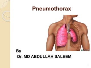
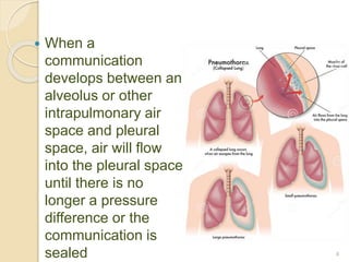
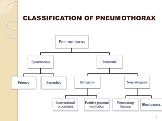
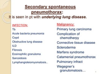
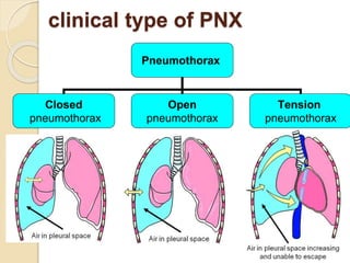
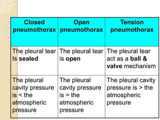
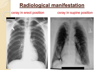
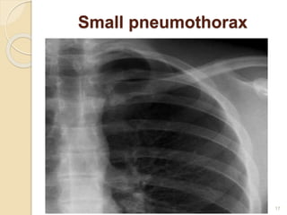
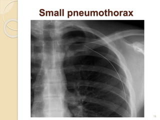
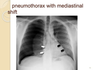
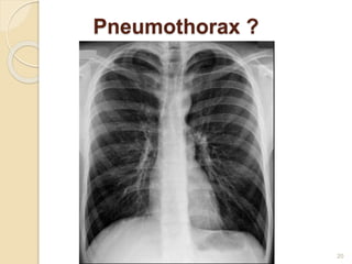
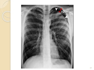
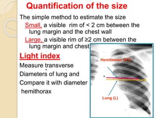
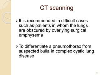
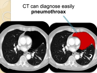
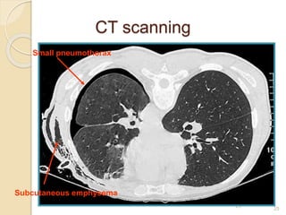
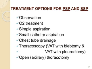
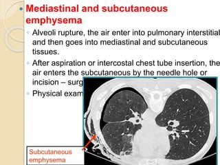
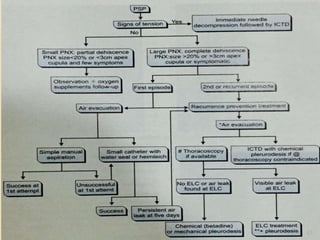
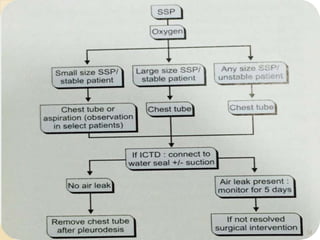
Pneumothorax is the presence of air or gas in the pleural space between the lung and chest wall. It occurs when there is a communication between an alveolus or air space and the pleural space, allowing air to enter the pleural space. Pneumothoraces are classified as spontaneous (primary or secondary), traumatic, or iatrogenic. Treatment depends on the type and severity but may include observation, oxygen therapy, needle aspiration, chest tube drainage, chemical pleurodesis, or surgery. Complications can include infection, bleeding, or mediastinal and subcutaneous emphysema.
there is the introduction part of the torso trauma,
check out my next ppts for further more about torso trauma.
contents are in following order...
introduction
mechanism of injury
junctional zones of torso
tension pneumothorax
cardiac temponade
massive hemothorax
etc.
check out all slides
Pulmonary edema is often caused by congestive heart failure. When the heart is not able to pump efficiently, blood can back up into the veins that take blood through the lungs. As the pressure in these blood vessels increases, fluid is pushed into the air spaces (alveoli) in the lungs.
This PowerPoint presentation provides an in-depth overview of pneumothorax, a medical condition that occurs when air leaks into the pleural cavity, causing the lung to collapse. The presentation covers the causes, symptoms, and diagnostic procedures for pneumothorax, including chest x-rays and CT scans.
The presentation also discusses the various treatment options available for pneumothorax, such as thoracentesis, chest tube insertion, and surgery. The benefits and risks of each treatment are also explained in detail, providing the audience with a comprehensive understanding of the condition and its management.
In addition, the presentation includes several case studies and real-life examples to help illustrate the impact of pneumothorax on patients and the importance of early diagnosis and treatment. It is an ideal resource for medical professionals, students, and anyone interested in learning more about this common medical condition.
Overall, this PowerPoint presentation provides a valuable resource for understanding pneumothorax, its causes, symptoms, and treatment options, helping to improve patient outcomes and quality of care.
Flu Vaccine Alert in Bangalore Karnatakaaddon Scans
As flu season approaches, health officials in Bangalore, Karnataka, are urging residents to get their flu vaccinations. The seasonal flu, while common, can lead to severe health complications, particularly for vulnerable populations such as young children, the elderly, and those with underlying health conditions.
Dr. Vidisha Kumari, a leading epidemiologist in Bangalore, emphasizes the importance of getting vaccinated. "The flu vaccine is our best defense against the influenza virus. It not only protects individuals but also helps prevent the spread of the virus in our communities," he says.
This year, the flu season is expected to coincide with a potential increase in other respiratory illnesses. The Karnataka Health Department has launched an awareness campaign highlighting the significance of flu vaccinations. They have set up multiple vaccination centers across Bangalore, making it convenient for residents to receive their shots.
To encourage widespread vaccination, the government is also collaborating with local schools, workplaces, and community centers to facilitate vaccination drives. Special attention is being given to ensuring that the vaccine is accessible to all, including marginalized communities who may have limited access to healthcare.
Residents are reminded that the flu vaccine is safe and effective. Common side effects are mild and may include soreness at the injection site, mild fever, or muscle aches. These side effects are generally short-lived and far less severe than the flu itself.
Healthcare providers are also stressing the importance of continuing COVID-19 precautions. Wearing masks, practicing good hand hygiene, and maintaining social distancing are still crucial, especially in crowded places.
Protect yourself and your loved ones by getting vaccinated. Together, we can help keep Bangalore healthy and safe this flu season. For more information on vaccination centers and schedules, residents can visit the Karnataka Health Department’s official website or follow their social media pages.
Stay informed, stay safe, and get your flu shot today!
The prostate is an exocrine gland of the male mammalian reproductive system
It is a walnut-sized gland that forms part of the male reproductive system and is located in front of the rectum and just below the urinary bladder
Function is to store and secrete a clear, slightly alkaline fluid that constitutes 10-30% of the volume of the seminal fluid that along with the spermatozoa, constitutes semen
A healthy human prostate measures (4cm-vertical, by 3cm-horizontal, 2cm ant-post ).
It surrounds the urethra just below the urinary bladder. It has anterior, median, posterior and two lateral lobes
It’s work is regulated by androgens which are responsible for male sex characteristics
Generalised disease of the prostate due to hormonal derangement which leads to non malignant enlargement of the gland (increase in the number of epithelial cells and stromal tissue)to cause compression of the urethra leading to symptoms (LUTS
micro teaching on communication m.sc nursing.pdfAnurag Sharma
Microteaching is a unique model of practice teaching. It is a viable instrument for the. desired change in the teaching behavior or the behavior potential which, in specified types of real. classroom situations, tends to facilitate the achievement of specified types of objectives.
These lecture slides, by Dr Sidra Arshad, offer a quick overview of physiological basis of a normal electrocardiogram.
Learning objectives:
1. Define an electrocardiogram (ECG) and electrocardiography
2. Describe how dipoles generated by the heart produce the waveforms of the ECG
3. Describe the components of a normal electrocardiogram of a typical bipolar leads (limb II)
4. Differentiate between intervals and segments
5. Enlist some common indications for obtaining an ECG
Study Resources:
1. Chapter 11, Guyton and Hall Textbook of Medical Physiology, 14th edition
2. Chapter 9, Human Physiology - From Cells to Systems, Lauralee Sherwood, 9th edition
3. Chapter 29, Ganong’s Review of Medical Physiology, 26th edition
4. Electrocardiogram, StatPearls - https://www.ncbi.nlm.nih.gov/books/NBK549803/
5. ECG in Medical Practice by ABM Abdullah, 4th edition
6. ECG Basics, http://www.nataliescasebook.com/tag/e-c-g-basics
TEST BANK for Operations Management, 14th Edition by William J. Stevenson, Ve...kevinkariuki227
TEST BANK for Operations Management, 14th Edition by William J. Stevenson, Verified Chapters 1 - 19, Complete Newest Version.pdf
MANAGEMENT OF ATRIOVENTRICULAR CONDUCTION BLOCK.pdfJim Jacob Roy
Cardiac conduction defects can occur due to various causes.
Atrioventricular conduction blocks ( AV blocks ) are classified into 3 types.
This document describes the acute management of AV block.
HOT NEW PRODUCT! BIG SALES FAST SHIPPING NOW FROM CHINA!! EU KU DB BK substit...GL Anaacs
Contact us if you are interested:
Email / Skype : kefaya1771@gmail.com
Threema: PXHY5PDH
New BATCH Ku !!! MUCH IN DEMAND FAST SALE EVERY BATCH HAPPY GOOD EFFECT BIG BATCH !
Contact me on Threema or skype to start big business!!
Hot-sale products:
NEW HOT EUTYLONE WHITE CRYSTAL!!
5cl-adba precursor (semi finished )
5cl-adba raw materials
ADBB precursor (semi finished )
ADBB raw materials
APVP powder
5fadb/4f-adb
Jwh018 / Jwh210
Eutylone crystal
Protonitazene (hydrochloride) CAS: 119276-01-6
Flubrotizolam CAS: 57801-95-3
Metonitazene CAS: 14680-51-4
Payment terms: Western Union,MoneyGram,Bitcoin or USDT.
Deliver Time: Usually 7-15days
Shipping method: FedEx, TNT, DHL,UPS etc.Our deliveries are 100% safe, fast, reliable and discreet.
Samples will be sent for your evaluation!If you are interested in, please contact me, let's talk details.
We specializes in exporting high quality Research chemical, medical intermediate, Pharmaceutical chemicals and so on. Products are exported to USA, Canada, France, Korea, Japan,Russia, Southeast Asia and other countries.
Ethanol (CH3CH2OH), or beverage alcohol, is a two-carbon alcohol
that is rapidly distributed in the body and brain. Ethanol alters many
neurochemical systems and has rewarding and addictive properties. It
is the oldest recreational drug and likely contributes to more morbidity,
mortality, and public health costs than all illicit drugs combined. The
5th edition of the Diagnostic and Statistical Manual of Mental Disorders
(DSM-5) integrates alcohol abuse and alcohol dependence into a single
disorder called alcohol use disorder (AUD), with mild, moderate,
and severe subclassifications (American Psychiatric Association, 2013).
In the DSM-5, all types of substance abuse and dependence have been
combined into a single substance use disorder (SUD) on a continuum
from mild to severe. A diagnosis of AUD requires that at least two of
the 11 DSM-5 behaviors be present within a 12-month period (mild
AUD: 2–3 criteria; moderate AUD: 4–5 criteria; severe AUD: 6–11 criteria).
The four main behavioral effects of AUD are impaired control over
drinking, negative social consequences, risky use, and altered physiological
effects (tolerance, withdrawal). This chapter presents an overview
of the prevalence and harmful consequences of AUD in the U.S.,
the systemic nature of the disease, neurocircuitry and stages of AUD,
comorbidities, fetal alcohol spectrum disorders, genetic risk factors, and
pharmacotherapies for AUD.
Title: Sense of Smell
Presenter: Dr. Faiza, Assistant Professor of Physiology
Qualifications:
MBBS (Best Graduate, AIMC Lahore)
FCPS Physiology
ICMT, CHPE, DHPE (STMU)
MPH (GC University, Faisalabad)
MBA (Virtual University of Pakistan)
Learning Objectives:
Describe the primary categories of smells and the concept of odor blindness.
Explain the structure and location of the olfactory membrane and mucosa, including the types and roles of cells involved in olfaction.
Describe the pathway and mechanisms of olfactory signal transmission from the olfactory receptors to the brain.
Illustrate the biochemical cascade triggered by odorant binding to olfactory receptors, including the role of G-proteins and second messengers in generating an action potential.
Identify different types of olfactory disorders such as anosmia, hyposmia, hyperosmia, and dysosmia, including their potential causes.
Key Topics:
Olfactory Genes:
3% of the human genome accounts for olfactory genes.
400 genes for odorant receptors.
Olfactory Membrane:
Located in the superior part of the nasal cavity.
Medially: Folds downward along the superior septum.
Laterally: Folds over the superior turbinate and upper surface of the middle turbinate.
Total surface area: 5-10 square centimeters.
Olfactory Mucosa:
Olfactory Cells: Bipolar nerve cells derived from the CNS (100 million), with 4-25 olfactory cilia per cell.
Sustentacular Cells: Produce mucus and maintain ionic and molecular environment.
Basal Cells: Replace worn-out olfactory cells with an average lifespan of 1-2 months.
Bowman’s Gland: Secretes mucus.
Stimulation of Olfactory Cells:
Odorant dissolves in mucus and attaches to receptors on olfactory cilia.
Involves a cascade effect through G-proteins and second messengers, leading to depolarization and action potential generation in the olfactory nerve.
Quality of a Good Odorant:
Small (3-20 Carbon atoms), volatile, water-soluble, and lipid-soluble.
Facilitated by odorant-binding proteins in mucus.
Membrane Potential and Action Potential:
Resting membrane potential: -55mV.
Action potential frequency in the olfactory nerve increases with odorant strength.
Adaptation Towards the Sense of Smell:
Rapid adaptation within the first second, with further slow adaptation.
Psychological adaptation greater than receptor adaptation, involving feedback inhibition from the central nervous system.
Primary Sensations of Smell:
Camphoraceous, Musky, Floral, Pepperminty, Ethereal, Pungent, Putrid.
Odor Detection Threshold:
Examples: Hydrogen sulfide (0.0005 ppm), Methyl-mercaptan (0.002 ppm).
Some toxic substances are odorless at lethal concentrations.
Characteristics of Smell:
Odor blindness for single substances due to lack of appropriate receptor protein.
Behavioral and emotional influences of smell.
Transmission of Olfactory Signals:
From olfactory cells to glomeruli in the olfactory bulb, involving lateral inhibition.
Primitive, less old, and new olfactory systems with different path
2. What is pneumothorax
Pneumothorax is defined as
presence of air or gas in the
pleural space
(OR)
air between the lung and chest wall,
in other term, air between the
visceral pleura and the parietal
pleura
2
3. Mechanism
In normal people, the pressure in
pleural space is negative during the
entire respiratory cycle.
Two opposite forces result in negative
pressure in pleural space.
(outward pull of the chest wall and
elastic recoil of the lung)
The negative pressure will
be disappeared if any communication
develops .
3
4. When a
communication
develops between an
alveolus or other
intrapulmonary air
space and pleural
space, air will flow
into the pleural space
until there is no
longer a pressure
difference or the
communication is
sealed 4
5. Pathophysiology
Negative pressure eliminated
The lung recoil-& lung-volume decrease
V/Q low –anatomic shunt
hypoxia
Positive pressure
◦ Compress blood vessels and heart
◦ Decreased cardiac output
◦ Impaired venous return
◦ Hypotension
Result in
◦ A decrease in vital capacity
◦ A decrease in PaO2
5
7. Primary spontaneous
pneumothorax
It occurs in young healthy individuals
without underlying lung disease.
It is due to rupture of apical
sub-pleural bleb or bullae
Predisposing factors:
Smoking.
Tall, thin male.
Airway inflammation (distal)
Structural abnormalities of bronchial tree
Genetic contribution
7
8. Secondary spontaneous
pneumothorax:
It is seen in pt with underlying lung disease.
INFECTION:
Tb
Acute bacteria pneumonia
Copd
Obstructive lung disease
ILD
Fibrosis
Eosinophilic granuloma
Sarcoidosis
Lymphangioleiomyomatosis..
Malignancy:
Primary lung carcinoma
Complication of
chemotherpy
Connective tissue disease
Scleroderma
Marfans syndrome
Catamenial pneumothorax
Pulmonary infract
Wegegner’s
granulomatosis… 8
11. 11
clinical type of PNX
Pneumothorax
Closed
pneumothorax
Open
pneumothorax
Tension
pneumothorax
12. 12
Closed
pneumothorax
Open
pneumothorax
Tension
pneumothorax
The pleural tear
Is sealed
The pleural tear
is open
The pleural tear
act as a ball &
valve mechanism
The pleural
cavity pressure
is < the
atmospheric
pressure
The pleural
cavity pressure
is = the
atmospheric
pressure
The pleural cavity
pressure is > the
atmospheric
pressure
13. Clinical manifestation
Tension pneumothorax
◦ Distressed with rapid labored respiration
◦ Cyanosis
◦ Marked tachycardia
Patient who suddenly deteriorate
clinically,
be suspected in the patient with
◦ Mechanical ventilation
◦ Cardiopulmonary resuscitation
13
14. Physical examination
◦ Depend on size of pneumothorax
◦ The vital signs usually normal
◦ Unilateral Chest movements
◦ The trachea may be shifted toward the
contralateral side if the pneumothorax is
large
◦ Tactile fremitus is absent
◦ The percussion note is hypersonant
◦ The breath sounds are reduced or absent
on the affected side
◦ The lower edge of the liver may be shifted
inferiorly with a right-side pneumothorax
14
22. Quantification of the size
The simple method to estimate the size
Small, a visible rim of < 2 cm between the
lung margin and the chest wall
Large, a visible rim of ≥2 cm between the
lung margin and chest wall
Light index
Measure transverse
Diameters of lung and
Compare it with diameter
hemithorax
22
22
Hemithorax (HT)
Lung (L)
23. CT scanning
It is recommended in difficult cases
such as patients in whom the lungs
are obscured by overlying surgical
emphysema
To differentiate a pneumothorax from
suspected bulla in complex cystic lung
disease
23
26. MANAGMENT
Goals
◦ To promote lung expansion
◦ To eliminate the pathogenesis
◦ To decrease pneumothorax recurrence
Treatment options according to
◦ Classification of pneumothorax
◦ Pathogenesis
◦ The extension of lung collapse
◦ Severity of disease
◦ Complication and concomitant underlying
diseases
26
27. TREATMENT OPTIONS FOR PSP AND SSP
Observation
O2 treatment
Simple aspiration
Small catheter aspiration
Chest tube drainage
Thoracoscopy (VAT with blebtomy &
VAT with pleurectomy)
Open (axillary) thoracotomy
27
28. Observation - PSP
Small, closed mildly symptomatic
spontaneous pneumothorax.
Do not require hospital admission
It should be stressed to patient that they
should be return directly to hospital in
the event of developing breathlessness.
28
29. Observation - SSP
Observation along is only recommend
in patients with small SSP of less than
1 cm depth or isolated apical
pneumothorax in asymptomatic
patients
Hospitalization is recommended in
these cases
All other cases will require active
intervention ( aspiration or chest drain
insertion)
29
30. 30
Marked breathlessness in a patient with a
small (<2 cm) PSP may develop tension
pneumothorax
Observation is inappropriate and active
intervation is required
If a patient is hospitalised for observation,
supplemental high flow (10 l/min) oxygen
should be given where feasible
Observation - PSP or SSP
31. 31
Inhalation of high concentration of oxygen
may reduce the total pressure of gases in
pleural capillaries by reducing the partial
pressure of nitrogen
This should increase the pressure gradient
between the pleural capillaries and the
pleural cavity
Thereby increasing absorption of air from
the pleural cavity
O2 TREATMENT-- PSP or
SSP
32. 32
The rate of resolution/reabsorption of
spontaneous pneumothorax is 1.25 –
1.8% of volume of hemithorax every 24
hours
The addition of high flow oxygen
therapy has been shown to result in a
4-fold increase in the rate of
peumothorax reabsorption during the
periods of oxygen supplementation
33. 33
Simple aspiration
Simple aspiration is recommended as first line
treatment for all PSP requiring intervention
Simple aspiration is less likely to succeed in
secondary pneumothoraces and in this situation,
is only recommended as an initial treatment in
small (<2 cm) pneumothoraces in minimally
breathless patients under the age of 50 years
Patients with secondary pneumothoraces
treated successfully with simple aspiration
should be admitted to hospital and observed for
at least 24 hours before discharge
34. 34
Repeated aspiration is reasonable for
primary pneumothorax when the first
aspiration has been unsuccessful
A volume of < 2.5 L has been aspirated on
the first attempt
The aspiration can be used by needle or
catheter
Catheter aspiration
35. Intercostal tube drainage
INDICATIONS
◦ Unstable
pneumothorax
◦ Severe dyspnea
◦ Large lung collapse
◦ Open or tension
pneumothorax
◦ Recurrent
pneumothorax
◦ Simple aspiration or
catheter aspiration
drainage is
unsuccessful in
controlling symptoms 35
36. 36
36
The site of chest tube
insertion is in the
midclavicular line of
second and third
intercostal
or anterior axillary line
of fifth and sixth
intercostal
Intercostal tube drainage
37. 37
37
Intercostal tube drainage
Fix the catheter and cover with gauze
Making a small incision
Using a forceps to extend the hole
Inserting a catheter into pleural cavity
40. 4
Operative tube thoracostomy
The physician’s index
finger is used to
enlarge the opening
and to explore the
pleural space
Placement of chest
tube
Intrapleurally using
large hemostat
41. 41
Observation of drainage
No bubble released
◦ The lung reexpansion
◦ The chest tube is obstructed by secretion or blood clot
◦ The chest tube shift to chest wall, the hole of the chest
tube is located in the chest wall
If the lung reexpansion, removing the chest tube 24
hours after reexpansion.
Otherwise, the chest tube will be inserted again or
regulated the position.
42. 42
Complications of intercostal tube
drainage
Penetration of major organs
◦ Lung, stomach, spleen, liver, heart and great
vessels
◦ It occurs more commonly when a sharp metal
trocar is inappropriately applied
Pleural infection
◦ Empyema, the rate of 1%
Surgical emphysema
◦ Subcutaneous emphysema
43. 43
Chemical pleurodesis
Goals
◦ To prevent pneumothorax recurrence
◦ To produce inflammation of pleura and
adhesions
Indications
◦ Persist air leak and repeated pneumothorax
◦ Bilateral pneumothorax
◦ Complicated with bullae
◦ Lung dysfunction, not tolerate to operation
44. 44
Chemical pleurodesis
Sclerosing agents
◦ Tetracycline
◦ Doxycline
◦ Talc
◦ Erythromycin
The instillation of sclerosing agents into the pleural
space lead to an aseptic inflammation with dense
adhesions.
45. 45
Methods
◦ Via chest tube or by surgical mean
◦ Administration of intrapleural local anaesthesia, 200 – 300
mg lidocaine intrapleurally injection
◦ Agents diluted by 60 – 100 ml saline
◦ Injected to pleural space
◦ Clamp the tube 4hours
◦ Drainage again
◦ Observed by chest X-ray film, if air of pleural space is
absorption, remove the chest tube
◦ If pneumothorax still exist, repeated pleurodesis.
Chemical pleurodesis
47. 47
Surgical treatment
Indications
◦ No response to medical treatment
◦ Persist air leak
◦ Hemopneumothorax
◦ Bilateral pneumothoraces
◦ Recurrent pneumothorax
◦ Tension pneumothorax failed to dainage
◦ Thicken pleura makes lung unable to
reexpansion
◦ Multiple blebs or bullae
48. Thoracoscopy- VATs
It is being increasly used in ot with
recurrent psp-ssp
Recent studies show VATs it may be
the procedure of choice and it has
very less complication when compare
to other open surgery
Pleural belbs causes pneumothorax
which can be treated by a method
called endostapling or
suturefollowed by pleurodosis
48
49. Axillary (open) thoracotomy
Transaxillary minithoracotomy is preferred
.when VATs isnot available
Open thotacotomy with suturing of blebs
and pleural abrasion is done
Recurrence
High risk professions
B/L or tension pneumothorax
49
INDICATIONS
50. 50
Complications of pneumothorax
Pyopneumothorax
◦ Caused by aspiration or intercostal chest tube
insertion (iatrogenic)
◦ Also results from necrotic pneumonia, lung
abscess, or caseous pneumonia
Hydropneumothorax.
Hemopneumotorax
◦ Bleeding in pleural space.
◦ Common cause is rupture of vessels in
adhesions.
◦ When lung re-expansion, bleeding will stop.
◦ When bleeding persists, surgical ligation will
be needed.
51. 51
Mediastinal and subcutaneous
emphysema
◦ Alveoli rupture, the air enter into pulmonary interstitial
and then goes into mediastinal and subcutaneous
tissues.
◦ After aspiration or intercostal chest tube insertion, the
air enters the subcutaneous by the needle hole or
incision – surgical emphysema
◦ Physical exam – crepitus is present.
Subcutaneous
emphysema