Downloaded 993 times
![Chalya P.L. 1
THORACIC EMPYEMA
Dr Phillipo Leo Chalya
M.D. [Dar]; M.MED surg [Mak]
Surgeon Specialist - BMC](https://image.slidesharecdn.com/thoracicempyema-150427102035-conversion-gate01/85/Thoracic-empyema-1-320.jpg)
![Chalya P.L. 1
THORACIC EMPYEMA
Dr Phillipo Leo Chalya
M.D. [Dar]; M.MED surg [Mak]
Surgeon Specialist - BMC](https://image.slidesharecdn.com/thoracicempyema-150427102035-conversion-gate01/75/Thoracic-empyema-1-2048.jpg)
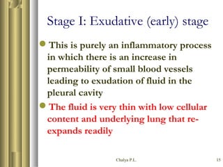
![Chalya P.L. 14
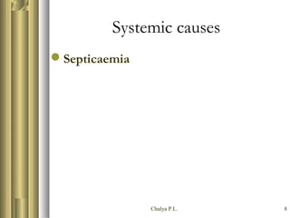
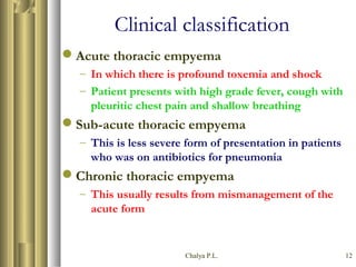
PATHOPHYSIOLOGY
According to the American Thoracic
Society [1962], the development of
thoracic empyema passes through 3
stages:-
– Exudative stage
– Fibrino-purulent stage
– Organizing stage](https://image.slidesharecdn.com/thoracicempyema-150427102035-conversion-gate01/85/Thoracic-empyema-14-320.jpg)


This document provides an overview of thoracic empyema, including its definition as pus in the pleural cavity secondary to underlying diseases. It discusses the historical background, etiology, classification, pathophysiology, clinical presentation, workup, treatment, and complications of thoracic empyema. The treatment section outlines both non-surgical options like antibiotics and needle aspiration as well as surgical procedures including closed chest drainage, open chest drainage, decortication, and thoracoplasty. The document serves as a guide to thoracic empyema covering its key aspects in detail across 31 pages.


