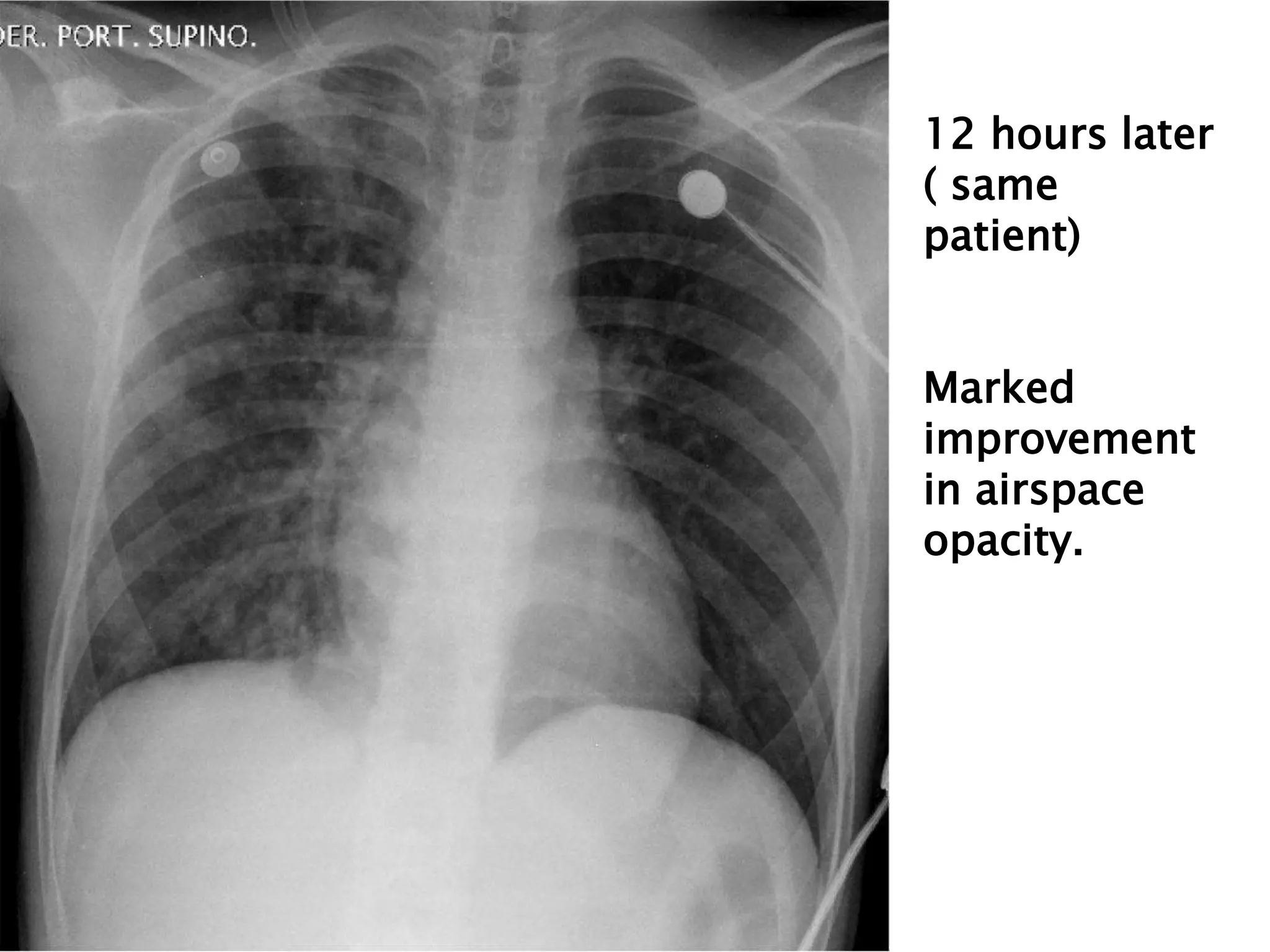
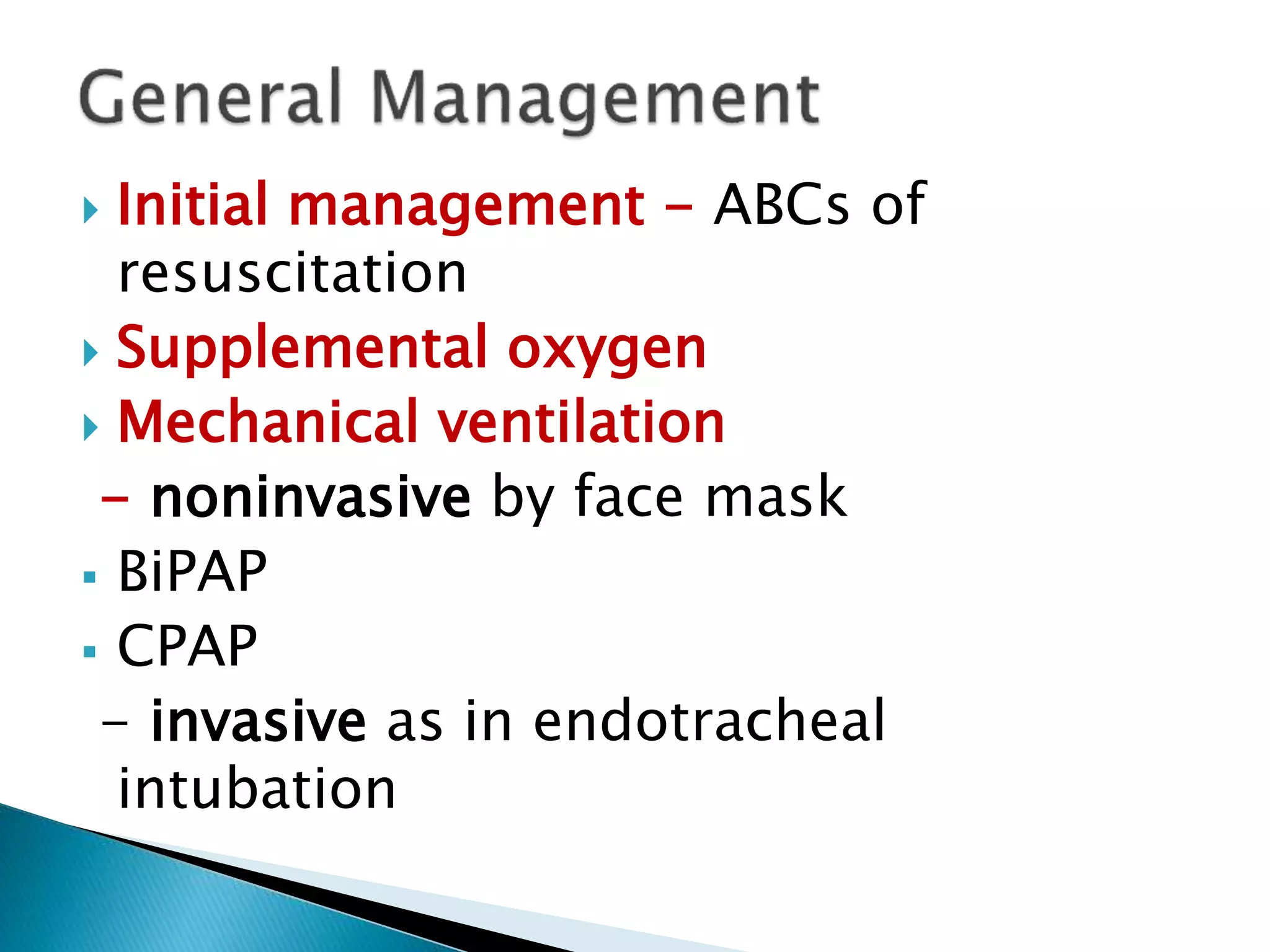
Dr. Amna Akram
CMH, Multan
 Pulmonary edema is a condition characterized
by fluid accumulation in the lungs caused by
extravasation of fluid from pulmonary
vasculature into the interstitium and alveoli of
the lungs
 Imbalance of starling force
-Increase pulmonary capillary
pressure
-decrease plasma oncotic pressure
-increase negative interstitial
pressure
 Damage to alveolar – capillary barrier
 Lymphatic obstruction
 Disruption of endothelial barrier
 Based on underlying cause
 Cardiogenic pulmonary edema
 Non-cardiogenic pulmonary edema
 Neurogenic PE
 High Altitude PE
 Post Aspiration PE
 Re-expansion PE
 Other ( inhaled toxins, lymphatic obstruction,
post lung transplant, etc.)
 Due to cardiac abnormalities, pulmonary
capillary pressure is increased that increases
the pulmonary venous pressure.
 CAUSES :
 LV failure is the most common
 Dysrhythmia
 LV hypertrophy and cardiomyopathy
 LV volume overload
 Myocardial infarction
 LV outflow obstruction
Left sided heart failure
Decreased pumping ability to the systemic
circulation
Congestion and accumulation of blood in
pulmonary area
Fluid leaks out of intravascular space to the
interstitium
Accumulation of fluid
Pulmonary edema
 Neurogenic PE
Patients with CNS disorders and
without apparent preexisting LV
dysfunction
 Re-expansion PE
Develops after removal of air or fluid,
post-thoracocentesis
 High Altitude PE
 Occurs in young people who have
quickly ascended to altitudes above
2700m and who then engage in
strenuous physical exercise at that
altitude, before they have become
acclimatized
 Reversible (in less than 48 hours)
 Based on the degree of fluid
accumulation
 Stage- 1:all excess fluid can still be
cleared by lymphatic drainage
 Stage- 2 presence of interstitial
edema
 Stage- 3 alveolar edema
 Mild: Only engorgement of pulmonary
vasculature.
 Moderate: Extravasation of fluid into the
interstitial space due to changes in oncotic
pressure
 Severe: Alveolar filling occurs
Symptoms:
ACUTE
 Severe shortness of breath
 Cough- with pink frothy sputum
 Profuse sweating
 Cyanosis
 Anxiety, restlessness
 Palpitation
 Chest pain
 LONG TERM (CHRONIC)
 Paroxysmal nocturnal dyspnea
 Orthopnea
 Rapid weight gain
 Loss of appetite
 Fatigue
 Ankle and leg swelling
 Tachycardia
 Tachypnea
 Confusion
 Agitation, anxious
 Diaphoriesis
 Hypertension
 Cool extremities
 Crepitant rales, ronchi or wheeze
 CVS findings: S3, accentuation of pulmonic
component of S2, jugular venous distension
 leg edema
 Ascites
 Pleural effusion
 Congestion and swelling of liver
 Myocardial infarction
 Cardiogenic shock
 Arrythmias
 Electrolyte disturbances
 Mesenteric insufficiency
 Protein enteropathy
 Respiratory arrest and death
Cardiogenic PE Non
cardiogenic PE
CVS findings:
- S3 gallop
- Elevated JVP
- Peripheral edema
- relatively normal in early
stages
cardiomegaly Heart size is normal
Engorgement of
vasculature to the apices
No engorgement
Pleural effusion is
common
uncommon
Cardiogenic PE Non
cardiogenic PE
Perihilar alveolar infiltrate Uniform alveolar infiltrate
Kerley B lines Not present
Hypoxemia due to
ventilation perfusion miss
match
Hypoxemia due to
intrapulmonary shunting
Responds to
administration of oxygen
Persist despite oxygen
supplementation
 CBC – severe anemia
 Serum electrolytes – Hypokalemia,
Hypomagnesemia
 Pulse oximetry – assess
 Hypoxia
 Response to supplemental oxygenation
 ABGs – Initially hypoxia and hypocapnia with
respi. Alkalosis
- Later Hypercapnia with respi and
metabolic acidosis
 ECG- tachydysrhythmia
- bradydysrhythmia
- acute MI
 Ultrasonography – B lines
sensitivity of 94.1%
specificity of 92.4%
 Chest x-ray- 1. enlarged heart
2.inverted blood flow
3. Kerley lines
4. Basilar edema (vs diffuse edema)
5. Absence of air bronchograms
6. bilateral and symmetrical pleural effusions
Bat wing
edema in a
71-year-old
woman with
fluid overload
and cardiac
failure.
Neurogenic
PE in a
patient with
subdural
hematoma.
Bilateral
alveolar
filling
process and
a normal-
sized heart.
Patient
admitted with
progressive
respiratory
distress 24
hours after
arriving at town
at 2700 meters
above sea level.
Right greater
than left
indistinct
airspace opacity
12 hours later
( same
patient)
Marked
improvement
in airspace
opacity.
Echocardiography –
 acute papillary muscle rupture
 acute VSD
 cardiac temponade
 acute severe mitral regurgitation
 aortic regurgitation.
 Pulmonary arterial catheterization :
(Swan-Ganz Catheter)
 PCWP >18mmHg indicates CPE.
 PCWP <18mmHg indicates NCPE
 Initial management - ABCs of
resuscitation
 Supplemental oxygen
 Mechanical ventilation
- noninvasive by face mask
 BiPAP
 CPAP
- invasive as in endotracheal
intubation
 3 main goals
 1. preload reduction:
 (a) Nitroglycerin (sublingual or
intravenous) IV NTG
-10mcg/min, rapidly uptitrated to
more than 100mcg/min
- 3mg IV boluses every 5 minutes
 (b) Diuretics (loop diuretics)
Furosemide
 (c) Nesiritide (recombinant human BNP)
 2. Afterload reduction:
 (a) ACE inhibitors –
 enalapril 1.25mg IV
 captopril 25mg sublingually
 (b) Angiotensin II receptor blockers –
 Valsartan and candesartan
 (c) Nitroprusside-
 Avoided in acute MI
 Prolonged use causes cyanide toxicity ,
tolerance and reflex tachycardia
 (3)Inotropic Support :
(a) Dobutamine
(b) Dopamine
(c) Norepinephrine
 Intra-aortic Balloon pumping –
 reduces afterload
 Increases cardiac output
 Reduces LA pressure and improves
CPE
 Resolves within 48-72 hours in majority
of patients
 Medical care:
 oxygen supplementation
 Diuretics
 Inotropic support
 Surgical Care : directed at the
neurological insult (e.g., intracerebral
hemorrhage, subdural hematoma, etc.)
 Descent and supplemental O2
 Tab nifedipine 10mg sublingual
or 20mg sustained release 6hrly.
 Hydralazine
 inhaled nitrous oxide
 acetazolamide
Pulmonary edema

Pulmonary edema

  • 1.
  • 2.
     Pulmonary edemais a condition characterized by fluid accumulation in the lungs caused by extravasation of fluid from pulmonary vasculature into the interstitium and alveoli of the lungs
  • 3.
     Imbalance ofstarling force -Increase pulmonary capillary pressure -decrease plasma oncotic pressure -increase negative interstitial pressure  Damage to alveolar – capillary barrier  Lymphatic obstruction  Disruption of endothelial barrier
  • 4.
     Based onunderlying cause  Cardiogenic pulmonary edema  Non-cardiogenic pulmonary edema  Neurogenic PE  High Altitude PE  Post Aspiration PE  Re-expansion PE  Other ( inhaled toxins, lymphatic obstruction, post lung transplant, etc.)
  • 5.
     Due tocardiac abnormalities, pulmonary capillary pressure is increased that increases the pulmonary venous pressure.  CAUSES :  LV failure is the most common  Dysrhythmia  LV hypertrophy and cardiomyopathy  LV volume overload  Myocardial infarction  LV outflow obstruction
  • 6.
    Left sided heartfailure Decreased pumping ability to the systemic circulation Congestion and accumulation of blood in pulmonary area Fluid leaks out of intravascular space to the interstitium Accumulation of fluid Pulmonary edema
  • 7.
     Neurogenic PE Patientswith CNS disorders and without apparent preexisting LV dysfunction  Re-expansion PE Develops after removal of air or fluid, post-thoracocentesis
  • 8.
     High AltitudePE  Occurs in young people who have quickly ascended to altitudes above 2700m and who then engage in strenuous physical exercise at that altitude, before they have become acclimatized  Reversible (in less than 48 hours)
  • 9.
     Based onthe degree of fluid accumulation  Stage- 1:all excess fluid can still be cleared by lymphatic drainage  Stage- 2 presence of interstitial edema  Stage- 3 alveolar edema
  • 10.
     Mild: Onlyengorgement of pulmonary vasculature.  Moderate: Extravasation of fluid into the interstitial space due to changes in oncotic pressure  Severe: Alveolar filling occurs
  • 11.
    Symptoms: ACUTE  Severe shortnessof breath  Cough- with pink frothy sputum  Profuse sweating  Cyanosis  Anxiety, restlessness  Palpitation  Chest pain
  • 12.
     LONG TERM(CHRONIC)  Paroxysmal nocturnal dyspnea  Orthopnea  Rapid weight gain  Loss of appetite  Fatigue  Ankle and leg swelling
  • 13.
     Tachycardia  Tachypnea Confusion  Agitation, anxious  Diaphoriesis  Hypertension  Cool extremities  Crepitant rales, ronchi or wheeze  CVS findings: S3, accentuation of pulmonic component of S2, jugular venous distension
  • 14.
     leg edema Ascites  Pleural effusion  Congestion and swelling of liver  Myocardial infarction  Cardiogenic shock  Arrythmias  Electrolyte disturbances  Mesenteric insufficiency  Protein enteropathy  Respiratory arrest and death
  • 15.
    Cardiogenic PE Non cardiogenicPE CVS findings: - S3 gallop - Elevated JVP - Peripheral edema - relatively normal in early stages cardiomegaly Heart size is normal Engorgement of vasculature to the apices No engorgement Pleural effusion is common uncommon
  • 16.
    Cardiogenic PE Non cardiogenicPE Perihilar alveolar infiltrate Uniform alveolar infiltrate Kerley B lines Not present Hypoxemia due to ventilation perfusion miss match Hypoxemia due to intrapulmonary shunting Responds to administration of oxygen Persist despite oxygen supplementation
  • 17.
     CBC –severe anemia  Serum electrolytes – Hypokalemia, Hypomagnesemia  Pulse oximetry – assess  Hypoxia  Response to supplemental oxygenation  ABGs – Initially hypoxia and hypocapnia with respi. Alkalosis - Later Hypercapnia with respi and metabolic acidosis
  • 18.
     ECG- tachydysrhythmia -bradydysrhythmia - acute MI  Ultrasonography – B lines sensitivity of 94.1% specificity of 92.4%  Chest x-ray- 1. enlarged heart 2.inverted blood flow 3. Kerley lines 4. Basilar edema (vs diffuse edema) 5. Absence of air bronchograms 6. bilateral and symmetrical pleural effusions
  • 19.
    Bat wing edema ina 71-year-old woman with fluid overload and cardiac failure.
  • 20.
    Neurogenic PE in a patientwith subdural hematoma. Bilateral alveolar filling process and a normal- sized heart.
  • 21.
    Patient admitted with progressive respiratory distress 24 hoursafter arriving at town at 2700 meters above sea level. Right greater than left indistinct airspace opacity
  • 22.
    12 hours later (same patient) Marked improvement in airspace opacity.
  • 23.
    Echocardiography –  acutepapillary muscle rupture  acute VSD  cardiac temponade  acute severe mitral regurgitation  aortic regurgitation.  Pulmonary arterial catheterization : (Swan-Ganz Catheter)  PCWP >18mmHg indicates CPE.  PCWP <18mmHg indicates NCPE
  • 24.
     Initial management- ABCs of resuscitation  Supplemental oxygen  Mechanical ventilation - noninvasive by face mask  BiPAP  CPAP - invasive as in endotracheal intubation
  • 25.
     3 maingoals  1. preload reduction:  (a) Nitroglycerin (sublingual or intravenous) IV NTG -10mcg/min, rapidly uptitrated to more than 100mcg/min - 3mg IV boluses every 5 minutes  (b) Diuretics (loop diuretics) Furosemide
  • 26.
     (c) Nesiritide(recombinant human BNP)  2. Afterload reduction:  (a) ACE inhibitors –  enalapril 1.25mg IV  captopril 25mg sublingually  (b) Angiotensin II receptor blockers –  Valsartan and candesartan  (c) Nitroprusside-  Avoided in acute MI  Prolonged use causes cyanide toxicity , tolerance and reflex tachycardia
  • 27.
     (3)Inotropic Support: (a) Dobutamine (b) Dopamine (c) Norepinephrine  Intra-aortic Balloon pumping –  reduces afterload  Increases cardiac output  Reduces LA pressure and improves CPE
  • 28.
     Resolves within48-72 hours in majority of patients  Medical care:  oxygen supplementation  Diuretics  Inotropic support  Surgical Care : directed at the neurological insult (e.g., intracerebral hemorrhage, subdural hematoma, etc.)
  • 29.
     Descent andsupplemental O2  Tab nifedipine 10mg sublingual or 20mg sustained release 6hrly.  Hydralazine  inhaled nitrous oxide  acetazolamide