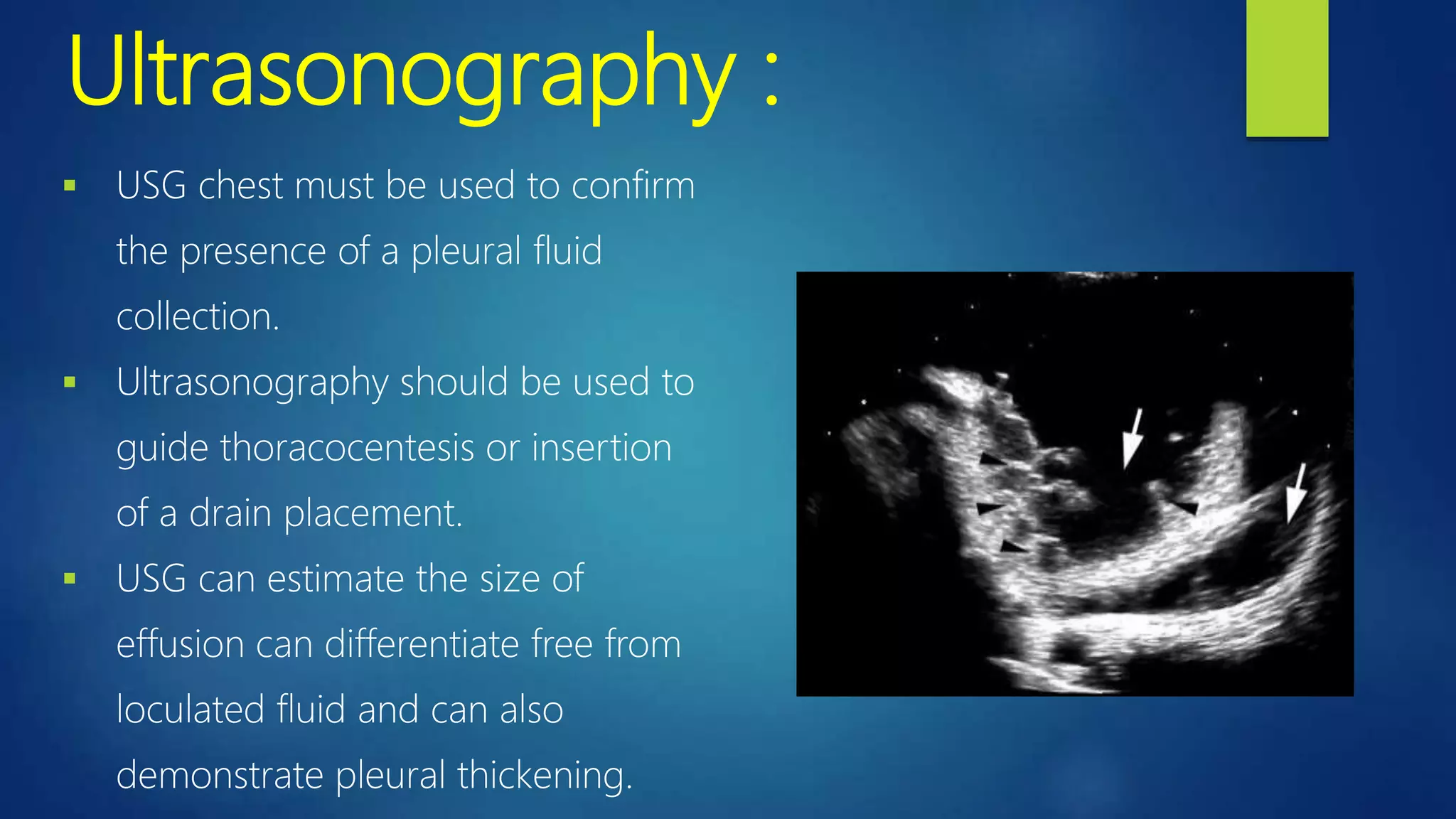
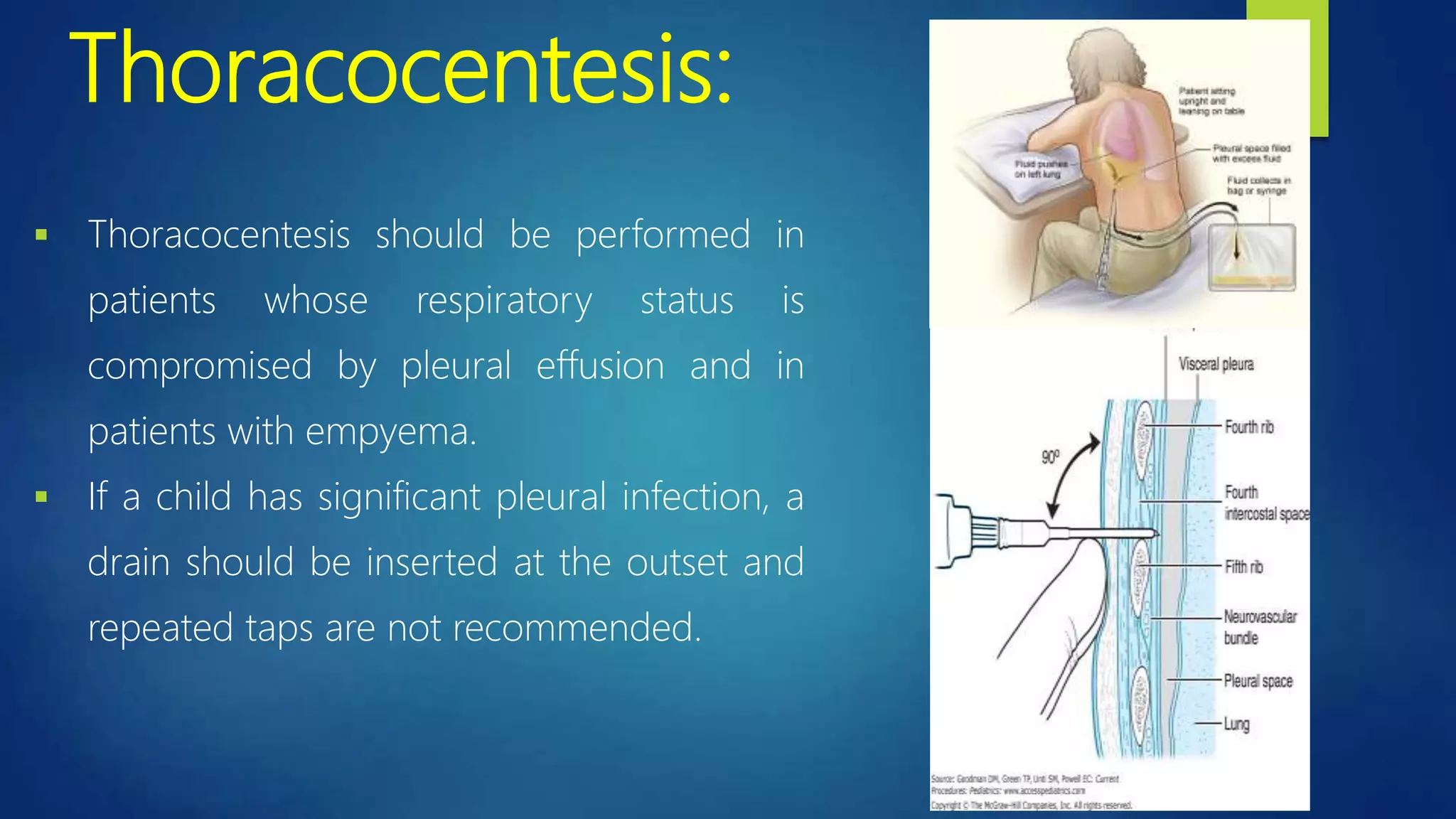
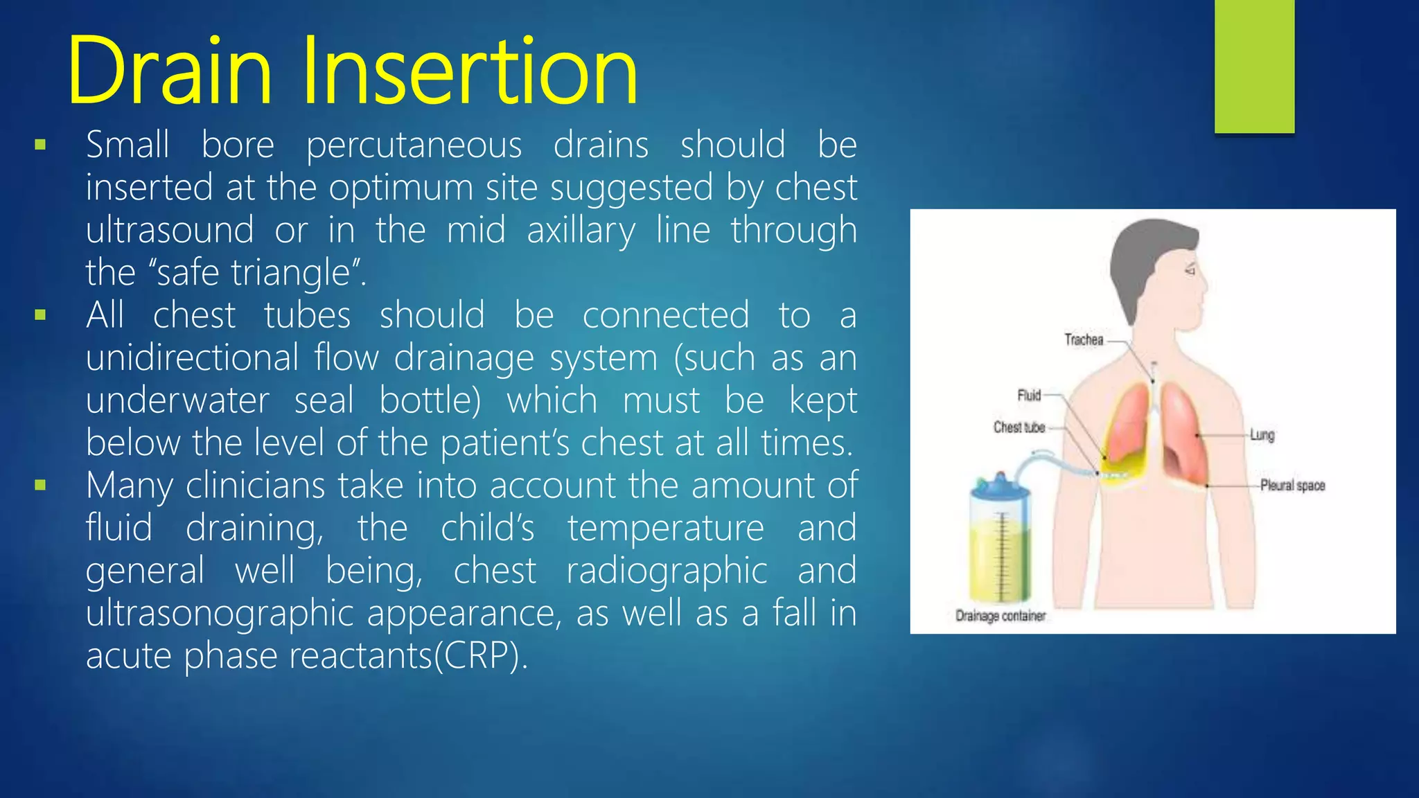
SlideShare a Scribd company logo
Approach to a patient of
Empyema
DR.ASIA NOUREEN
PGR PEDIATRIC MEDICINE
Objectives:
 Definition
 Epidemiology
 Etiology
 Pathophysiology
 Clinical menifestations
 Investigations
 Treatment options
Definition:
 Empyema is an accumulation of pus in the
pleural space.
 An effusion is called an Empyema when the
concentration of leucocytes becomes
macroscopically evident as a thick and turbid
fluid(pus).
Epidemiology:
 It has an incidence of 3.3 per 100,000 children.
 5-10% of children with bacterial pneumonia.
 86% of children with Necrotizing pneumonia.
 Infants and young children.
 more common in boys than girls.
 Winter and spring.
Etiology:
 In a previously well child, pleural effusion and empyema are usually
secondary to acute bacterial pneumonia and less often due to chronic
infections such as pulmonary tuberculosis.
Streptococcus pneumonia
Staphylococcus aureus
Haemophilus Influenzae type b
Group A Streptococcus,Gram negative organisms, tuberculosis, Fungi, Viruses and
Malignancy
Rupture of a lung abscess into the pleural space
Trauma and thoracic surgery
Extension of intra abdominal abscesses
Stages of Empyema:
3 Stages
 the exudative stage
 the fibrinopurulent stage
 the organizing stage
The exudative stage:
 In this stage, the inflammatory process associated with the
underlying pneumonia leads to the accumulation of clear fluid with a
low white cell count (simple parapneumonic effusion).
 At this stage, the effusion is thin, contains neutrophils, has normal pH
and glucose levels, and is often sterile. LDH level is <1,000 IU.
The fibrinopurulent stage:
 This stage is characterized by deposition of fibrin in pleural space leading to
septation and formation of loculations and significant polymorphonuclear
(PMN) leukocyte invasion.
 Inflammation is characterized by progressive decreases in the pleural fluid
glucose and pH levels and increased protein and lactate dehydrogenase
(LDH) levels.
The organizing stage:
 fibroblasts infiltration and formation of intrapleural membranes which are
thick and non-elastic (“the peels”). The pleural peels may prevent lung re-
expansion (trapped lung).
 spontaneous healing may occur or a chronic empyema
 Further complications include Broncho-pleural fistula, lung abscess or even
perforation through the chest wall. (empyema necessitatis).
Pathophysiology:
 Pleural fluid < 1 mL and it makes a thin layer about 10 μL.
 There is a continuous circulation and is absorbed by lymphatic vessels. An
imbalance will result in a pleural effusion.
 The protein concentration is similar to that of the interstitial fluid but it has a
higher level of bicarbonate, a lower level of LDH, and a similar level of
glucose.
 The cells are mostly macrophages with few lymphocytes and RBCs.
 Absorption enhanced by chest wall and diaphragmatic movements.
 In disease state, vascular permeability allows migration of inflammatory cells
and activation of cytokines. The results in the exudative stage of pleural
effusion. This progresses to the fibropurulant stage due to increased fluid
accumulation and bacterial invasion. Deposition of the fibrin in the pleural
space leads to septation or loculation. The pleural fluid pH and glucose levels
fall while LDH levels increase.
Clinical Features:
 Two common presentation.
 In the first, the child has classic symptoms of pneumonia such as fever,
cough, breathlessness, exercise intolerance, poor appetite, abdominal pain,
halitosis, lethargy and malaise. They may have pleuritic chest pain and may
lie on the effected side (splinting) to provide temporary analgesia.
 The second scenario is of the child who has been diagnosed with
pneumonia but does not respond to usual and appropriate treatment .
Diagnosis:
Clinical History
• Fever
• Cough
• Dyspnea
• Malaise
• Loss of Appetite
• Pleuritic chest pain
• Abdominal Pain
Physical Examination
• Dec. chest expansion
• Dullness to percussion
• Reduced or absent breath
sounds
• Assessment of hydration,
height and weight
Initial Investigation
• Chest radiograph
• USG of chest
• Blood Culture
• Full blood count (for anemia,
TLC, PLT count)
• CRP (A useful marker of
progress)
Imaging studies:
 Radiologic evaluation is the primary tool for the diagnosis of a parapneumonic effusion
 Frontal, decubitus and lateral radiographs
 Chest radiograph : Obliteration of CP angle is the earliest sign of an effusion and a rim of
fluid may be seen ascending the lateral chest wall (meniscus sign).
 A lateral decubitus film is the most sensitive view and can detect as little as 5-10 mL of free
fluid.
 A fluid layer of more than 1 cm on decubitus film is amenable to thoracentesis.
 Non-shifting fluid suggests either thick fluid or loculation.
Ultrasonography :
 USG chest must be used to confirm
the presence of a pleural fluid
collection.
 Ultrasonography should be used to
guide thoracocentesis or insertion
of a drain placement.
 USG can estimate the size of
effusion can differentiate free from
loculated fluid and can also
demonstrate pleural thickening.
CT scan:
 CT chest should not be performed
routinely but it may provide additional
information in complicated cases such as
loculated pleural fluid, parenchymal lung
abnormalities or lung abscess.
 In addition, CT scan guidance may also be
useful in interventions in which effusions
are difficult to access.
Blood test:
 Blood Culture should be performed in all cases with parapneumonic
effusion.
 Acute phase reactant such as TLC, CRP and ESR help in distinguishing
bacterial from viral infections. Clinical practice has shown that serial
measurements of CRP and the TLC can help to identify the effectiveness of
treatment.
 When available, sputum should be sent for bacterial culture.
Pleural fluid analysis :
Conventionally, the initial evaluation of pleural fluid is directed at
determining whether the effusion is an exudate or a transudate.
Pleural fluid analysis:
 Grossly purulent fluid indicates an empyema.
 Effusion is an empyema if
bacteria on Gram Stain
pH <7.2
>100,000 neutrophils/ul
Treatment:
Initial Treatment
 Oxygen if necessary (SpaO2 <92%)
 Fluid therapy if child dehydrated or unable/unwilling to drink
 Initiate intravenous antibiotics (empirical)
 Analgesia and antipyretics
 Physiotherapy is not indicated
 Patients with empyema should receive a longer course of therapy analogous
to necrotizing pneumonia, but the response to therapy determines the
duration of treatment. The patient should receive systemic antibiotics for 4
weeks or should receive treatment until he or she is afebrile, off supplemental
oxygen, and appropriately responds to therapy.
 Continuation of oral antibiotics may be recommended for 1-3 weeks after
discharge but is not required for less complicated infections.
Intrapleural fibrinolytics:
 Intrapleural fibrinolytics promote drainage,decrease the fever, shorten
hospital stay and are recommended for any complicated parapneumonic
effusion (thick fluid with loculations) or empyema (overt pus).
 Streptokinase, Urokinase and Alteplase.
 There is no evidence that any of the three fibrinolytics are more effective
than the others, but only urokinase has been studied in a randomized
controlled trial in children so is recommended.
 Urokinase should be given twice daily for 3 days (6 doses in total) using
40,000 units in 40 ml 0.9% saline for children aged 1 year or above, and
10,000 units in 10 ml 0.9% saline for children aged under 1 year.
 There is a risk of anaphylaxis with streptokinase and all three drugs can be
associated with hemorrhage and other complication.
Thoracocentesis:
 Thoracocentesis should be performed in
patients whose respiratory status is
compromised by pleural effusion and in
patients with empyema.
 If a child has significant pleural infection, a
drain should be inserted at the outset and
repeated taps are not recommended.
Drain Insertion
 Small bore percutaneous drains should be
inserted at the optimum site suggested by chest
ultrasound or in the mid axillary line through
the ‘‘safe triangle’’.
 All chest tubes should be connected to a
unidirectional flow drainage system (such as an
underwater seal bottle) which must be kept
below the level of the patient’s chest at all times.
 Many clinicians take into account the amount of
fluid draining, the child’s temperature and
general well being, chest radiographic and
ultrasonographic appearance, as well as a fall in
acute phase reactants(CRP).
Surgical Care
Indications:
 Pleural thickening
 Loculated empyema
 Non-expansion of lungs with chest drain
 Broncho-pleural fistula
VATS:
 Video-assisted thoracoscopic surgery (VATS)
achieves debridement of fibrinous pyogenic
material, breakdown of loculations, and drainage of
pus from the pleural cavity under direct vision. It
leaves three small scars.
 This treatment regimen is very effective, with
a reported 95% success rate for patients
with fibrinopurulent effusions.
Open surgery:
 Mini-thoracotomy achieves debridement and evacuation in a similar manner
to VATS but it is an open procedure leaving a small linear scar along the rib
line.
 Open decortication involves an open posterolateral thoracotomy and
excision of the thick fibrous pleural rind with evacuation of pyogenic
material. It is a longer and more complicated procedur
Prognosis:
 The long-term clinical prognosis for adequately treated empyema is
excellent, and follow-up pulmonary function studies suggest that residual
restrictive disease is uncommon, with or without surgical intervention.
Paediatric empyema case presentation

More Related Content

What's hot

Respiratory failure paediatrics
Respiratory failure  paediatricsRespiratory failure  paediatrics
Respiratory failure paediatrics
Arvinthran Suguna Seelan
 
Empyma in children
Empyma in childrenEmpyma in children
Empyma in children
Gopakumar Hariharan
 
Acute bronchitis in children
Acute bronchitis in childrenAcute bronchitis in children
Acute bronchitis in childrenFabio Grubba
 
Paediatric respiratory problems
Paediatric respiratory problemsPaediatric respiratory problems
Paediatric respiratory problems
medicostest
 
Bronchiolitis final 1
Bronchiolitis final 1Bronchiolitis final 1
Bronchiolitis final 1
HabibKhan132
 
Approaches to pleural effusion
Approaches to pleural effusionApproaches to pleural effusion
Approaches to pleural effusion
Dr Slayer
 
Empyema
EmpyemaEmpyema
Empyema
Dr Ravi Dosi
 
Paediatric Cystic Fibrosis
Paediatric Cystic FibrosisPaediatric Cystic Fibrosis
Paediatric Cystic Fibrosis
jayatheeswaranvijayakumar
 
pediatric air way obstruction
pediatric air way obstructionpediatric air way obstruction
pediatric air way obstruction
million negasa
 
Approach to Respiratory Distress in Children By Essam Sidqi
Approach to Respiratory Distress in Children By Essam SidqiApproach to Respiratory Distress in Children By Essam Sidqi
Approach to Respiratory Distress in Children By Essam Sidqi
Essam Sidqi Yaqoob
 
Approach to wheeze
Approach to wheezeApproach to wheeze
Approach to wheeze
Silah Aysha
 
Pediatric pneumonia sadeghpour
Pediatric pneumonia  sadeghpourPediatric pneumonia  sadeghpour
Pediatric pneumonia sadeghpour
saba sadeghpour
 
Wheeze in Children
Wheeze in ChildrenWheeze in Children
Wheeze in Childrendivyaanair
 
Bronchiolitis in children
Bronchiolitis in childrenBronchiolitis in children
Bronchiolitis in children
Azad Haleem
 
Persistant pneumonia
Persistant pneumoniaPersistant pneumonia
Persistant pneumonia
Soumya Ranjan Parida
 
Rsv bronchiolitis ppt
Rsv bronchiolitis pptRsv bronchiolitis ppt
Rsv bronchiolitis ppt
Pediatrics
 
Foreign body aspiration dr yusuf imran
Foreign body aspiration  dr yusuf imranForeign body aspiration  dr yusuf imran
Foreign body aspiration dr yusuf imran
University college of Medical Sciences, Delhi
 

What's hot (20)

Respiratory failure paediatrics
Respiratory failure  paediatricsRespiratory failure  paediatrics
Respiratory failure paediatrics
 
Empyema dr yusuf imran
Empyema dr yusuf imranEmpyema dr yusuf imran
Empyema dr yusuf imran
 
Empyma in children
Empyma in childrenEmpyma in children
Empyma in children
 
Acute bronchitis in children
Acute bronchitis in childrenAcute bronchitis in children
Acute bronchitis in children
 
Paediatric respiratory problems
Paediatric respiratory problemsPaediatric respiratory problems
Paediatric respiratory problems
 
Bronchiolitis final 1
Bronchiolitis final 1Bronchiolitis final 1
Bronchiolitis final 1
 
Foreign body aspiration
Foreign body aspirationForeign body aspiration
Foreign body aspiration
 
Approaches to pleural effusion
Approaches to pleural effusionApproaches to pleural effusion
Approaches to pleural effusion
 
Empyema
EmpyemaEmpyema
Empyema
 
Paediatric Cystic Fibrosis
Paediatric Cystic FibrosisPaediatric Cystic Fibrosis
Paediatric Cystic Fibrosis
 
pediatric air way obstruction
pediatric air way obstructionpediatric air way obstruction
pediatric air way obstruction
 
Pneumomediastinum
PneumomediastinumPneumomediastinum
Pneumomediastinum
 
Approach to Respiratory Distress in Children By Essam Sidqi
Approach to Respiratory Distress in Children By Essam SidqiApproach to Respiratory Distress in Children By Essam Sidqi
Approach to Respiratory Distress in Children By Essam Sidqi
 
Approach to wheeze
Approach to wheezeApproach to wheeze
Approach to wheeze
 
Pediatric pneumonia sadeghpour
Pediatric pneumonia  sadeghpourPediatric pneumonia  sadeghpour
Pediatric pneumonia sadeghpour
 
Wheeze in Children
Wheeze in ChildrenWheeze in Children
Wheeze in Children
 
Bronchiolitis in children
Bronchiolitis in childrenBronchiolitis in children
Bronchiolitis in children
 
Persistant pneumonia
Persistant pneumoniaPersistant pneumonia
Persistant pneumonia
 
Rsv bronchiolitis ppt
Rsv bronchiolitis pptRsv bronchiolitis ppt
Rsv bronchiolitis ppt
 
Foreign body aspiration dr yusuf imran
Foreign body aspiration  dr yusuf imranForeign body aspiration  dr yusuf imran
Foreign body aspiration dr yusuf imran
 

Similar to Paediatric empyema case presentation

EMPYEMA THORACIS.pdf
EMPYEMA THORACIS.pdfEMPYEMA THORACIS.pdf
EMPYEMA THORACIS.pdf
Shapi. MD
 
Bts guidelines for the management of pleural infection in
Bts guidelines for the management of pleural infection inBts guidelines for the management of pleural infection in
Bts guidelines for the management of pleural infection in
abdullah alzahrani
 
Bts guidelines for the management of pleural infection in children
Bts guidelines for the management of pleural infection in children Bts guidelines for the management of pleural infection in children
Bts guidelines for the management of pleural infection in children
abdullah alzahrani
 
Aetiopathogenesis and management of empyema thoracis
Aetiopathogenesis and management of  empyema thoracisAetiopathogenesis and management of  empyema thoracis
Aetiopathogenesis and management of empyema thoracis
DR.NABAJYOTI HAZARIKA
 
Empyema
EmpyemaEmpyema
Empyema
KararSurgery
 
Pleural Emphysema
Pleural EmphysemaPleural Emphysema
Pleural Emphysema
Eneutron
 
Empyema- Pus in Pleura
Empyema- Pus in PleuraEmpyema- Pus in Pleura
Empyema- Pus in Pleura
Sharmin Susiwala
 
Pleural effusion by nurse peter
Pleural effusion by nurse peterPleural effusion by nurse peter
Pleural effusion by nurse peterPeter Shirima
 
Empyema Guidelines
Empyema GuidelinesEmpyema Guidelines
Empyema Guidelines
drpreethamp1
 
Empyema .ppt
Empyema .pptEmpyema .ppt
Empyema .ppt
KiranMahadevappa2
 
MALIGNANT PLEURAL EFFUSION..pptx
MALIGNANT PLEURAL EFFUSION..pptxMALIGNANT PLEURAL EFFUSION..pptx
MALIGNANT PLEURAL EFFUSION..pptx
JibinJames35
 
Empyema presentation
Empyema presentationEmpyema presentation
Empyema presentation
mohammad tailakh
 
Role of medical thoracoscopy in treatment of parapneumonic
Role of medical thoracoscopy in treatment of parapneumonicRole of medical thoracoscopy in treatment of parapneumonic
Role of medical thoracoscopy in treatment of parapneumonicMohamed M.Kamel MBBCh, MSc, MD
 
Pleural effusion
Pleural effusionPleural effusion
Pleural effusion
Nirav Dhinoja
 
Parapneumonic effusion and Pneumothorax
Parapneumonic effusion and PneumothoraxParapneumonic effusion and Pneumothorax
Parapneumonic effusion and Pneumothorax
Pratap Tiwari
 
Pleural Effusion in Children-converted.pptx
Pleural Effusion in Children-converted.pptxPleural Effusion in Children-converted.pptx
Pleural Effusion in Children-converted.pptx
LadderGroup
 
Updates in Parapneumonic Effusion and Empyema
Updates in Parapneumonic Effusion and EmpyemaUpdates in Parapneumonic Effusion and Empyema
Updates in Parapneumonic Effusion and EmpyemaGamal Agmy
 
Neonatal respiratory diseases
Neonatal respiratory diseasesNeonatal respiratory diseases
Neonatal respiratory diseasesTheva Thy
 
Pleural effusion
Pleural effusionPleural effusion
Pleural effusion
Sanket Patel
 
Aproach To Diagnosis of Pleural Effusion
Aproach To Diagnosis of Pleural EffusionAproach To Diagnosis of Pleural Effusion
Aproach To Diagnosis of Pleural Effusion
AmitKalne1
 

Similar to Paediatric empyema case presentation (20)

EMPYEMA THORACIS.pdf
EMPYEMA THORACIS.pdfEMPYEMA THORACIS.pdf
EMPYEMA THORACIS.pdf
 
Bts guidelines for the management of pleural infection in
Bts guidelines for the management of pleural infection inBts guidelines for the management of pleural infection in
Bts guidelines for the management of pleural infection in
 
Bts guidelines for the management of pleural infection in children
Bts guidelines for the management of pleural infection in children Bts guidelines for the management of pleural infection in children
Bts guidelines for the management of pleural infection in children
 
Aetiopathogenesis and management of empyema thoracis
Aetiopathogenesis and management of  empyema thoracisAetiopathogenesis and management of  empyema thoracis
Aetiopathogenesis and management of empyema thoracis
 
Empyema
EmpyemaEmpyema
Empyema
 
Pleural Emphysema
Pleural EmphysemaPleural Emphysema
Pleural Emphysema
 
Empyema- Pus in Pleura
Empyema- Pus in PleuraEmpyema- Pus in Pleura
Empyema- Pus in Pleura
 
Pleural effusion by nurse peter
Pleural effusion by nurse peterPleural effusion by nurse peter
Pleural effusion by nurse peter
 
Empyema Guidelines
Empyema GuidelinesEmpyema Guidelines
Empyema Guidelines
 
Empyema .ppt
Empyema .pptEmpyema .ppt
Empyema .ppt
 
MALIGNANT PLEURAL EFFUSION..pptx
MALIGNANT PLEURAL EFFUSION..pptxMALIGNANT PLEURAL EFFUSION..pptx
MALIGNANT PLEURAL EFFUSION..pptx
 
Empyema presentation
Empyema presentationEmpyema presentation
Empyema presentation
 
Role of medical thoracoscopy in treatment of parapneumonic
Role of medical thoracoscopy in treatment of parapneumonicRole of medical thoracoscopy in treatment of parapneumonic
Role of medical thoracoscopy in treatment of parapneumonic
 
Pleural effusion
Pleural effusionPleural effusion
Pleural effusion
 
Parapneumonic effusion and Pneumothorax
Parapneumonic effusion and PneumothoraxParapneumonic effusion and Pneumothorax
Parapneumonic effusion and Pneumothorax
 
Pleural Effusion in Children-converted.pptx
Pleural Effusion in Children-converted.pptxPleural Effusion in Children-converted.pptx
Pleural Effusion in Children-converted.pptx
 
Updates in Parapneumonic Effusion and Empyema
Updates in Parapneumonic Effusion and EmpyemaUpdates in Parapneumonic Effusion and Empyema
Updates in Parapneumonic Effusion and Empyema
 
Neonatal respiratory diseases
Neonatal respiratory diseasesNeonatal respiratory diseases
Neonatal respiratory diseases
 
Pleural effusion
Pleural effusionPleural effusion
Pleural effusion
 
Aproach To Diagnosis of Pleural Effusion
Aproach To Diagnosis of Pleural EffusionAproach To Diagnosis of Pleural Effusion
Aproach To Diagnosis of Pleural Effusion
 

Recently uploaded

Report Back from SGO 2024: What’s the Latest in Cervical Cancer?
Report Back from SGO 2024: What’s the Latest in Cervical Cancer?Report Back from SGO 2024: What’s the Latest in Cervical Cancer?
Report Back from SGO 2024: What’s the Latest in Cervical Cancer?
bkling
 
Surat @ℂall @Girls ꧁❤8527049040❤꧂@ℂall @Girls Service Vip Top Model Safe
Surat @ℂall @Girls ꧁❤8527049040❤꧂@ℂall @Girls Service Vip Top Model SafeSurat @ℂall @Girls ꧁❤8527049040❤꧂@ℂall @Girls Service Vip Top Model Safe
Surat @ℂall @Girls ꧁❤8527049040❤꧂@ℂall @Girls Service Vip Top Model Safe
Savita Shen $i11
 
micro teaching on communication m.sc nursing.pdf
micro teaching on communication m.sc nursing.pdfmicro teaching on communication m.sc nursing.pdf
micro teaching on communication m.sc nursing.pdf
Anurag Sharma
 
The Normal Electrocardiogram - Part I of II
The Normal Electrocardiogram - Part I of IIThe Normal Electrocardiogram - Part I of II
The Normal Electrocardiogram - Part I of II
MedicoseAcademics
 
How to Give Better Lectures: Some Tips for Doctors
How to Give Better Lectures: Some Tips for DoctorsHow to Give Better Lectures: Some Tips for Doctors
How to Give Better Lectures: Some Tips for Doctors
LanceCatedral
 
POST OPERATIVE OLIGURIA and its management
POST OPERATIVE OLIGURIA and its managementPOST OPERATIVE OLIGURIA and its management
POST OPERATIVE OLIGURIA and its management
touseefaziz1
 
The hemodynamic and autonomic determinants of elevated blood pressure in obes...
The hemodynamic and autonomic determinants of elevated blood pressure in obes...The hemodynamic and autonomic determinants of elevated blood pressure in obes...
The hemodynamic and autonomic determinants of elevated blood pressure in obes...
Catherine Liao
 
Phone Us ❤85270-49040❤ #ℂall #gIRLS In Surat By Surat @ℂall @Girls Hotel With...
Phone Us ❤85270-49040❤ #ℂall #gIRLS In Surat By Surat @ℂall @Girls Hotel With...Phone Us ❤85270-49040❤ #ℂall #gIRLS In Surat By Surat @ℂall @Girls Hotel With...
Phone Us ❤85270-49040❤ #ℂall #gIRLS In Surat By Surat @ℂall @Girls Hotel With...
Savita Shen $i11
 
ARTIFICIAL INTELLIGENCE IN HEALTHCARE.pdf
ARTIFICIAL INTELLIGENCE IN  HEALTHCARE.pdfARTIFICIAL INTELLIGENCE IN  HEALTHCARE.pdf
ARTIFICIAL INTELLIGENCE IN HEALTHCARE.pdf
Anujkumaranit
 
New Directions in Targeted Therapeutic Approaches for Older Adults With Mantl...
New Directions in Targeted Therapeutic Approaches for Older Adults With Mantl...New Directions in Targeted Therapeutic Approaches for Older Adults With Mantl...
New Directions in Targeted Therapeutic Approaches for Older Adults With Mantl...
i3 Health
 
For Better Surat #ℂall #Girl Service ❤85270-49040❤ Surat #ℂall #Girls
For Better Surat #ℂall #Girl Service ❤85270-49040❤ Surat #ℂall #GirlsFor Better Surat #ℂall #Girl Service ❤85270-49040❤ Surat #ℂall #Girls
For Better Surat #ℂall #Girl Service ❤85270-49040❤ Surat #ℂall #Girls
Savita Shen $i11
 
KDIGO 2024 guidelines for diabetologists
KDIGO 2024 guidelines for diabetologistsKDIGO 2024 guidelines for diabetologists
KDIGO 2024 guidelines for diabetologists
د.محمود نجيب
 
Charaka Samhita Sutra sthana Chapter 15 Upakalpaniyaadhyaya
Charaka Samhita Sutra sthana Chapter 15 UpakalpaniyaadhyayaCharaka Samhita Sutra sthana Chapter 15 Upakalpaniyaadhyaya
Charaka Samhita Sutra sthana Chapter 15 Upakalpaniyaadhyaya
Dr KHALID B.M
 
BENIGN PROSTATIC HYPERPLASIA.BPH. BPHpdf
BENIGN PROSTATIC HYPERPLASIA.BPH. BPHpdfBENIGN PROSTATIC HYPERPLASIA.BPH. BPHpdf
BENIGN PROSTATIC HYPERPLASIA.BPH. BPHpdf
DR SETH JOTHAM
 
Couples presenting to the infertility clinic- Do they really have infertility...
Couples presenting to the infertility clinic- Do they really have infertility...Couples presenting to the infertility clinic- Do they really have infertility...
Couples presenting to the infertility clinic- Do they really have infertility...
Sujoy Dasgupta
 
basicmodesofventilation2022-220313203758.pdf
basicmodesofventilation2022-220313203758.pdfbasicmodesofventilation2022-220313203758.pdf
basicmodesofventilation2022-220313203758.pdf
aljamhori teaching hospital
 
Charaka Samhita Sutra Sthana 9 Chapter khuddakachatuspadadhyaya
Charaka Samhita Sutra Sthana 9 Chapter khuddakachatuspadadhyayaCharaka Samhita Sutra Sthana 9 Chapter khuddakachatuspadadhyaya
Charaka Samhita Sutra Sthana 9 Chapter khuddakachatuspadadhyaya
Dr KHALID B.M
 
heat stroke and heat exhaustion in children
heat stroke and heat exhaustion in childrenheat stroke and heat exhaustion in children
heat stroke and heat exhaustion in children
SumeraAhmad5
 
Surgical Site Infections, pathophysiology, and prevention.pptx
Surgical Site Infections, pathophysiology, and prevention.pptxSurgical Site Infections, pathophysiology, and prevention.pptx
Surgical Site Infections, pathophysiology, and prevention.pptx
jval Landero
 
MANAGEMENT OF ATRIOVENTRICULAR CONDUCTION BLOCK.pdf
MANAGEMENT OF ATRIOVENTRICULAR CONDUCTION BLOCK.pdfMANAGEMENT OF ATRIOVENTRICULAR CONDUCTION BLOCK.pdf
MANAGEMENT OF ATRIOVENTRICULAR CONDUCTION BLOCK.pdf
Jim Jacob Roy
 

Recently uploaded (20)

Report Back from SGO 2024: What’s the Latest in Cervical Cancer?
Report Back from SGO 2024: What’s the Latest in Cervical Cancer?Report Back from SGO 2024: What’s the Latest in Cervical Cancer?
Report Back from SGO 2024: What’s the Latest in Cervical Cancer?
 
Surat @ℂall @Girls ꧁❤8527049040❤꧂@ℂall @Girls Service Vip Top Model Safe
Surat @ℂall @Girls ꧁❤8527049040❤꧂@ℂall @Girls Service Vip Top Model SafeSurat @ℂall @Girls ꧁❤8527049040❤꧂@ℂall @Girls Service Vip Top Model Safe
Surat @ℂall @Girls ꧁❤8527049040❤꧂@ℂall @Girls Service Vip Top Model Safe
 
micro teaching on communication m.sc nursing.pdf
micro teaching on communication m.sc nursing.pdfmicro teaching on communication m.sc nursing.pdf
micro teaching on communication m.sc nursing.pdf
 
The Normal Electrocardiogram - Part I of II
The Normal Electrocardiogram - Part I of IIThe Normal Electrocardiogram - Part I of II
The Normal Electrocardiogram - Part I of II
 
How to Give Better Lectures: Some Tips for Doctors
How to Give Better Lectures: Some Tips for DoctorsHow to Give Better Lectures: Some Tips for Doctors
How to Give Better Lectures: Some Tips for Doctors
 
POST OPERATIVE OLIGURIA and its management
POST OPERATIVE OLIGURIA and its managementPOST OPERATIVE OLIGURIA and its management
POST OPERATIVE OLIGURIA and its management
 
The hemodynamic and autonomic determinants of elevated blood pressure in obes...
The hemodynamic and autonomic determinants of elevated blood pressure in obes...The hemodynamic and autonomic determinants of elevated blood pressure in obes...
The hemodynamic and autonomic determinants of elevated blood pressure in obes...
 
Phone Us ❤85270-49040❤ #ℂall #gIRLS In Surat By Surat @ℂall @Girls Hotel With...
Phone Us ❤85270-49040❤ #ℂall #gIRLS In Surat By Surat @ℂall @Girls Hotel With...Phone Us ❤85270-49040❤ #ℂall #gIRLS In Surat By Surat @ℂall @Girls Hotel With...
Phone Us ❤85270-49040❤ #ℂall #gIRLS In Surat By Surat @ℂall @Girls Hotel With...
 
ARTIFICIAL INTELLIGENCE IN HEALTHCARE.pdf
ARTIFICIAL INTELLIGENCE IN  HEALTHCARE.pdfARTIFICIAL INTELLIGENCE IN  HEALTHCARE.pdf
ARTIFICIAL INTELLIGENCE IN HEALTHCARE.pdf
 
New Directions in Targeted Therapeutic Approaches for Older Adults With Mantl...
New Directions in Targeted Therapeutic Approaches for Older Adults With Mantl...New Directions in Targeted Therapeutic Approaches for Older Adults With Mantl...
New Directions in Targeted Therapeutic Approaches for Older Adults With Mantl...
 
For Better Surat #ℂall #Girl Service ❤85270-49040❤ Surat #ℂall #Girls
For Better Surat #ℂall #Girl Service ❤85270-49040❤ Surat #ℂall #GirlsFor Better Surat #ℂall #Girl Service ❤85270-49040❤ Surat #ℂall #Girls
For Better Surat #ℂall #Girl Service ❤85270-49040❤ Surat #ℂall #Girls
 
KDIGO 2024 guidelines for diabetologists
KDIGO 2024 guidelines for diabetologistsKDIGO 2024 guidelines for diabetologists
KDIGO 2024 guidelines for diabetologists
 
Charaka Samhita Sutra sthana Chapter 15 Upakalpaniyaadhyaya
Charaka Samhita Sutra sthana Chapter 15 UpakalpaniyaadhyayaCharaka Samhita Sutra sthana Chapter 15 Upakalpaniyaadhyaya
Charaka Samhita Sutra sthana Chapter 15 Upakalpaniyaadhyaya
 
BENIGN PROSTATIC HYPERPLASIA.BPH. BPHpdf
BENIGN PROSTATIC HYPERPLASIA.BPH. BPHpdfBENIGN PROSTATIC HYPERPLASIA.BPH. BPHpdf
BENIGN PROSTATIC HYPERPLASIA.BPH. BPHpdf
 
Couples presenting to the infertility clinic- Do they really have infertility...
Couples presenting to the infertility clinic- Do they really have infertility...Couples presenting to the infertility clinic- Do they really have infertility...
Couples presenting to the infertility clinic- Do they really have infertility...
 
basicmodesofventilation2022-220313203758.pdf
basicmodesofventilation2022-220313203758.pdfbasicmodesofventilation2022-220313203758.pdf
basicmodesofventilation2022-220313203758.pdf
 
Charaka Samhita Sutra Sthana 9 Chapter khuddakachatuspadadhyaya
Charaka Samhita Sutra Sthana 9 Chapter khuddakachatuspadadhyayaCharaka Samhita Sutra Sthana 9 Chapter khuddakachatuspadadhyaya
Charaka Samhita Sutra Sthana 9 Chapter khuddakachatuspadadhyaya
 
heat stroke and heat exhaustion in children
heat stroke and heat exhaustion in childrenheat stroke and heat exhaustion in children
heat stroke and heat exhaustion in children
 
Surgical Site Infections, pathophysiology, and prevention.pptx
Surgical Site Infections, pathophysiology, and prevention.pptxSurgical Site Infections, pathophysiology, and prevention.pptx
Surgical Site Infections, pathophysiology, and prevention.pptx
 
MANAGEMENT OF ATRIOVENTRICULAR CONDUCTION BLOCK.pdf
MANAGEMENT OF ATRIOVENTRICULAR CONDUCTION BLOCK.pdfMANAGEMENT OF ATRIOVENTRICULAR CONDUCTION BLOCK.pdf
MANAGEMENT OF ATRIOVENTRICULAR CONDUCTION BLOCK.pdf
 

Paediatric empyema case presentation

  • 1.
  • 2. Approach to a patient of Empyema DR.ASIA NOUREEN PGR PEDIATRIC MEDICINE
  • 3. Objectives:  Definition  Epidemiology  Etiology  Pathophysiology  Clinical menifestations  Investigations  Treatment options
  • 4. Definition:  Empyema is an accumulation of pus in the pleural space.  An effusion is called an Empyema when the concentration of leucocytes becomes macroscopically evident as a thick and turbid fluid(pus).
  • 5. Epidemiology:  It has an incidence of 3.3 per 100,000 children.  5-10% of children with bacterial pneumonia.  86% of children with Necrotizing pneumonia.  Infants and young children.  more common in boys than girls.  Winter and spring.
  • 6. Etiology:  In a previously well child, pleural effusion and empyema are usually secondary to acute bacterial pneumonia and less often due to chronic infections such as pulmonary tuberculosis. Streptococcus pneumonia Staphylococcus aureus Haemophilus Influenzae type b Group A Streptococcus,Gram negative organisms, tuberculosis, Fungi, Viruses and Malignancy Rupture of a lung abscess into the pleural space Trauma and thoracic surgery Extension of intra abdominal abscesses
  • 7. Stages of Empyema: 3 Stages  the exudative stage  the fibrinopurulent stage  the organizing stage
  • 8. The exudative stage:  In this stage, the inflammatory process associated with the underlying pneumonia leads to the accumulation of clear fluid with a low white cell count (simple parapneumonic effusion).  At this stage, the effusion is thin, contains neutrophils, has normal pH and glucose levels, and is often sterile. LDH level is <1,000 IU.
  • 9. The fibrinopurulent stage:  This stage is characterized by deposition of fibrin in pleural space leading to septation and formation of loculations and significant polymorphonuclear (PMN) leukocyte invasion.  Inflammation is characterized by progressive decreases in the pleural fluid glucose and pH levels and increased protein and lactate dehydrogenase (LDH) levels.
  • 10. The organizing stage:  fibroblasts infiltration and formation of intrapleural membranes which are thick and non-elastic (“the peels”). The pleural peels may prevent lung re- expansion (trapped lung).  spontaneous healing may occur or a chronic empyema  Further complications include Broncho-pleural fistula, lung abscess or even perforation through the chest wall. (empyema necessitatis).
  • 11. Pathophysiology:  Pleural fluid < 1 mL and it makes a thin layer about 10 μL.  There is a continuous circulation and is absorbed by lymphatic vessels. An imbalance will result in a pleural effusion.  The protein concentration is similar to that of the interstitial fluid but it has a higher level of bicarbonate, a lower level of LDH, and a similar level of glucose.  The cells are mostly macrophages with few lymphocytes and RBCs.  Absorption enhanced by chest wall and diaphragmatic movements.
  • 12.  In disease state, vascular permeability allows migration of inflammatory cells and activation of cytokines. The results in the exudative stage of pleural effusion. This progresses to the fibropurulant stage due to increased fluid accumulation and bacterial invasion. Deposition of the fibrin in the pleural space leads to septation or loculation. The pleural fluid pH and glucose levels fall while LDH levels increase.
  • 13. Clinical Features:  Two common presentation.  In the first, the child has classic symptoms of pneumonia such as fever, cough, breathlessness, exercise intolerance, poor appetite, abdominal pain, halitosis, lethargy and malaise. They may have pleuritic chest pain and may lie on the effected side (splinting) to provide temporary analgesia.  The second scenario is of the child who has been diagnosed with pneumonia but does not respond to usual and appropriate treatment .
  • 14. Diagnosis: Clinical History • Fever • Cough • Dyspnea • Malaise • Loss of Appetite • Pleuritic chest pain • Abdominal Pain Physical Examination • Dec. chest expansion • Dullness to percussion • Reduced or absent breath sounds • Assessment of hydration, height and weight Initial Investigation • Chest radiograph • USG of chest • Blood Culture • Full blood count (for anemia, TLC, PLT count) • CRP (A useful marker of progress)
  • 15. Imaging studies:  Radiologic evaluation is the primary tool for the diagnosis of a parapneumonic effusion  Frontal, decubitus and lateral radiographs  Chest radiograph : Obliteration of CP angle is the earliest sign of an effusion and a rim of fluid may be seen ascending the lateral chest wall (meniscus sign).  A lateral decubitus film is the most sensitive view and can detect as little as 5-10 mL of free fluid.  A fluid layer of more than 1 cm on decubitus film is amenable to thoracentesis.  Non-shifting fluid suggests either thick fluid or loculation.
  • 16.
  • 17.
  • 18.
  • 19. Ultrasonography :  USG chest must be used to confirm the presence of a pleural fluid collection.  Ultrasonography should be used to guide thoracocentesis or insertion of a drain placement.  USG can estimate the size of effusion can differentiate free from loculated fluid and can also demonstrate pleural thickening.
  • 20. CT scan:  CT chest should not be performed routinely but it may provide additional information in complicated cases such as loculated pleural fluid, parenchymal lung abnormalities or lung abscess.  In addition, CT scan guidance may also be useful in interventions in which effusions are difficult to access.
  • 21. Blood test:  Blood Culture should be performed in all cases with parapneumonic effusion.  Acute phase reactant such as TLC, CRP and ESR help in distinguishing bacterial from viral infections. Clinical practice has shown that serial measurements of CRP and the TLC can help to identify the effectiveness of treatment.  When available, sputum should be sent for bacterial culture.
  • 22. Pleural fluid analysis : Conventionally, the initial evaluation of pleural fluid is directed at determining whether the effusion is an exudate or a transudate.
  • 23. Pleural fluid analysis:  Grossly purulent fluid indicates an empyema.  Effusion is an empyema if bacteria on Gram Stain pH <7.2 >100,000 neutrophils/ul
  • 24. Treatment: Initial Treatment  Oxygen if necessary (SpaO2 <92%)  Fluid therapy if child dehydrated or unable/unwilling to drink  Initiate intravenous antibiotics (empirical)  Analgesia and antipyretics  Physiotherapy is not indicated
  • 25.  Patients with empyema should receive a longer course of therapy analogous to necrotizing pneumonia, but the response to therapy determines the duration of treatment. The patient should receive systemic antibiotics for 4 weeks or should receive treatment until he or she is afebrile, off supplemental oxygen, and appropriately responds to therapy.  Continuation of oral antibiotics may be recommended for 1-3 weeks after discharge but is not required for less complicated infections.
  • 26. Intrapleural fibrinolytics:  Intrapleural fibrinolytics promote drainage,decrease the fever, shorten hospital stay and are recommended for any complicated parapneumonic effusion (thick fluid with loculations) or empyema (overt pus).  Streptokinase, Urokinase and Alteplase.  There is no evidence that any of the three fibrinolytics are more effective than the others, but only urokinase has been studied in a randomized controlled trial in children so is recommended.  Urokinase should be given twice daily for 3 days (6 doses in total) using 40,000 units in 40 ml 0.9% saline for children aged 1 year or above, and 10,000 units in 10 ml 0.9% saline for children aged under 1 year.  There is a risk of anaphylaxis with streptokinase and all three drugs can be associated with hemorrhage and other complication.
  • 27. Thoracocentesis:  Thoracocentesis should be performed in patients whose respiratory status is compromised by pleural effusion and in patients with empyema.  If a child has significant pleural infection, a drain should be inserted at the outset and repeated taps are not recommended.
  • 28. Drain Insertion  Small bore percutaneous drains should be inserted at the optimum site suggested by chest ultrasound or in the mid axillary line through the ‘‘safe triangle’’.  All chest tubes should be connected to a unidirectional flow drainage system (such as an underwater seal bottle) which must be kept below the level of the patient’s chest at all times.  Many clinicians take into account the amount of fluid draining, the child’s temperature and general well being, chest radiographic and ultrasonographic appearance, as well as a fall in acute phase reactants(CRP).
  • 29. Surgical Care Indications:  Pleural thickening  Loculated empyema  Non-expansion of lungs with chest drain  Broncho-pleural fistula
  • 30. VATS:  Video-assisted thoracoscopic surgery (VATS) achieves debridement of fibrinous pyogenic material, breakdown of loculations, and drainage of pus from the pleural cavity under direct vision. It leaves three small scars.  This treatment regimen is very effective, with a reported 95% success rate for patients with fibrinopurulent effusions.
  • 31. Open surgery:  Mini-thoracotomy achieves debridement and evacuation in a similar manner to VATS but it is an open procedure leaving a small linear scar along the rib line.  Open decortication involves an open posterolateral thoracotomy and excision of the thick fibrous pleural rind with evacuation of pyogenic material. It is a longer and more complicated procedur
  • 32. Prognosis:  The long-term clinical prognosis for adequately treated empyema is excellent, and follow-up pulmonary function studies suggest that residual restrictive disease is uncommon, with or without surgical intervention.