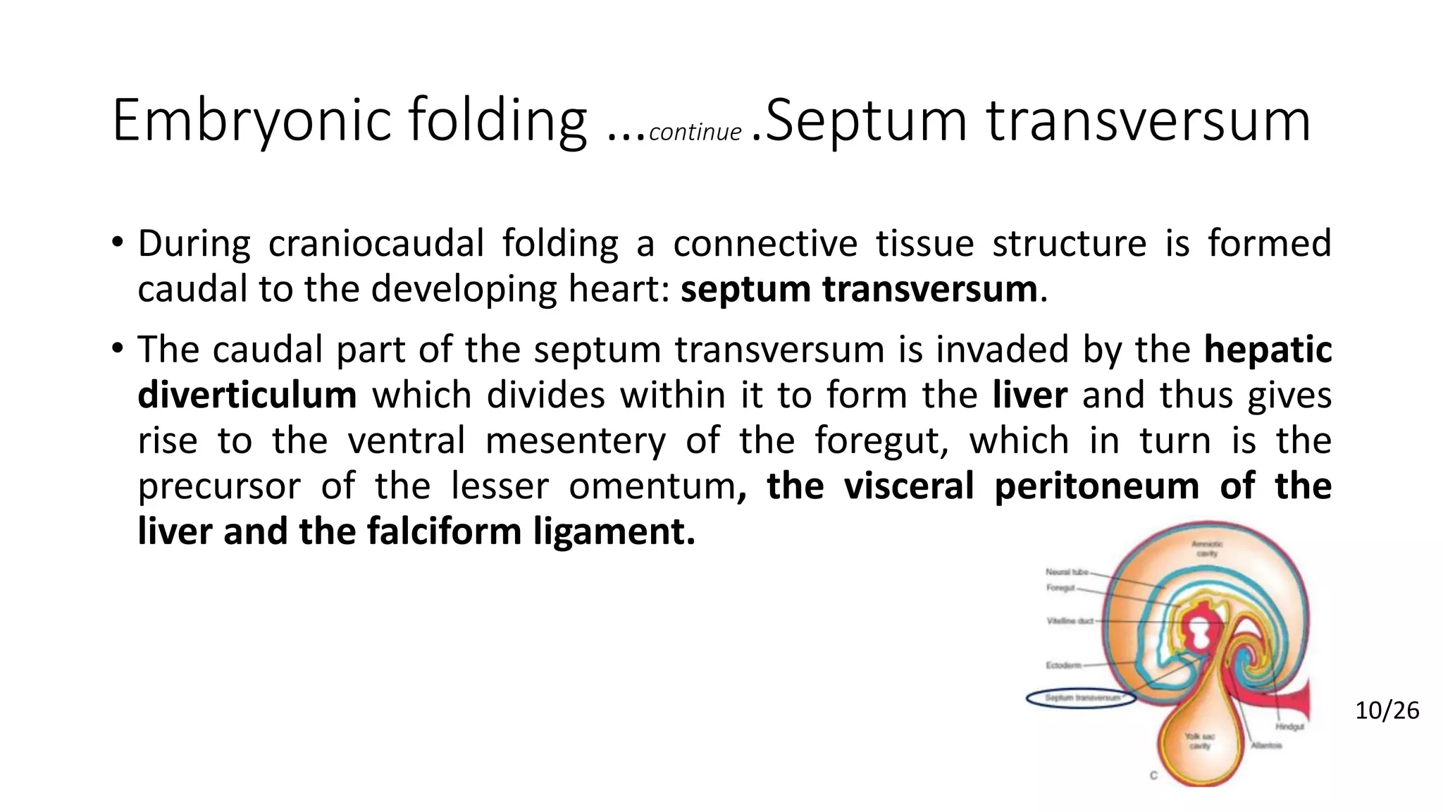
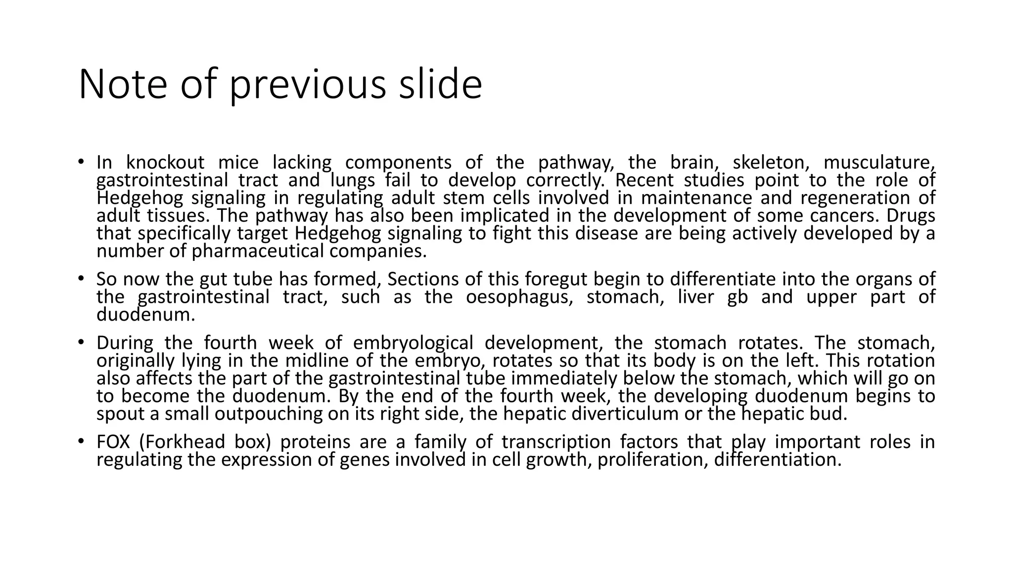
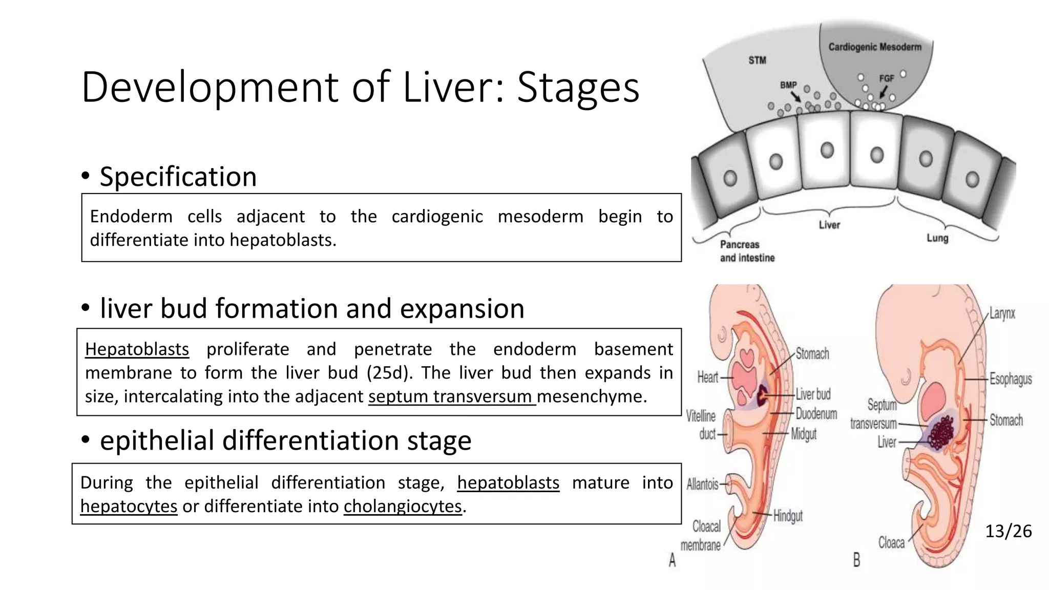
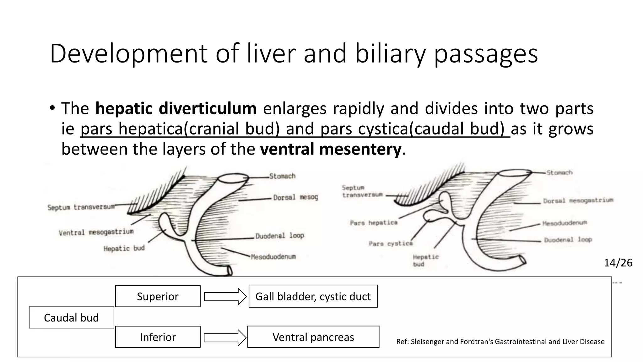
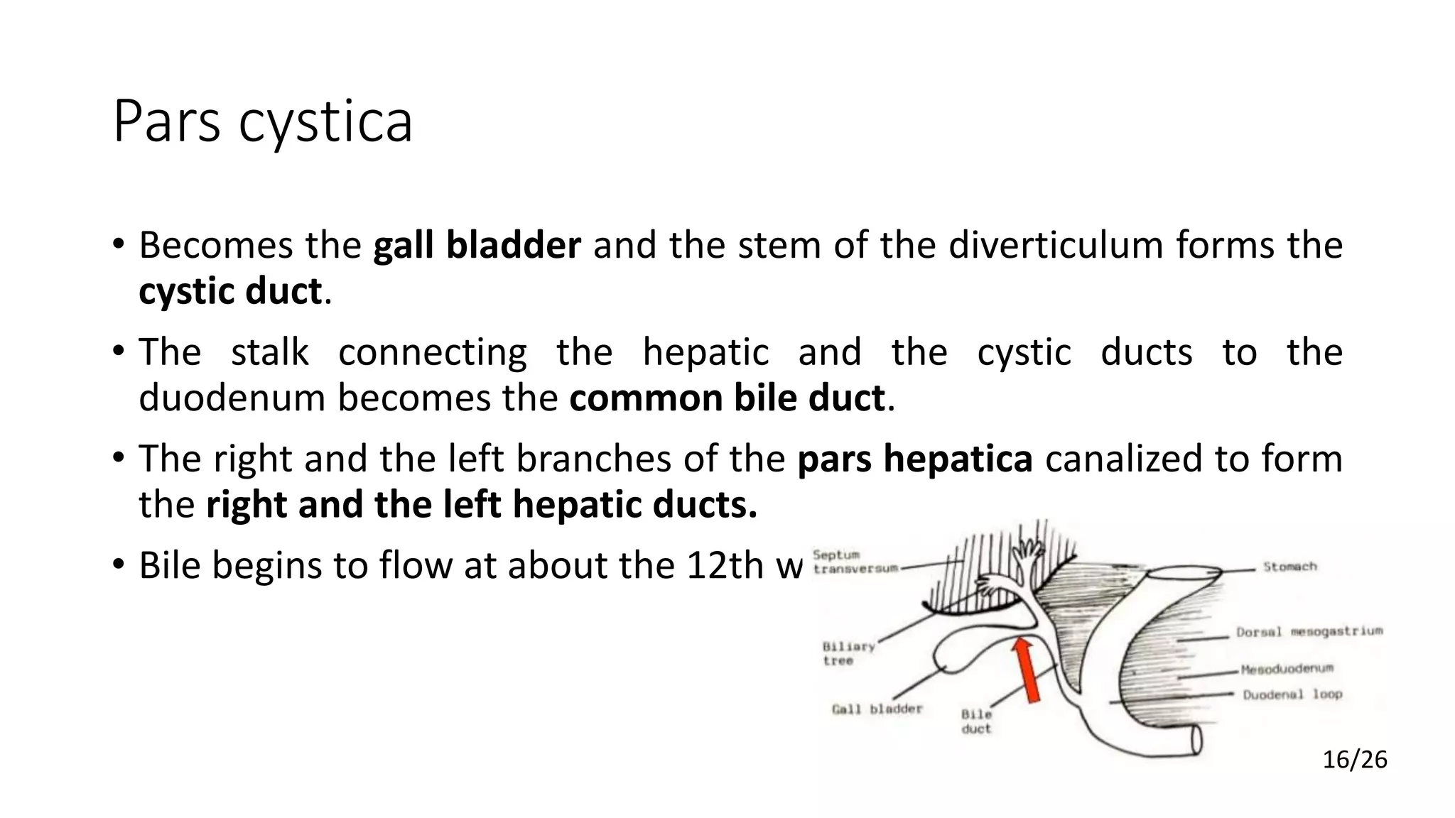
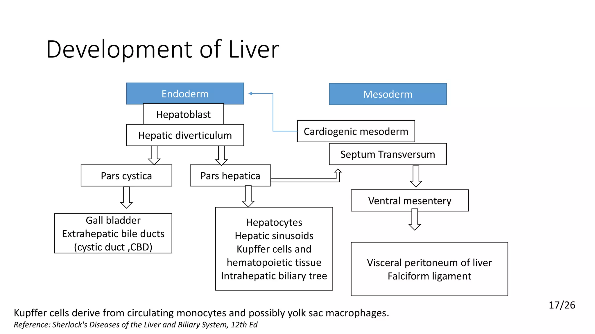
The liver develops from the endoderm of the foregut. During the 4th week, the hepatic diverticulum buds off from the foregut and divides into the pars hepatica and pars cystica. The pars hepatica gives rise to the liver parenchyma of hepatocytes and bile ducts. It expands between the layers of the septum transversum mesenchyme. The pars cystica develops into the gallbladder and cystic duct. By week 8, the basic structure of the liver and biliary tree is established.
In this presentation the development of Small intestine and Pancreas has been discussed. The viewer would be able to understand the concept of physiological herniation and rotation of the Primary intestinal loop with in the connecting stalk.
Week Wise Pregnancy and Fetus DevelopmentVirtue Baby
What My BABY Looks Like in WOMB !!! Follow your baby's development week by week. Find out the changes you can expect to your body, your emotions and your life as your pregnancy progresses.
Growth & development of face/certified fixed orthodontic courses by India...Indian dental academy
The Indian Dental Academy is the Leader in continuing dental education , training dentists in all aspects of dentistry and
offering a wide range of dental certified courses in different formats.for more details please visit
www.indiandentalacademy.com
The skin is divided into two parts: the superficial part, the
epidermis; and the deep part, the dermis (Fig. 1.4). The
epidermis is a stratified epithelium whose cells become flat
tened as they mature and rise to the surface. On the palms of
the hands and the soles of the feet, the epidermis is extremely
thick, to withstand the wear and tear that occurs in these
regions. In other areas of the body, for example, on the ante
rior surface of the arm and forearm, it is thin. The dermis is
composed of dense connective tissue containing many blood
vessels, lymphatic vessels, and nerves. It shows considerable
variation in thickness in different parts of the body, tending
to be thinner on the anterior than on the posterior surface.
It is thinner in women than in men. The dermis of the skin
is connected to the underlying deep fascia or bones by the
superficial fascia, otherwise known as subcutaneous tissue.
The skin over joints always folds in the same place, the
SKIN CREASES (Fig. 1.5). At these sites, the skin is thinner
than elsewhere and is firmly tethered to underlying struc
tures by strong bands of fibrous tissue.
The appendages of the skin are the nails, hair follicles,
sebaceous glands, and sweat glands.
The nails are keratinized plates on the dorsal surfaces of
the tips of the fingers and toes. The proximal edge of the
plate is the root of the nail (see Fig. 1.5). With the exception
of the distal edge of the plate, the nail is surrounded and
overlapped by folds of skin known as nail folds. The sur
face of skin covered by the nail is the nail bed (see Fig. 1.5).
Hairs grow out of follicles, which are invaginations
of the epidermis into the dermis (see Fig. 1.4). The folli
cles lie obliquely to the skin surface, and their expanded
extremities, called hair bulbs, penetrate to the deeper part
of the dermis. Each hair bulb is concave at its end, and
Functionally the urogenital system can be divided into two entirely different components:
The urinary system
The genital system.
Embryologically and anatomically they are intimately interwoven.
Both develop from a common mesodermal ridge (intermediate mesoderm) along the posterior wall of the abdominal cavity,
Initially the excretory ducts of both systems enter a common cavity, the cloaca.
Three slightly overlapping kidney systems are formed in a cranial to caudal sequence during intrauterine life in humans:
The pronephros, (rudimentary and nonfunctional).
The mesonephros, (function for a short time during the early fetal period).
The metanephros, (forms the permanent kidney)
This is a slide presentation for MBBS students. a brief overview of hemochromatosis, an iron overload condition. overview of hemochromatosis, pathophysiology, clinical features, approach, and management
Liver transplantation; notes of DM/DNB/SpecialistsPratap Tiwari
Liver transplantation; extensive notes of DM/DNB/Specialists. This was my notes for my exam compiled from several sources, credit goes to original authors. This is just for quick revision
This is a lecture note for 5th-semester MBBS students. Lecture notes on hepatology, liver disease, and liver abscess. Introduction to a liver abscess, pyogenic liver abscess, causes, approach and management of liver abscess.
This is a lecture note for 5th semester MBBS students. Lecture notes on hepatology, liver disease, alcoholic liver disease, alcohol-related liver disease, portal hypertension, hepatic encephalopathy, and acute liver failure. Introduction to acute liver failure, causes, approach, and management of acute liver failure .
This is a lecture note for 5th semester MBBS students. Lecture notes on hepatology, liver disease, alcoholic liver disease, alcohol-related liver disease, portal hypertension, and hepatic encephalopathy. Introduction to hepatic encephalopathy, causes, differentials, approach, and management of hepatic encephalopathy .
This is a lecture note for 5th semester MBBS students. Lecture notes on hepatology, liver disease, alcoholic liver disease, alcohol-related liver disease, alcoholic hepatitis, portal hypertension, ascites. Introduction to ascites and management of ascites.
This is a lecture note for 5th semester MBBS students. Lecture notes on hepatology, liver disease, alcoholic liver disease, alcohol-related liver disease, portal hypertension, ascites. Introduction to ascites and management of ascites.
brief lecture notes for 5th sem MBBS, on portal hypertension and varices. Introduction to portal hypertension and esophageal and gastric varices and management of variceal bleeding.
Chronic liver disease, lecture presentation for 5th sem MBBS students. Introduction to chronic liver disease, notes on liver fibrosis, alcoholic hepatitis, liver histology and overview.
HOT NEW PRODUCT! BIG SALES FAST SHIPPING NOW FROM CHINA!! EU KU DB BK substit...GL Anaacs
Contact us if you are interested:
Email / Skype : kefaya1771@gmail.com
Threema: PXHY5PDH
New BATCH Ku !!! MUCH IN DEMAND FAST SALE EVERY BATCH HAPPY GOOD EFFECT BIG BATCH !
Contact me on Threema or skype to start big business!!
Hot-sale products:
NEW HOT EUTYLONE WHITE CRYSTAL!!
5cl-adba precursor (semi finished )
5cl-adba raw materials
ADBB precursor (semi finished )
ADBB raw materials
APVP powder
5fadb/4f-adb
Jwh018 / Jwh210
Eutylone crystal
Protonitazene (hydrochloride) CAS: 119276-01-6
Flubrotizolam CAS: 57801-95-3
Metonitazene CAS: 14680-51-4
Payment terms: Western Union,MoneyGram,Bitcoin or USDT.
Deliver Time: Usually 7-15days
Shipping method: FedEx, TNT, DHL,UPS etc.Our deliveries are 100% safe, fast, reliable and discreet.
Samples will be sent for your evaluation!If you are interested in, please contact me, let's talk details.
We specializes in exporting high quality Research chemical, medical intermediate, Pharmaceutical chemicals and so on. Products are exported to USA, Canada, France, Korea, Japan,Russia, Southeast Asia and other countries.
Anti ulcer drugs and their Advance pharmacology ||
Anti-ulcer drugs are medications used to prevent and treat ulcers in the stomach and upper part of the small intestine (duodenal ulcers). These ulcers are often caused by an imbalance between stomach acid and the mucosal lining, which protects the stomach lining.
||Scope: Overview of various classes of anti-ulcer drugs, their mechanisms of action, indications, side effects, and clinical considerations.
Prix Galien International 2024 Forum ProgramLevi Shapiro
June 20, 2024, Prix Galien International and Jerusalem Ethics Forum in ROME. Detailed agenda including panels:
- ADVANCES IN CARDIOLOGY: A NEW PARADIGM IS COMING
- WOMEN’S HEALTH: FERTILITY PRESERVATION
- WHAT’S NEW IN THE TREATMENT OF INFECTIOUS,
ONCOLOGICAL AND INFLAMMATORY SKIN DISEASES?
- ARTIFICIAL INTELLIGENCE AND ETHICS
- GENE THERAPY
- BEYOND BORDERS: GLOBAL INITIATIVES FOR DEMOCRATIZING LIFE SCIENCE TECHNOLOGIES AND PROMOTING ACCESS TO HEALTHCARE
- ETHICAL CHALLENGES IN LIFE SCIENCES
- Prix Galien International Awards Ceremony
Title: Sense of Taste
Presenter: Dr. Faiza, Assistant Professor of Physiology
Qualifications:
MBBS (Best Graduate, AIMC Lahore)
FCPS Physiology
ICMT, CHPE, DHPE (STMU)
MPH (GC University, Faisalabad)
MBA (Virtual University of Pakistan)
Learning Objectives:
Describe the structure and function of taste buds.
Describe the relationship between the taste threshold and taste index of common substances.
Explain the chemical basis and signal transduction of taste perception for each type of primary taste sensation.
Recognize different abnormalities of taste perception and their causes.
Key Topics:
Significance of Taste Sensation:
Differentiation between pleasant and harmful food
Influence on behavior
Selection of food based on metabolic needs
Receptors of Taste:
Taste buds on the tongue
Influence of sense of smell, texture of food, and pain stimulation (e.g., by pepper)
Primary and Secondary Taste Sensations:
Primary taste sensations: Sweet, Sour, Salty, Bitter, Umami
Chemical basis and signal transduction mechanisms for each taste
Taste Threshold and Index:
Taste threshold values for Sweet (sucrose), Salty (NaCl), Sour (HCl), and Bitter (Quinine)
Taste index relationship: Inversely proportional to taste threshold
Taste Blindness:
Inability to taste certain substances, particularly thiourea compounds
Example: Phenylthiocarbamide
Structure and Function of Taste Buds:
Composition: Epithelial cells, Sustentacular/Supporting cells, Taste cells, Basal cells
Features: Taste pores, Taste hairs/microvilli, and Taste nerve fibers
Location of Taste Buds:
Found in papillae of the tongue (Fungiform, Circumvallate, Foliate)
Also present on the palate, tonsillar pillars, epiglottis, and proximal esophagus
Mechanism of Taste Stimulation:
Interaction of taste substances with receptors on microvilli
Signal transduction pathways for Umami, Sweet, Bitter, Sour, and Salty tastes
Taste Sensitivity and Adaptation:
Decrease in sensitivity with age
Rapid adaptation of taste sensation
Role of Saliva in Taste:
Dissolution of tastants to reach receptors
Washing away the stimulus
Taste Preferences and Aversions:
Mechanisms behind taste preference and aversion
Influence of receptors and neural pathways
Impact of Sensory Nerve Damage:
Degeneration of taste buds if the sensory nerve fiber is cut
Abnormalities of Taste Detection:
Conditions: Ageusia, Hypogeusia, Dysgeusia (parageusia)
Causes: Nerve damage, neurological disorders, infections, poor oral hygiene, adverse drug effects, deficiencies, aging, tobacco use, altered neurotransmitter levels
Neurotransmitters and Taste Threshold:
Effects of serotonin (5-HT) and norepinephrine (NE) on taste sensitivity
Supertasters:
25% of the population with heightened sensitivity to taste, especially bitterness
Increased number of fungiform papillae
MANAGEMENT OF ATRIOVENTRICULAR CONDUCTION BLOCK.pdfJim Jacob Roy
Cardiac conduction defects can occur due to various causes.
Atrioventricular conduction blocks ( AV blocks ) are classified into 3 types.
This document describes the acute management of AV block.
Title: Sense of Smell
Presenter: Dr. Faiza, Assistant Professor of Physiology
Qualifications:
MBBS (Best Graduate, AIMC Lahore)
FCPS Physiology
ICMT, CHPE, DHPE (STMU)
MPH (GC University, Faisalabad)
MBA (Virtual University of Pakistan)
Learning Objectives:
Describe the primary categories of smells and the concept of odor blindness.
Explain the structure and location of the olfactory membrane and mucosa, including the types and roles of cells involved in olfaction.
Describe the pathway and mechanisms of olfactory signal transmission from the olfactory receptors to the brain.
Illustrate the biochemical cascade triggered by odorant binding to olfactory receptors, including the role of G-proteins and second messengers in generating an action potential.
Identify different types of olfactory disorders such as anosmia, hyposmia, hyperosmia, and dysosmia, including their potential causes.
Key Topics:
Olfactory Genes:
3% of the human genome accounts for olfactory genes.
400 genes for odorant receptors.
Olfactory Membrane:
Located in the superior part of the nasal cavity.
Medially: Folds downward along the superior septum.
Laterally: Folds over the superior turbinate and upper surface of the middle turbinate.
Total surface area: 5-10 square centimeters.
Olfactory Mucosa:
Olfactory Cells: Bipolar nerve cells derived from the CNS (100 million), with 4-25 olfactory cilia per cell.
Sustentacular Cells: Produce mucus and maintain ionic and molecular environment.
Basal Cells: Replace worn-out olfactory cells with an average lifespan of 1-2 months.
Bowman’s Gland: Secretes mucus.
Stimulation of Olfactory Cells:
Odorant dissolves in mucus and attaches to receptors on olfactory cilia.
Involves a cascade effect through G-proteins and second messengers, leading to depolarization and action potential generation in the olfactory nerve.
Quality of a Good Odorant:
Small (3-20 Carbon atoms), volatile, water-soluble, and lipid-soluble.
Facilitated by odorant-binding proteins in mucus.
Membrane Potential and Action Potential:
Resting membrane potential: -55mV.
Action potential frequency in the olfactory nerve increases with odorant strength.
Adaptation Towards the Sense of Smell:
Rapid adaptation within the first second, with further slow adaptation.
Psychological adaptation greater than receptor adaptation, involving feedback inhibition from the central nervous system.
Primary Sensations of Smell:
Camphoraceous, Musky, Floral, Pepperminty, Ethereal, Pungent, Putrid.
Odor Detection Threshold:
Examples: Hydrogen sulfide (0.0005 ppm), Methyl-mercaptan (0.002 ppm).
Some toxic substances are odorless at lethal concentrations.
Characteristics of Smell:
Odor blindness for single substances due to lack of appropriate receptor protein.
Behavioral and emotional influences of smell.
Transmission of Olfactory Signals:
From olfactory cells to glomeruli in the olfactory bulb, involving lateral inhibition.
Primitive, less old, and new olfactory systems with different path
Ozempic: Preoperative Management of Patients on GLP-1 Receptor Agonists Saeid Safari
Preoperative Management of Patients on GLP-1 Receptor Agonists like Ozempic and Semiglutide
ASA GUIDELINE
NYSORA Guideline
2 Case Reports of Gastric Ultrasound
Flu Vaccine Alert in Bangalore Karnatakaaddon Scans
As flu season approaches, health officials in Bangalore, Karnataka, are urging residents to get their flu vaccinations. The seasonal flu, while common, can lead to severe health complications, particularly for vulnerable populations such as young children, the elderly, and those with underlying health conditions.
Dr. Vidisha Kumari, a leading epidemiologist in Bangalore, emphasizes the importance of getting vaccinated. "The flu vaccine is our best defense against the influenza virus. It not only protects individuals but also helps prevent the spread of the virus in our communities," he says.
This year, the flu season is expected to coincide with a potential increase in other respiratory illnesses. The Karnataka Health Department has launched an awareness campaign highlighting the significance of flu vaccinations. They have set up multiple vaccination centers across Bangalore, making it convenient for residents to receive their shots.
To encourage widespread vaccination, the government is also collaborating with local schools, workplaces, and community centers to facilitate vaccination drives. Special attention is being given to ensuring that the vaccine is accessible to all, including marginalized communities who may have limited access to healthcare.
Residents are reminded that the flu vaccine is safe and effective. Common side effects are mild and may include soreness at the injection site, mild fever, or muscle aches. These side effects are generally short-lived and far less severe than the flu itself.
Healthcare providers are also stressing the importance of continuing COVID-19 precautions. Wearing masks, practicing good hand hygiene, and maintaining social distancing are still crucial, especially in crowded places.
Protect yourself and your loved ones by getting vaccinated. Together, we can help keep Bangalore healthy and safe this flu season. For more information on vaccination centers and schedules, residents can visit the Karnataka Health Department’s official website or follow their social media pages.
Stay informed, stay safe, and get your flu shot today!
Lung Cancer: Artificial Intelligence, Synergetics, Complex System Analysis, S...Oleg Kshivets
RESULTS: Overall life span (LS) was 2252.1±1742.5 days and cumulative 5-year survival (5YS) reached 73.2%, 10 years – 64.8%, 20 years – 42.5%. 513 LCP lived more than 5 years (LS=3124.6±1525.6 days), 148 LCP – more than 10 years (LS=5054.4±1504.1 days).199 LCP died because of LC (LS=562.7±374.5 days). 5YS of LCP after bi/lobectomies was significantly superior in comparison with LCP after pneumonectomies (78.1% vs.63.7%, P=0.00001 by log-rank test). AT significantly improved 5YS (66.3% vs. 34.8%) (P=0.00000 by log-rank test) only for LCP with N1-2. Cox modeling displayed that 5YS of LCP significantly depended on: phase transition (PT) early-invasive LC in terms of synergetics, PT N0—N12, cell ratio factors (ratio between cancer cells- CC and blood cells subpopulations), G1-3, histology, glucose, AT, blood cell circuit, prothrombin index, heparin tolerance, recalcification time (P=0.000-0.038). Neural networks, genetic algorithm selection and bootstrap simulation revealed relationships between 5YS and PT early-invasive LC (rank=1), PT N0—N12 (rank=2), thrombocytes/CC (3), erythrocytes/CC (4), eosinophils/CC (5), healthy cells/CC (6), lymphocytes/CC (7), segmented neutrophils/CC (8), stick neutrophils/CC (9), monocytes/CC (10); leucocytes/CC (11). Correct prediction of 5YS was 100% by neural networks computing (area under ROC curve=1.0; error=0.0).
CONCLUSIONS: 5YS of LCP after radical procedures significantly depended on: 1) PT early-invasive cancer; 2) PT N0--N12; 3) cell ratio factors; 4) blood cell circuit; 5) biochemical factors; 6) hemostasis system; 7) AT; 8) LC characteristics; 9) LC cell dynamics; 10) surgery type: lobectomy/pneumonectomy; 11) anthropometric data. Optimal diagnosis and treatment strategies for LC are: 1) screening and early detection of LC; 2) availability of experienced thoracic surgeons because of complexity of radical procedures; 3) aggressive en block surgery and adequate lymph node dissection for completeness; 4) precise prediction; 5) adjuvant chemoimmunoradiotherapy for LCP with unfavorable prognosis.
- Video recording of this lecture in English language: https://youtu.be/lK81BzxMqdo
- Video recording of this lecture in Arabic language: https://youtu.be/Ve4P0COk9OI
- Link to download the book free: https://nephrotube.blogspot.com/p/nephrotube-nephrology-books.html
- Link to NephroTube website: www.NephroTube.com
- Link to NephroTube social media accounts: https://nephrotube.blogspot.com/p/join-nephrotube-on-social-media.html
Report Back from SGO 2024: What’s the Latest in Cervical Cancer?bkling
Are you curious about what’s new in cervical cancer research or unsure what the findings mean? Join Dr. Emily Ko, a gynecologic oncologist at Penn Medicine, to learn about the latest updates from the Society of Gynecologic Oncology (SGO) 2024 Annual Meeting on Women’s Cancer. Dr. Ko will discuss what the research presented at the conference means for you and answer your questions about the new developments.
Report Back from SGO 2024: What’s the Latest in Cervical Cancer?
Embryology of liver
1. Embryology of the Liver
Pratap Sagar Tiwari
DM resident, Department of Hepatology, NAMS
2. Introduction: Liver
• The liver is largest internal organ & the largest gland in the
human body .
• It lies under the diaphragm in the right upper abdomen and
midabdomen and extends to the left upper abdomen. The
liver has the general shape of a wedge, with its base to the
right and its apex to the left .1
• The normal liver extends from the 5th ICS in the Rt MCL
down to the costal margin.
• It measures 12–15 cm coronally and 15–20 cm transversely.1
• The median liver weight is 1,800 g in m and 1,400 g in F.1
1. Wanless I R. Physioanatomic Considerations. In: Schiff ER, Maddrey WC, Sorrell MF, editors. Diseases of the liver. 11th edition. John Wiley & Sons Ltd;2012.
Pic source: http://www.medicinenet.com/drug_induced_liver_disease/article.htm
1/26
4. Notes of the previous slide
• This slide shows the quick review of the embryogenesis. The embryonic period in humans begins at
fertilization and continues until the end of the 10th week of gestation (8th week by embryonic age).
• It starts out as formation of a single cell zygote and the process of cleavage begins, the cell divides several
times to form a ball of cells called a morula covered by zona pellucida. The cells start to bind firmly together
in the process of caompaction and then their cleavage continue as cellular differentiation.
• Further cellular division is accompanied by the formation of a small cavity between the cells. This stage is
called a blastocyst. Cells differentiate in outer layer collectively called the trophoblast and the inner cell
mass.
• The iner cell mass with later give rise to embryo proper, the amnion, yolk sac and allantois and the outer
trophoblast layer will form the fetal parts of the placenta.
• The blastocyst reaches the uterus at roughly the 5th day after fertilization. It is here that lysis of the zona
pellucida occurs,This allows the trophectoderm cells of the blastocyst to come into contact with, and adhere
to, the endometrial cells of the uterus initiaing the implantation process on day 7.
• The inner cell mass or the embryoblast is the source of embryonic stem cells which are pluripotent cells and
can develop into any of the germ layers.
5. Formation of trilamminar disk
Primitive streak
Bilaminar disk
Hypoblast
Epiblast
Ectoderm
Endoderm
mesoderm
Third wk: Formation of Primitive streak at midline causing
the disk to have right and left halves.
Movement of some cells from the epiblast
towards the hypoblast forms the mesoderm
layer
The embryoblast forms an embryonic disc which is bilaminar disc with upper layer the epiblast ie the primitive ectderm
and the inner layer which is the hypoblast :the primitive endoderm.
3/26
8. Extra note
Retroperitoneal SADPUCKER
S = Suprarenal (adrenal) glands
A = Aorta/Inferior Vena Cava
D = Duodenum (second and third segments)
P = Pancreas
U = Ureters/
C = Colon (ascending and descending only)
K = Kidneys
E = Esophagus
R = Rectum
Intraperitoneal
S = Stomach
A = Appendix
L = Liver
T = Transverse colon
D = duodenum (only the 1st part, though)
S = Small intestines
P = Pancreas (only the tail though)
R = Rectum (only the upper 3rd)
S = Sigmoid colon
S = Spleen
9. Embryonic folding
During the 4th week of development a period of
rapid growth begins in the embryo, the embryo
begins to change shape from flat trilamminar
disc into a cylinder ,a process known as
embryonic folding. 6/26
Embryonic folding occurs in two planes the horizontal plane and the median plane or the cephalocaudal folding .
Folding of the embryo in horizontal plane results in development of two lateral body folds .
Folding in medial results in development of cranial and caudal fold. Folding in both planes takes place simultaneously resulting in rapid development of the
embryo.
10. Embryonic folding
The endoderm is mainly responsible for development of GI tract. As the craniocaudal and the lateral folding continues the
endoderm moves towards the midline and fuses incorporating the dorsal part of yolk sac to create the primitive gut tube.
The primitive gut tube differentiates into three main parts the foregut, mid gut and the hind gut.
7/26
11. Embryonic folding.. • The foregut can be seen at the cranial end of the
embryo.
• It is temporarily closed by the orophayngeal
membrane which at the end of the 4th week of
development ruptures to form the mouth.
• The midgut lies between the forgut and the hind
gut and remains connected to the yolk sac until
the fifth week of development. As embryonic
folding continues the connection to the yolk sac
narrows into a stalk called vitelline duct.
• The hind gut lies at the caudal end of the embryo
,it is temporarily closed by the cloacal membrane
which during the 7th wk of development ruptures
to form urogenital and anal opening.
• As a result of embryonic folding ,the major body
plan is established and the three germ layers
continues to differentiate giving rise to their
specific tissues and their organ systems.
Pic source: Copyright 2009, John Wiley & Sons, Inc. Embryonic folding
8/26
12. Note:Vitelline duct
• In the human embryo, the vitelline duct, also known as the
omphalomesenteric duct, is a long narrow tube that joins the yolk sac
to the midgut lumen of the developing fetus.[.Generally, the duct fully
obliterates (narrows and disappears) during the 5–6th week of
fertilization age (9th week of gestational age), but a failure of the duct
to close is termed a vitelline fistula. This results in discharge of
meconium from the umbilicus.
• A Meckel's diverticulum, a true congenital diverticulum, a vestigial
remnanat of the omphalomesenteric duct (also called the vitelline
duct or yolk stalk). It is the most common malformation of the
gastrointestinal tract and is present in approximately 2% of the
population.
13. Lateral Plate mesoderm
• Lateral plate mesoderm is a type of mesoderm that is found at the
periphery.
• It will split into 2 layers, the somatic layer/mesoderm and the splanchnic
layer/mesoderm
1. The somatopleuric layer associates with ectoderm.
contributes to connective tissue of body wall & limbs.
1. The splanchnopleuric layer associates with endoderm,
circulatory system ie heart, blood vessels
• Spaces within the lateral plate are enclosed and forms the intraembryonic
coelom.
In the 4th week the coelom divides into pericardial, pleural and peritoneal
cavities.
9/26
14. Embryonic folding …continue .Septum transversum
• During craniocaudal folding a connective tissue structure is formed
caudal to the developing heart: septum transversum.
• The caudal part of the septum transversum is invaded by the hepatic
diverticulum which divides within it to form the liver and thus gives
rise to the ventral mesentery of the foregut, which in turn is the
precursor of the lesser omentum, the visceral peritoneum of the
liver and the falciform ligament.
10/26
15. Derivatives of gut tube
• 11/26 Differentiation of the gut and its derivatives
depends upon reciprocal interactions
between the gut endoderm and its
surrounding mesoderm.
Hox genes in the mesoderm are induced by a
Hedgehog signaling pathway secreted by gut
endoderm and regulate the craniocaudal
organization of the gut and its derivatives.
The endoderm cells that will form the liver
express Foxa proteins, identifying it as “future
liver” and labelling foregut as different from
midgut and hindgut .
11/26
16. Note of previous slide
So now the gut tube has formed, Sections of this gut begin to differentiate into the organs of the
gastrointestinal tract.
• Hox genes which are a group of related genes that control the body plan of an embryo along the
cranio-caudal axis. After the embryonic segments have formed, the Hox proteins determine the
type of structures that will form on a given segment. Hox proteins thus confer segmental identity,
but do not form the actual segments themselves.and which are further influenced or induced bu
• The Hedgehog signaling pathway which is a signaling pathway that transmits information to
embryonic cells required for proper cell differentiation. Different parts of the embryo have
different concentrations of hedgehog signaling proteins.
• The pathway also has roles in the adult. Diseases associated with the malfunction of this pathway
include basal cell carcinoma.
• The Hedgehog (Hh) signaling pathway has numerous roles in the control of cell proliferation,
tissue patterning, stem cell maintenance and development. Mammals have three Hedgehog
homologues, Desert (DHH), Indian (IHH), and Sonic (SHH), of which Sonic is the best studied.
17. Note of previous slide
• In knockout mice lacking components of the pathway, the brain, skeleton, musculature,
gastrointestinal tract and lungs fail to develop correctly. Recent studies point to the role of
Hedgehog signaling in regulating adult stem cells involved in maintenance and regeneration of
adult tissues. The pathway has also been implicated in the development of some cancers. Drugs
that specifically target Hedgehog signaling to fight this disease are being actively developed by a
number of pharmaceutical companies.
• So now the gut tube has formed, Sections of this foregut begin to differentiate into the organs of
the gastrointestinal tract, such as the oesophagus, stomach, liver gb and upper part of
duodenum.
• During the fourth week of embryological development, the stomach rotates. The stomach,
originally lying in the midline of the embryo, rotates so that its body is on the left. This rotation
also affects the part of the gastrointestinal tube immediately below the stomach, which will go on
to become the duodenum. By the end of the fourth week, the developing duodenum begins to
spout a small outpouching on its right side, the hepatic diverticulum or the hepatic bud.
• FOX (Forkhead box) proteins are a family of transcription factors that play important roles in
regulating the expression of genes involved in cell growth, proliferation, differentiation.
18. Development of Liver
Liver development requires two linked
processes:
1. Differentiation of the various hepatic cell
types from their embryonic progenitors.
2. The arrangement of those cells into
structures that permit the distinctive
circulatory, metabolic and excretory
functions of the liver.
Mediated by many essential regulators
which include signaling molecules, and
transcription factors.
12/26
19. Notes of the previous slide
• So summarizing the events until now…
• Following gastrulation, embryos are composed of three germ layers: the ectoderm, mesoderm,
and endoderm.
• The initial liver bud is mainly composed of hepatoblasts. Hepatoblasts are a liver stem cell that
can later be turned into hepatocytes or cholangiocytes, cells which line bile ducts.
• The major epithelial cells of the liver – hepatocytes and cholangiocytes – are derived from the
endoderm. However, these cells represent only about two-thirds of the liver volume. The
remaining one-third consists of a variety of cells derived primarily from the mesoderm, including
vascular cell types: Kupffer cells, stellate cells, fibroblasts, and leukocytes. Therefore, liver
development requires the coordinated integration of the cells from distinct embryonic layers.
• Liver development requires two linked processes: differentiation of the various hepatic cell types
from their embryonic progenitors and the arrangement of those cells into structures that permit
the distinctive circulatory, metabolic, and excretory functions of the liver and these are further
controlled or mediated by many essential regulators which include several signaling molecules
and transcription factors.
20. Development of Liver: Stages
• Specification
• liver bud formation and expansion
• epithelial differentiation stage
Endoderm cells adjacent to the cardiogenic mesoderm begin to
differentiate into hepatoblasts.
Hepatoblasts proliferate and penetrate the endoderm basement
membrane to form the liver bud (25d). The liver bud then expands in
size, intercalating into the adjacent septum transversum mesenchyme.
During the epithelial differentiation stage, hepatoblasts mature into
hepatocytes or differentiate into cholangiocytes.
13/26
21. Notes to the previous slide
• The first stage of liver development is specification, during which endoderm cells after
receiving inductive signals from the adjacent cardiogenic mesoderm and the septum
transversum mesenchyme (STM) via BMP, bone morphogenetic protein &FGF, fibroblast
growth factor begin to differentiate into hepatoblasts, These factors are important in
development. For example, in the absence of FGF signaling from the cardiac mesoderm,
the ventral endoderm develops into pancreas, but too high a concentration of FGF
results in differentiation toward lung.
• This is followed by liver bud formation and expansion: the hepatoblasts proliferate and
penetrate the endoderm basement membrane (of the most caudal portion of foregut) to
form the liver bud or also called hepatic diverticulum . In humans, this occurs at
approximately day 25 . Initially, the liver bud is separated from the mesenchyme of the
septum transversum by basement membrane. Shortly, however, the basement
membrane surrounding the liver bud is lost,and cells delaminate from the bud and
invade the septum transversum as cords of hepatoblasts—bipotential cells that
differentiate into hepatocytes and cholangiocytes.
22. Development of liver and biliary passages
• The hepatic diverticulum enlarges rapidly and divides into two parts
ie pars hepatica(cranial bud) and pars cystica(caudal bud) as it grows
between the layers of the ventral mesentery.
Caudal bud
Inferior
Superior Gall bladder, cystic duct
Ventral pancreas Ref: Sleisenger and Fordtran's Gastrointestinal and Liver Disease
14/26
23. Pars hepatica
• It is the larger cranial part of the hepatic diverticulum.
• Gives rise to:
1. Hepatocytes
2. Hepatic sinusoids
3. Kupffer cells and hematopoietic tissue
4. Intrahepatic bile ducts
• The liver grows rapidly to fill a large part of the abdominal cavity.
• At first, the 2 lobes are of the same size but soon the right become
larger.
15/26
24. Pars cystica
• Becomes the gall bladder and the stem of the diverticulum forms the
cystic duct.
• The stalk connecting the hepatic and the cystic ducts to the
duodenum becomes the common bile duct.
• The right and the left branches of the pars hepatica canalized to form
the right and the left hepatic ducts.
• Bile begins to flow at about the 12th week.
16/26
25. Development of Liver
Pars cystica
Endoderm
Pars hepatica
Hepatocytes
Hepatic sinusoids
Kupffer cells and
hematopoietic tissue
Intrahepatic biliary tree
Gall bladder
Extrahepatic bile ducts
(cystic duct ,CBD)
Ventral mesentery
Mesoderm
Visceral peritoneum of liver
Falciform ligament
Septum Transversum
Cardiogenic mesoderm
Hepatoblast
Hepatic diverticulum
Kupffer cells derive from circulating monocytes and possibly yolk sac macrophages.
Reference: Sherlock's Diseases of the Liver and Biliary System, 12th Ed
17/26
26. Formation of the capsule & ligaments of the
liver
• As the septum transversum is penetrated by
the growing pars hepatica:
• The mesoderm of the septum transversum
between the liver and the anterior
abdominal wall becomes the FALCIFORM
LIGAMENT.
• The mesoderm of the septum transversum
between the liver and the foregut (stomach
and duodenum) forms the LESSER
OMENTUM.
• The mesoderm on the surface of the liver
differentiates into CAPSULE AND
PERITONEAL COVERING.
18/26
27. Fate of ventral & dorsal mesentery
Derived from the septum transversum is the ventral
mesentery and the growth of the liver divides the ventral
mesentery into lesser omentum and falciform ligament.
19/26
28. Vascular Development related to Liver
Extraembryonic
Major Venous system
Intraembryonic
Vitelline veins Cardinal VeinsUmbilical Veins
Sinus venosus
20/26
29. Notes to the previous slide
• During early development, there are 3 major venous systems in the
embryo—2 extraembryonic and 1 intraembryonic.
• The extraembryonic venous systems are the omphalomesenteric
(vitelline) and umbilical (placental) veins, and the intraembryonic
system includes the cardinal veins that drain the venous blood of the
embryo to the heart.
• All of these systems converge into the sinus venosus, a cavity that is
incorporated into the heart.
30. Development of Vitelline & umbilical veins
A: 4th week B. 5th week. Note the plexus around the duodenum, formation of the hepatic
sinusoids, and initiation of left to right shunt between the vitelline veins. 21/26
Before entering the sinus venosus, the vitelline
veins form a plexus around the duodenum and
pass through the septum transversum.
The liver cords growing into the septum interrupt
the course of the veins and an extensive vascular
network, the hepatic sinusoids,forms. There is
also initiation of the left to right shunting
between the vitelline veins
31. Development of Vitelline & umbilical veins
A. 2nd mnth B.3rd mnth. Note formation of the ductus venosus,
portal vein , hepatic portion of IVC. The splenic & SMV enter the PV.
22/26
Blood from the left side of the liver is rechanneled
towards the right, resulting in an enlargement of the
right vitelline vein (right hepatocardiac channel).
The right hepatocardiac channel forms the
hepatocardiac portion of the inferior venacava.
The proximal part of the left vitelline vein disappears.
The anastomotic network around the duodenum
develops into a single vessel, the portal vein.
The superior mesenteric vein derives from the right
vitelline vein.
The distal portion of the left vitelline vein also
disappears.
The superior segment of vitelline veins becomes the
hepatic veins
32. Development of Vitelline & umbilical veins
A. 2nd mnth B.3rd mnth. Note formation of the ductus venosus,
portal vein , hepatic portion of IVC. The splenic & SMV enter the
PV. 22/26
The umbilical veins run from the placenta to the heart
and during fetal life are the predominant afferent vessels
that supply the liver.
Initially the umbilical veins pass on each side of the liver
but some connect to the hepatic sinusoids.
The proximal part of both umbilical veins and the
remainder of the right umbilical vein then disappear.
The left vein is the only one to carry blood from the
placenta to the liver.
With the increase of the placental circulation, a direct
communication forms between the left umbilical vein
and the right hepatocardiac channel, the ductus
venosus.
The ductus venosus bypasses the sinusoidal plexus of
the liver.
After birth the left umbilical vein and the ductus venosus
are obliterated and form the ligamentum teres hepatic
or the round ligament and ligamentum venosum
respectively.
33. Arterial supply of liver
• The arterial supply of the liver begins as an offshoot from the celiac
trunk at around the eighth week of gestation.
• By the 10th week, the first arterial radicles are visible in the central
portion of the liver, and by the fifteenth week, they reach the
periphery of the liver.
23/26
34. Congenital anomalies of liver
Riedel lobe is a tongue-like, inferior projection of
the right lobe of the liver beyond the level of the
most inferior costal cartilage . It is not considered a
true accessory lobe of the liver but an anatomical
variant of the right lobe of the liver.
• Congenital solitary nonparasitic cysts of the
liver
• Congenital Hepatic Fibrosis
• Congenital vascular malformation of the liver
• Intrahepatic Biliary Atresia
• Mesenchymal Hamartoma
• Accessory and Ectopic Lobes of the Liver 24/26
36. Ductul plate malformations
• Intrahepatic bile ducts (IHBDs) develop from bi-potential liver
progenitor cells(hepatoblasts) in contact with the mesenchyme of the
portal vein and thus form the “ductal plates.”
• The ductal plates are remodeled into mature tubular ducts. Lack of
remodeling results in “ductal plate malformation”.
• A proposal is that virtually all congenital diseases of IHBDs represent
examples of DPM.
• DPM are developmental anomalies considered to result from lack of
ductal plate remodeling during bile duct morphogenesis.
37. Classification of DPM
• Autosomal recessive polycystic kidney disease (hepatic ARPKD) (50% of
children, 70% of families): DPM of interlobular bile ducts associated with
tubular dilatation of collecting renal tubules
• Caroli disease : DPM of the larger IHBDs
• Caroli syndrome: Caroli disease + congenital hepatic fibrosis
• Von Meyenburg complexes: DPM of smaller interlobular ducts (liver cysts
in autosomal dominant polycystic kidney disease)
• Mesenchymal hamartoma
• Meckel syndrome
• Non-syndromal ductal plate malformation
38. End of slides…………..TO BE CONTINUED
References:
• Harrison & Gastroenterology & Hepatology
• Schiff's Diseases of the Liver, 11th Edition
• Sherlock's Diseases of the Liver and Biliary System, 12th Edition
• Sleisenger and Fordtran's Gastrointestinal and Liver Disease, Review and Assessment- Ninth Edition
• Suchy 2014
26/26